SlideShare a Scribd company logo
1 of 146
The Arabidopsis Book ©2002 American Society of Plant
Biologists
Pseudomonas syringae is a Gram-negative, rod-shaped
bacterium with polar flagella (Figure 1; Agrios, 1997).
Strains of P. syringae collectively infect a wide variety of
plants. Different strains of P. syringae, however, are
known for their diverse and host-specific interactions
with plants (Hirano and Upper, 2000). A specific strain
may be assigned to one of at least 40 pathovars based
on its host range among different plant species (Gardan
et al., 1999) and then further assigned to a race based on
differential interactions among cultivars of the host plant.
Understanding the molecular basis of this high level of
host specificity has been a driving force in using P.
syringae as a model for the study of host-pathogen
interactions. In crop fields, infected seeds are often an
important source of primary inoculum in P. syringae
diseases, and epiphytic bacterial growth on leaf surfaces
often precedes disease development (Hirano and Upper,
2000). P. syringae enters the host tissues (usually leaves)
through wounds or natural openings such as stomata,
and in a susceptible plant it multiplies to high population
levels in intercellular spaces. Infected leaves show
water-soaked patches, which eventually become
necrotic. Depending on P. syringae strains, necrotic
lesions may be surrounded by diffuse chlorosis. Some
strains of P. syringae also cause cankers and galls
(Agrios, 1997). In resistant plants, on the other hand, P.
syringae triggers the hypersensitive response (HR), a
rapid, defense-associated death of plant cells in contact
with the pathogen (Klement, 1963; Klement et al., 1964;
Bent, 1996; Greenberg, 1996; Dangl et al., 1996;
Hammond-Kosack and Jones, 1997). In this situation, P.
syringae fails to multiply to high population levels and
causes no disease symptoms.
In the late 1980s, several strains belonging to pathovars
tomato, maculicola, pisi, and atropurpurea of
Pseudomonas syringae were discovered to infect the
model plant Arabidopsis thaliana (reviewed by Crute et al.,
1994). The establishment of the Arabidopsis-P. syringae
pathosystem triggered a period of highly productive
research that has contributed to the elucidation of the
fascinating mechanisms underlying plant recognition of
pathogens, signal transduction pathways controlling plant
defense responses, host susceptibility, and pathogen
virulence and avirulence determinants. In this chapter we
trace the discovery of this pathosystem, overview the most
salient aspects of this interaction, and point out the gaps
in our knowledge. At the end of this chapter we will also
provide a glossary of relevant pathology-related technical
terms (Appendix I), a list of people who are studying this
interaction so readers can seek help if they have further
The Arabidopsis Thaliana-Pseudomonas Syringae
Interaction
Fumiaki Katagiria, Roger Thilmonyb, and Sheng Yang Heb
aPlant Health Department, Torrey Mesa Research Institute,
3115 Merryfield Row, San Diego, CA 92121, USA
bDepartment of Energy Plant Research Laboratory, Michigan
State University, East Lansing, MI 48824, USA.
Corresponding Author:
Sheng Yang He
206 Plant Biology Bldg.
Plant Research Laboratory
Michigan State University
East Lansing, MI 48824, USA
Tel: (517) 353-9181
Fax: (517) 353 –9168
E-mail: [email protected]
Introduction
The Arabidopsis Book ©2002 American Society of Plant
Biologists
First published on March 27, 2002; doi: 10.1199/tab.0039
The Arabidopsis Book 2 of 35
questions about the Arabidopsis-P. syringae interaction
(Appendix II), and several experimental procedures
commonly used in the study of the Arabidopsis-P. syringae
interaction (Appendix III).
1. EARLY DEVELOPMENT OF THE ARABIDOPSIS-
PSEUDOMONAS SYRINGAE SYSTEM
1.1. Beginning: Are there any Arabidopsis pathogens?
In the 1980s, P. syringae was the first pathogen to be
demonstrated to infect Arabidopsis and to cause disease
symptoms in the laboratory setting. This was achieved by
screening many P. syringae strains on various Arabidopsis
accessions (Dong et al., 1991; Whalen et al., 1991; Dangl
et al., 1992). The two virulent strains most widely used
today, P. syringae pv. tomato DC3000 and P. syringae pv.
maculicola ES4326, originated from these early studies.
When a suspension of 108 bacteria/ml (a high dose of
bacteria) is sprayed with a surfactant onto susceptible
Arabidopsis plants (e.g., ecotype Columbia), the first sign
of disease is the appearance of “water-soaked” patches
on leaves on day 2. The water-soaked symptom results
from massive release of water and, presumably, nutrients
from infected Arabidopsis cells. The water-soaked
patches become necrotic and dark-colored on day 3, and
the surrounding leaf tissue shows extensive chlorosis,
giving the characteristic appearance of a ‘speck’ disease
(Figure 2, Figure 3E and 3F). In addition, closely related,
but avirulent strains (such as P. syringae pv. tomato
JL1965 and P. syringae pv. maculicola M2) that became
sources of some avirulence (avr) genes were also identified
(Dong et al., 1991; Whalen et al., 1991; Dangl et al., 1992).
The main reason for examining P. syringae strains as
potential pathogens of Arabidopsis was because P.
syringae had already been proven to be an excellent
genetically tractable pathogen of soybean, tomato, and
bean in the mid-1980s (Keen, 1990). Development of the
Arabidopsis-P. syringae pathosystem would provide a
system in which both the plant and the pathogen are
amenable to rigorous genetic analysis. However, it is
interesting to note that even after the demonstration of
disease symptoms caused by P. syringae on Arabidopsis,
not many people were convinced that this was a good
model system for two reasons: there was no report of
naturally occurring P. syringae disease in Arabidopsis in
the wild, and in the laboratory inoculation required artificial
methods (infiltration or use of surfactant).
1.2. Establishing the system: gene-for-gene
Figure 1. A transmission electron microscope image of
Pseudomonas syringae pv. tomato DC3000. Note that
DC3000 produces polar flagella (15 nm in diameter) and a
few Hrp pili (8 nm in diameter). The flagella and Hrp pili are
indicated with arrows. Flagella enable bacteria to swim
toward or away from specific chemical stimuli. Hrp pili are
involved in type III secretion of avirulence and virulence pro-
teins.
Figure 2. Disease symptoms in Arabidopsis leaves caused
by DC3000 infection. Leaves (indicated with arrows) were
syringe-infiltrated with 5 x 10⁵ cfu/mL of Pst DC3000 and
pictures were taken four days after inoculation. The whole
plant is shown in (A). A close-up of a diseased leaf is shown
in (B).
The Arabidopsis Thaliana-Pseudomonas Syringae Interaction 3
of 35
interactions
A significant milestone in the development of the
Arabidopsis-P. syringae system was the demonstration
that this pathosystem conforms to the gene-for-gene
relationship that underlies many well-known plant-
pathogen interactions in nature (such as the flax-rust
fungus interaction or the soybean-P. syringae interaction)
(Keen, 1990). The gene-for-gene hypothesis was
advanced by H.H. Flor, based on his work on the flax-rust
fungus interaction in the 1940s and 1950s (Flor, 1971).
This hypothesis states that when a pathogen (in this case
a P. syringae strain) has an avirulence (avr) gene, and a
plant host (in this case the Arabidopsis plant) has the
corresponding disease resistance (R) gene, the plant is
resistant to the pathogen (Table 1). It is defined by a single
plant R gene for a single pathogen avr gene, hence the
name gene-for-gene resistance. Table 2 defines the
terminology involved in gene-for-gene resistance. When
the plant is resistant, the pathogen is said to be avirulent
and the interaction is said to be incompatible. When the
plant is susceptible, the pathogen is said to be virulent and
the interaction is said to be compatible. The laboratories
of Fred Ausubel and Brian Staskawicz showed that an avr
gene, avrRpt2, from an avirulent P. syringae pv. tomato
strain, JL1065, converted DC3000 and ES4326 into
avirulent strains in the Arabidopsis ecotype Columbia
(Dong et al., 1991; Whalen et al., 1991). Subsequent
Arabidopsis mutagenesis and screening efforts led to the
identification of mutations in the Arabidopsis RPS2
disease resistance gene (Kunkel et al., 1993; Yu et al.,
1993). Thus, a demonstration of the avrRpt2-RPS2 gene-
for-gene interaction was completed. Similar efforts in the
laboratory of Jeff Dangl led to the identification of the
avrRpm1 gene in P. syringae pv. maculicola strain M2 and
the RPM1 resistance gene in Arabidopsis ecotype
Columbia (Dangl et al., 1992). Interestingly, the RPM1
resistance gene also recognizes avrB, which was isolated
initially from P. syringae pv. glycinea in the soybean-P.
syringae interaction (Bisgrove et al., 1994). These
pioneering efforts spurred subsequent research to identify
additional P. syringae avr genes and the corresponding
Arabidopsis resistance gene loci, the eventual cloning of
the first Arabidopsis resistance gene, RPS2 (Bent et al.,
1994; Mindrinos et al., 1994), and identification of non-R
gene plant components involved in gene-for-gene
resistance (see section 3.1.5).
Early concerns about the lack of natural infection and
artificial inoculation methods have still not been
addressed, but the Arabidopsis-P. syringae system has
flourished as a widely recognized model plant-pathogen
system. This fact presents us a lesson on what is
important in developing a new model system. A model
system cannot address every single aspect of a natural
system. For example, the Arabidopsis-P. syringae system
is probably not an appropriate system to model a bacterial
invasion process seen in P. syringae infection of beans in
the field because P. syringae infection of Arabidopsis
requires an artificial infection method. However, it has
been a great system in which to study broadly observed
phenomena, such as gene-for-gene interactions. Thus, we
have to define appropriate questions to ask in a model
system; it is not reasonable to dismiss a model system
simply because it cannot address every single aspect of
natural systems.
Success in the Arabidopsis-P. syringae system
encouraged development of other Arabidopsis pathogen
systems (see chapters by Dangl, Somerville, Innes,
Buell, and Crute). At the same time, comparisons with
other plant-pathogen systems, especially the
Arabidopsis-Peronospora parasitica system, have helped
advancement of the Arabidopsis-P. syringae system.
2. EARLY INTERACTIONS IN THE ARABIDOPSIS
INTERCELLULAR SPACE (APOPLAST)
The Arabidopsis Book 4 of 35
Figure 3. Disease symptom development in a susceptible
Arabidopsis plant 1, 2, 3 and 4 days after inoculation. Leaves
were
vacuum infiltrated with 1 x 10⁶ bacteria/ml of DC3000. A
picture was taken before inoculation (A) immediately after
vacuum infil-
tration (B) and every day for 4 days (C to F). To the right of
each picture is a plot of the level of bacteria present within the
leaves
The Arabidopsis Thaliana-Pseudomonas Syringae Interaction 5
of 35
at that particular time. Note, water-soaking symptoms,
appeared at 48 to 60 hours. Significant chlorosis and necrosis
occurred
at 72 to 96 hours after inoculation. Note that bacteria
multiplied to a near maximum level before chlorosis or massive
cell death
appeared.
The Arabidopsis Book 6 of 35
As mentioned above, P. syringae often first flourishes on
the surface of plants as an epiphyte in the wild before it
enters the intercellular space to initiate pathogenesis
(Hirano and Upper, 2000). The ability of P. syringae to grow
epiphytically is ecologically important for pathogen
survival and spread in the field and is a topic of intensive
study. In the laboratory, however, where the Arabidopsis-
P. syringae system is studied, we often bypass the
epiphytic growth phase and place P. syringae directly in
the intercellular space by syringe injection, or we artificially
facilitate the entry of P. syringae into the intercellular space
by spraying leaves with high doses of bacterial suspension
in the presence of a surfactant (e.g., Silwet L-77). In
addition, the commonly used strain DC3000 is a very poor
epiphyte in the field (Kyle Willis, University of Wisconsin,
Madison, personal communication) and therefore, the
Arabidopsis-P. syringae interaction is not a good model for
the study of epiphytic interaction. In this chapter, which is
focused on the Arabidopsis-P. syringae pathogenic
interaction, we therefore begin our discussion with the
events occurring immediately after P. syringae arrives in
the intercellular space, where bacteria are in direct contact
with Arabidopsis cells that are about 10,000 times larger,
are enveloped with a >100 nm thick cell wall, and are full
of exploitable photosynthates behind the wall (Figure 4).
The mesh size of the cell wall is in the order of 20 to 30 nm,
and this is too small for a bacterium of a few µm to simply
penetrate. Both P. syringae and Arabidopsis must react to
this situation quickly, obviously for different reasons.
Whatever happens in the first few hours of the encounter
will determine whether P. syringae will be successful in
becoming a virulent pathogen of Arabidopsis or the
Arabidopsis plant will effectively stop further infection of P.
syringae.
The precise details of the early events after P. syringae
enters the leaf apoplast are still not clear, but a few key
steps have been revealed or can be speculated about.
From the plant side, it is believed that Arabidopsis (and
presumably all other plants) have developed mechanisms
to detect the invasion of any microbe and respond with the
first line (a basal level) of general defense and that this
defense is effective enough to stop some microbes (e.g.,
saprophytes, which lack the ability to flourish in the living
tissues). The first line of defense is not well characterized
but presumably involves expression of some defense
genes (Jakobek et al., 1993). It is relatively benign – it does
not sacrifice the plant cell under attack. However, it is still
costly to the cell, and that is why it is regulated. How
Arabidopsis cells detect the presence of a microbe at this
stage is not clear, but it likely involves sensing of
constitutively expressed extracellular molecules or
structures of the microbe. One example may be the
bacterial flagellin protein, the structural protein of the
bacterial flagellum. Felix et al. (1999) showed that a
peptide whose sequence is well conserved among
flagellins of eubacteria, including Pseudomonas, can elicit
general defense responses in plants (Felix et al., 1999). An
Arabidopsis gene involved in the perception of this
peptide, FLS2, has been cloned and it encodes a leucine-
rich-repeat (LRR) receptor-like protein kinase (Gomez-
Gomez and Boller, 2000). Thus, the putative FLS2
receptor can potentially respond to a wide variety of
bacterial pathogens, including P. syringae, and activate a
general defense response. It is interesting that flagellins of
Agrobacterium and Rhizobium, which do not elicit strong
defense in plants, do not have this peptide sequence
conserved (Felix et al., 1999).
For P. syringae, the plant intercellular space is a potential
niche from which to exploit the bulk of photosynthates and
other nutrients hidden behind the host cell wall. The
normal apoplast is believed to be limited in water and
possibly nutrients (although theoretical considerations
argue for sufficient nutrients in the apoplast, see Hancock
and Huisman, 1981) and is a depository for some plant
defense compounds. In order to flourish and attain an
extremely high population density (typically 5x10⁷
bacteria/cm2) in the apoplast, P. syringae must produce
appropriate virulence factors to cause Arabidopsis cells to
‘leak’ nutrients and water into the intercellular space and at
the same time to suppress or evade Arabidopsis defense
aimed at inhibiting bacterial proliferation. Because no
massive host cell death occurs before P. syringae has
achieved a near maximum population in infected leaves
Figure 4. A scanning electron microscopic image of a cross
section of an Arabidopsis (ecotype Columbia; susceptible)
leaf infected with DC3000. HC: host cells. Ba: Bacteria.
Arrows indicate the direction of type III secretion from bacte-
ria in the apoplast into the host cell interior. Note that the
host cell wall remains intact, physically separating bacteria
and host cells until the very late stages of the interaction,
when host cells collapse.
The Arabidopsis Thaliana-Pseudomonas Syringae Interaction 7
of 35
(Figure 3), it is believed that nutrient and water release from
host cells during the early to mid (probably most critical)
stages of P. syringae infection is not caused by nonspecific
host cell rupture. The exact arsenal of P. syringae virulence
factors has not been determined, but two virulence
systems have been shown to play a key role: the type III
protein secretion system that delivers a battery of bacterial
avirulence and virulence proteins (type III effectors,
hereinafter) to the apoplast and also into the Arabidopsis
cells (Alfano and Collmer, 1997; Lindgren, 1997; He, 1998;
Preston, 2000) and a diffusible toxin coronatine that
partially mimics plant hormone methyl jasmonate (MeJA)
(Bender et al., 1999; Preston, 2000). Both of these
virulence systems are induced in plant tissues, presumably
because they are not needed before bacteria encounter
plant cells. A detailed description of these two virulence
systems will be presented in section 4, but it is important
to mention here that direct injection of bacterial virulence
proteins into host cells via the type III secretion system is
a widespread phenomenon in bacterial pathogens of
plants and animals, and is considered to be an
evolutionarily critical invention of bacterial pathogens (He,
1998; Hueck, 1998; Galan and Collmer, 1999).
Once P. syringae has injected type III effector proteins,
which include Avr proteins (see below for more discussion
of the relationship between Avr proteins and type III
effector proteins), into Arabidopsis cells via the type III
protein secretion system, there are two outcomes,
depending on the genotypes of the infecting P. syringae
and Arabidopsis plants. These two outcomes are most
elegantly (albeit in an oversimplified manner) explained by
the gene-for-gene hypothesis, i.e., if an infected
Arabidopsis plant has an R gene that recognizes a P.
syringae type III effector (i.e., an Avr protein in this
situation), a rapid defense mechanism of the plant will be
triggered. Alternatively, if the infected Arabidopsis plant
has no corresponding R gene and/or the P. syringae strain
has no avr gene, defense responses will be activated
slowly, the infection will continue, and the plant will
succumb to P. syringae and become diseased. In a given
Arabidopsis-P. syringae system, it is possible that more
than one specific combination of avr and R genes are
operating at the same time, and such different
combinations are often co-dominant (Table 3). For
example, it is possible to have a strain of P. syringae
carrying avr genes interacts with an Arabidopsis plant
carrying two corresponding R genes. The two
combinations, avr1/R1 and avr2/R2, may be co-dominant
in the system.
All known P. syringae avr genes (with the exception of
avrD; Keen et al., 1990) that trigger gene-for-gene
resistance encode type III effector proteins that are
apparently delivered by bacteria to the plant cell via the
type III secretion system (Mudgett and Staskawicz, 1998;
Collmer et al., 2000; Kjemtrup et al., 2000; White et al.,
2000). Why would P. syringae inject Avr proteins into
Arabidopsis cells to trigger host resistance, thus inhibiting
bacterial growth? The likely answer is that avr genes
actually function as virulence genes when host plants do
not carry the corresponding R genes. In fact, virulence
functions of avrRpm1 and avrRpt2 on plants lacking the
RPM1 and RPS2 resistance genes, respectively, have
been demonstrated (Ritter and Dangl, 1995; Chen et al.,
2000; Guttman and Greenberg, 2001). Thus, a more
appropriate view of avr genes is probably that these are
virulence genes first evolved to promote bacterial
parasitism and that plants then counter-evolved
surveillance systems to recognize virulence gene-based
molecules (effectively turning virulence genes into
avirulence genes). When P. syringae injects these proteins
into Arabidopsis cells with the original purpose of
parasitizing the Arabidopsis cells, it does not “know” that
the recipient Arabidopsis cells may already be armed with
one or more corresponding R genes, which would turn
these virulence-intended proteins into defense elicitors-an
elegant example of the adaptive co-evolution of pathogen
virulence and plant resistance traits.
3. P. SYRINGAE ATTACKS AND ARABIDOPSIS
COUNTER-ATTACKS
Tremendous progress has been made in understanding
how Arabidopsis recognizes P. syringae Avr proteins and
The Arabidopsis Book 8 of 35
mounts effective defense against P. syringae. We now
know that the pathogen recognition and defense signal
transduction mechanisms underlying the Arabidopsis-P.
syringae interaction share many common features with
those observed in other Arabidopsis-pathogen
interactions. Readers are encouraged to consult several
excellent reviews on this topic (Glazebrook, et al., 1997;
Dong, 1998; Innes, 1998; Bent, 2001; Thomma, et al.,
2001; Glazebrook, 2001; Dangl and Jones, 2001;
Staskawicz et al, 2001). In addition, several chapters of
this book describe other Arabidopsis-pathogen systems or
discuss pathogen recognition and disease signal
transduction. We therefore will highlight only examples
that particularly illustrate either the important contribution
of using P. syringae as a model or the uniqueness of the
Arabidopsis-P. syringae system.
3.1. Pathogen avirulence and plant resistance in
incompatible Arabidopsis-P. syringae interactions
3.1.1. Gene-for-gene resistance in the Arabidopsis-P.
syringae system
Gene-for-gene incompatibility is prevalent among various
plant-pathogen systems and one of the best characterized
genetic relationships between plant hosts and pathogens.
The prevalence of gene-for-gene resistance and
similarities in associated responses among different plant-
pathogen systems strongly suggest common underlying
molecular mechanisms (Bent, 1996; Hammond-Kosack
and Jones, 1997) and, therefore, gene-for-gene resistance
was chosen to be the first target of study for the use of this
model system. To study gene-for-gene resistance, first, a
single avr gene was isolated by introducing a cosmid
library made from an avirulent strain into a virulent strain.
A cosmid clone containing an avr gene transformed the
virulent strain to an avirulent strain. Use of such a strain
carrying only a single avr gene created a situation in which
only a single gene-for-gene interaction was operating in
the plant-pathogen system. This step was crucial because
different combinations of avr and R genes are usually co-
dominant. For identification of corresponding R genes
after genetic isolation of avr genes, r- plants were identified
either by mutational analysis or by screens of various
ecotypes. Genetically isolated avr-R gene combinations
include avrRpt2-RPS2 (Kunkel et al., 1993; Yu et al., 1993),
avrRpm1 (or nearly identical avrPpiA1)-RPM1 (Dangl et al.
1992), avrB-RPM1 (originally called RPS3, but later shown
to be identical to RPM1) (Bisgrove et al., 1994), avrRps4-
RPS4 (Hinsch and Staskawicz, 1996), avrPphB (formerly
known as avrPph3)-RPS5 (Simonich and Innes, 1995), and
avrPphB-PBS1 (Warren et al., 1999).
Breakdowns in the narrowly defined version of the gene-
for-gene concept are seen here. The R gene RPM1
corresponds to two avr genes, avrRpm1 and avrB. The avr
gene avrPphB corresponds to two R genes, RPS5 and
PBS1. AvrRpm1 and AvrB are not homologous, neither
are RPS5 and PBS1. We should remember that the gene-
for-gene hypothesis was forwarded by Flor, based on the
study of flax-flax rust fungus interactions in the 1940s and
1950s (Flor, 1971), when knowledge about how genes
function at the molecular level was almost non-existent.
With the knowledge we currently have about molecular
interaction mechanisms, we can easily imagine a number
of possible molecular interaction models to explain these
situations, and we should interpret the gene-for-gene
concept more broadly. In a broader interpretation of the
gene-for-gene resistance, the key concepts should be that
a plant has pathogen recognition mechanism(s) composed
of a repertoire of genetically definable recognition
specificities and that pathogen recognition by these
mechanism(s) leads to a successful deployment of
defense responses in the plant.
3.1.2. R genes in the Arabidopsis-P. syringae system
All of the above-mentioned Arabidopsis R genes, RPS2,
RPM1, RPS4, RPS5, and PBS1, have been isolated by a
map-based cloning approach (Bent et al., 1994; Mindrinos
et al., 1994; Grant et al., 1995; Gassmann et al., 1999;
Warren et al., 1998; Swiderski and Innes, 2001). All but
PBS1 belong to the nucleotide binding site-leucine rich
repeat (NBS-LRR) class of R genes. The protein encoded
by an R gene of the NBS-LRR class has one NBS structure
that is located N-terminal to large imperfect LRRs, which
are located close to the C-terminus (Bent, 1996; van der
Biezen and Jones, 1998b). NBS-LRR is the dominating
class among the R genes so far isolated from dicots and
monocots (Ellis et al., 2000). Although each class member
usually has a high specificity to a particular pathogen of a
particular genotype (i.e., with the corresponding avr gene),
R gene members of this class collectively can confer
resistance against all major types of plant pests, namely
bacteria, oomycetes, fungi, viruses, nematodes, and
aphids. This fact provides strong molecular support to the
notion of common underlying mechanisms responsible for
many gene-for-gene resistance phenomena.
RPS2, RPM1, and RPS5 proteins have coiled-coil (cc)
structures (such as a leucine zipper) at their N-termini, and
The Arabidopsis Thaliana-Pseudomonas Syringae Interaction 9
of 35
are classified in the cc subclass of NBS-LRR (cc-NBS-
LRR). RPS4 has a TIR (for Toll and Interleukin-1 Receptor)
homology at its N-terminus, which is conserved among the
cytoplasmic domains of Drosophila Toll protein,
mammalian interleukin-1 receptors, and other NBS-LRR R
proteins (Whitham et al., 1994). Thus, RPS4 belongs to the
TIR subclass of NBS-LRR (TIR-NBS-LRR). Arabidopsis
ecotype Col has ~140 NBS-LRR genes in its genome,
including ~ 50 cc-NBS-LRRs and ~ 90 TIR-NBS-LRRs.
Although it is generally assumed that the role of most, if
not all, functional NBS-LRR genes is as R genes, it is not
known how many of these NBS-LRR genes are functional
R genes. It is conceivable that some of the NBS-LRR
genes might function as a reservoir of sequence materials
to create new recognition specificities through
recombination.
NBS-LRR proteins are believed to be intracellular, based
on computer predictions from their primary sequences.
RPS2 is not secreted or membrane-integrated in a
heterologous in vitro system (Leister et al., 1996). RPM1 is
peripherally associated with the plasma membrane (Boyes
et al., 1998). The plasma membrane localization of RPM1
seems appropriate because the corresponding Avr
proteins, AvrRpm1 and AvrB, are also localized at the
plasma membrane (Nimchuk et al., 2000). It is possible
that NBS-LRR proteins are localized at different subcellular
compartments in the cell for optimal detection of signal
molecules generated by the corresponding avr genes. For
example, it will be interesting to see whether there are any
NBS-LRR proteins localized in the nucleus to detect
nuclear-transported pathogen signal molecules.
PBS1 encodes a predicted cytoplasmic protein
serine/threonine kinase. The tomato R gene PTO (Martin
et al., 1993) is so far the only other example of an R gene
of the cytoplasmic protein kinase class. Both PBS1 and
the RPS5 NBS-LRR genes are required for resistance
against a P. syringae strain carrying avrPphB (Warren et al.,
1999). This combination of protein kinase and NBS-LRR
genes is reminiscent of the tomato R genes PTO (a protein
kinase gene) and PRF (NBS-LRR), both of which are
required for resistance against a P. syringae strain carrying
avrPto (Salmeron et al., 1996). Although PBS1 and PTO
belong to a large subfamily of plant protein
serine/threonine kinase genes, within the subfamily they
are not very closely related. It is likely that the substrate
specificities of these kinases are significantly different
(Warren et al., 1998).
3.1.3. avr genes in the Arabidopsis-P. syringae system
Although direct demonstrations are still lacking, all the
above-mentioned avr gene products, AvrRpt2, AvrRpm1,
AvrB, AvrRps4, and AvrPphB, are believed to be delivered
from bacteria into the plant cell via the type III secretion
system for the following reasons: (i) These avr genes
require type III secretion genes (called hrp/hrc genes in P.
syringae and other pathogenic bacteria) to express their
avirulence functions when P. syringae strains carrying avr
genes are inoculated into plants carrying the
corresponding R genes (Pirhonen et al., 1996; Gopalan et
al., 1996). (ii) Direct expression of these avr genes in the
plant cell leads to the HR, which is dependent on the
corresponding R genes (Alfano et al., 1997; Gopalan et al.,
1996; Leister et al., 1996; Scofield et al., 1996; Tang et al.,
1996; McNeillis et al., 1998; Stevens et al., 1998; Nimchuk
et al., 2000; Chen et al., 2000; Figure 5) – when expressed
in the plant cell, these Avr proteins are predicted to stay in
the cytoplasm. (iii) All but AvrRps4 seem to be modified in
the plant cell in a host cell-specific manner (Mudgett and
Staskawicz, 1999; Nimchuk et al., 2000). Again, it should
be emphasized that P. syringae stays outside of plant cells
(i.e., in the intercellular space) until plant cells start to
disintegrate at a very late stage of the interaction.
Host cell-specific modifications of Avr proteins are
Figure 5. The RPM1 resistance gene-dependent HR induced
by the expression of the P. syringae avrB gene directly in
Arabidopsis. Left panel: An Arabidopsis rps3-1 (an
rpm1mutant; Columbia background) seedling expressing
avrB under the 35S promoter. No HR is present. Right panel:
An F1 seedling from a cross between the rps3-1/avrB plant
and a wild-type Columbia plant (RPM1+). Arrow indicates
dark HR necroses on the cotyledon leaf. This seedling died
before true leaves emerged because of systemic develop-
ment of the HR.
The Arabidopsis Book 10 of 35
intriguing in the light of evolution of virulence/avirulence
factors – evolving mechanisms that are dependent on host
cell functions. AvrRpt2 protein is cleaved at a specific site
when it is incubated with plant cell extracts or when it is
directly expressed in the plant cell, whereas it is not
cleaved when produced in P. syringae or E. coli (Mudgett
and Staskawicz, 1999). The cleaving activity was not
detected in Arabidopsis intercellular fluids, so the cleavage
event is predicted to occur inside the plant cell. When a P.
syringae strain carrying avrRpt2 is inoculated into the leaf,
the inoculated tissue accumulates the cleaved form of
AvrRpt2. Therefore, AvrRpt2 seems to be transported into
the plant cell and specifically cleaved inside the cell.
AvrRpm1, AvrB, and the processed form of AvrPphB
(AvrPphB is rapidly cleaved when expressed in bacteria or
plants) have canonical eukaryotic acylation sequences at
their N-termini (Nimchuk et al., 2000). Mutations in the
potential N-terminal myristoylation sites in AvrRpm1 and
AvrB dramatically decreased their Avr activities when they
were delivered by P. syringae (it is not known whether the
mutations affected translocation of the proteins) or when
they were expressed in the plant cell. When the proteins
were expressed in the plant cell, they were myristoylated
and localized to the plasma membrane in a myristoylation
site-dependent manner. Because overexpression of the
Avr proteins in the plant cell can overcome the requirement
for the myristoylation site, myristoylation seems to be a
mechanism to increase the concentration of the protein at
the site of action, which is probably the intracellular side of
the plasma membrane. AvrPphB protein expressed in the
plant cell is cleaved and plasma membrane-localized in a
myristoylation site-dependent manner. This observation
demonstrates that proteins with canonical sites can be
myristoylated post-translationally and supports the notion
that proteins with canonical sites can be myristoylated
after they are delivered from bacteria via the type III
system.
3.1.4. Models for molecular mechanisms of gene-for-
gene relationships
We have yet to determine how Avr-based signals are
recognized by the R-mediated mechanism. Below we
discuss two popular hypotheses.
Two observations led to the ligand-receptor model (or
elicitor-receptor model) (Gabriel and Rolfe, 1990): (i) both
avr and R genes are usually dominant, and (ii) in most
cases, genetic single gene-single gene correspondence
can be seen. In this model: (i) an avr gene generates a
specific molecular signal (elicitor) directly (with the Avr
protein as the signal) or indirectly (e.g., Avr protein is an
enzyme that makes the signal molecule; Keen et al., 1990);
(ii) the corresponding R gene encodes the receptor for the
molecular signal; and (iii) this ligand-receptor interaction
initiates signal transduction to induce downstream
responses (Figure 6). Because the general concept of
specific interactions between ligands and the cognate
receptors has been known in biology for a long time, this
model is an intuitively obvious one. Although the R protein
in Figure 6 is depicted as a membrane receptor, the model
based on genetic relationships does not specify the nature
of the receptor, and the R protein could be an intracellular
receptor. If an Avr protein itself is the specific molecular
signal, this model predicts that the Avr protein and the
corresponding R protein physically interact. The P.
syringae AvrPto protein and the corresponding tomato Pto
protein interact in the yeast two-hybrid assay, and the
specificity for this interaction tightly correlates with the
specificity in their avr and R functionalities (Scofield et al.,
1996; Tang et al., 1996; Frederick et al., 1998). PTO
belongs to a rare R gene class of protein kinase genes.
The rice blast fungus Avr-Pita protein and the
corresponding rice Pi-ta R protein interact in the yeast
two-hybrid assay and in vitro (Jia et al., 2000). Pi-ta
belongs to the NBS-LRR class, although its LRRs do not
have a typical consensus sequence (Bryan et al., 2000).
These observations of physical interactions between Avr
and R proteins support the ligand-receptor model.
Although the ligand-receptor model is an obvious one, it
does not provide simple explanations to some
observations. (i) Plants do not have an efficient
mechanism to create new pathogen recognition
specificities and select good ones, compared with the
vertebrate adaptive immune system, in which somatic
recombination creates a vast repertoire of recognition
specificities and clonal selection provides a way to select
good specificities. Given this limitation, how can plants
Figure 6. The ligand-receptor model of R gene and avr gene
interaction. A specific signal molecule is directly or indirectly
generated by the avr gene in P. syringae. The signal mole-
cule is recognized by the receptor encoded by the corre-
sponding R gene in Arabidopsis. This moleuclar recognition
leads to rapid induction of defense response.
The Arabidopsis Thaliana-Pseudomonas Syringae Interaction 11
of 35
have enough recognition specificities, which are limited by
the number of R genes in the genome, to effectively fend
off most potential pathogens, when pathogens, which are
in most cases microbes, can evolve much faster than
plants? For example, Arabidopsis has only ~140 NBS-
LRR genes. (ii) The same or very similar R genes can
confer resistance against very different types of
pathogens. The tomato Mi gene can confer resistance
against both nematodes and aphids (Rossi et al., 1998).
The potato Rx and Gpa2 genes confer resistance against
potato virus X and nematode, respectively, and are highly
homologous (Bendahmane et al., 1999; van der Vossen et
al., 2000). Similarly, the Arabidopsis RPP8 and HRT
genes, which are highly homologous, confer resistance
against the Peronospora parasitica and turnip crinkle virus,
respectively (McDowell et al., 1998; Kachroo et al., 2000).
How could molecular signals derived from very different
types of pathogens be recognized by the same or very
similar R genes? (iii) In many cases, cloned R genes
cannot function in different families of plants. The NBS-
LRR based mechanism, for example, apparently evolved
before major diversification of angiosperms. It is difficult to
imagine that the downstream mechanism has become
incompatible with the NBS-LRR upstream factors. (iv) Avr
proteins in general appear to be virulence factors when the
plant does not have the appropriate R genes. Is there any
reason that the factors to be recognized by plants as
signals of pathogen attack should have virulence functions
in nature?
To explain these phenomena, the “guard model” has
been put forward recently (van der Biezen and Jones,
1998a). According to this model: virulence factors
originating from pathogens have targets in the host to
express their virulence functions; the function of an R
protein is to guard such a target of a virulence factor;
when the target is attacked by the virulence factor, the R
protein somehow senses it and initiates signal
transduction to induce defense responses (Figure 7). The
guard model has been gaining popularity despite the lack
of directly supporting evidence, because it can provide
simple explanations to the above questions. (i) Assuming
that the number of targets for virulence factors is limited,
plants may not need to have a large number of R genes,
nor do they have to generate new specificities quickly. A
population genetic study of RPM1 alleles among
ecotypes supported the trench warfare hypothesis in the
evolution of avr and R genes, rather than the arms race
hypothesis (Stahl et al., 1999). The trench warfare
hypothesis images a battle between a host and its
pathogen in which one wins sometimes and loses other
times at the front line, but the overall situation does not
change drastically. The arms race hypothesis, on the
other hand, images a battle in which one acquires a new
weapon and almost eliminates the other, then the other
fights back with another new weapon. The RPM1 gene
was not defeated by the pathogen for a long time, and its
occurrence among the population fluctuates during this
time. This observation is consistent with the trench
warfare hypothesis and could be explained by assuming a
relatively limited number of potential virulence targets (so
that it is not easy for a pathogen to evolve a totally new
virulence factor) and a balance between benefit and cost
of resistance. (ii) If the same or very similar molecules are
targeted by virulence factors derived from different types
of pathogens (this situation is more likely to occur if the
number of potential virulence targets is limited), it is
conceivable that the same or very similar R proteins can
guard the same or very similar virulence target molecules.
(iii) A combination of a virulence factor target and the
guarding R protein can co-evolve and drift, so that it is
conceivable that after some evolutionary time, partners in
orthologous combinations in different taxa of plants
become unexchangeable. (iv) That virulence factors are
the molecules to be recognized as signals of pathogen
attack is a built-in assumption of the model.
From the viewpoint of molecular recognition
mechanisms, the guard model appears to be a small
extension of the ligand-receptor model. The combination
of the virulence factor target and the R protein can be
considered as a receptor complex. However, the guard
model adds more restrictions in this figure from the
viewpoint of biological functions – the ligand must be
intrinsically a virulence factor, and the receptor complex
must contain the virulence factor target in addition to the R
Figure 7. The guard model of R gene and avr gene interac-
tion. When a plant does not have an appropriate R gene (r-
background; left), a virulence factor derived from P. syringae
interacts with the plant virulence target molecule. The viru-
lence target molecule has a role in defense response induc-
tion in the plant cell, and this function is inhibited by the
interacting virulence factor. When a plant has the appropriate
R gene (R+ background; right), the virulence target is guard-
ed by the R protein. When the target is attacked by the viru-
lence factor, the R protein senses the attack and rapidly
induces defense response.
The Arabidopsis Book 12 of 35
protein. These restrictions are the reason that the guard
model can provide the above simple explanations.
The result from co-immunoprecipitation of AvrRpt2 and
RPS2 after expressing the proteins in Arabidopsis
protoplasts was consistent with the notion of receptor
complex (Leister and Katagiri, 2000). They were co-
immunoprecipitated together with some other plant
proteins from the plant extracts; they were not co-
immunoprecipitated by themselves in vitro. However, this
study does not tell whether the other complex
components are necessary for gene-for-gene recognition,
or whether one of the components is the AvrRpt2 virulence
factor target.
3.1.5. Other genes involved in gene-for-gene
resistance
A few Arabidopsis genes in which mutations affect a group
of R gene-mediated resistance are known. A mutation in
NDR1 strongly affects resistance mediated by some cc-
NBS-LRR R genes, whereas a mutation in EDS1 strongly
affects resistance mediated by TIR-NBS-LRR R genes
(Aarts et al., 1998). The most simple-minded model is that
cc-NBS-LRR and TIR-NBS-LRR use different signal
transduction pathways, and NDR1 and EDS1 are signal
transducers in each pathway. For gene-for-gene
resistance against P. syringae, resistance mediated by cc-
NBS-LRRs (namely RPS2, RPM1, and RPS5 R genes) is
affected by the ndr1mutation but not much by the
eds1mutation, whereas resistance mediated by the RPS4
TIR-NBS-LRR is affected by eds1 and not by ndr1. This
does not conflict with the model if we assume both
pathways can independently induce a set of defense
responses that are important for resistance against P.
syringae. Alternatively, these pathways may induce two
different sets of defense responses, and both sets are
effective against P. syringae. However, there are many
other simple models to explain the behavior of ndr1 and
eds1 mutations. In addition, there is no reason to believe
that NDR1 and EDS1 have comparable positions in the
sequence of events. For example, these proteins might be
needed to produce functional R proteins in a subclass-
specific manner (e.g., modification, localization); one of
them might be affecting pathogens directly but show
differential effects due to differences in the sensitivity of R-
mediated recognition (the other functions in a different
way); in the guard model, they might be the targets of
multiple virulence factors, guarded by multiple R proteins.
It should be emphasized that the effects of ndr1 and eds1
mutations on R gene-mediated resistance are not always
clear-cut. For example, RPS2-mediated resistance is
strongly suppressed by the ndr1 mutation, but RPM1-
mediated resistance is only partially suppressed (Century
et al., 1995). NDR1 might be a quantitative factor for NBS-
LRR R gene functions (Tao et al., 2000).
A mutation in PBS2 affects resistance mediated by
RPS2, RPM1, and RPS5, but does not affect resistance
mediated by RPS4. Within this set of R genes, it appears
that pbs2 suppresses cc-NBS-LRR mediated resistance,
although a larger set of R genes needs to be tested to
obtain a general conclusion about the R gene subclass
specificity. There are also NBS-LRR genes whose
functions are independent of any of NDR1 , EDS1 , PAD4,
and PBS2 (Bittner-Eddy and Beynon, 2001). Some of such
complications could stem from a signal transduction
network (involving not only divergent pathways but also
convergent pathways) and quantitative dynamics of the
network.
3.2. General resistance in the Arabidopsis-P. syringae
interaction
Here we use the term “general resistance” as resistance
that contributes to reduction in growth of virulent and
avirulent pathogens to a similar extent. As you will see in
this section, even the growth of a virulent pathogen is
limited by general resistance of the plant. Note that the
growth of an avirulent pathogen is limited by the gene-
for-gene resistance in addition to general resistance.
Even when the gene-for-gene resistance component is
intact, a defect in general resistance component may
allow an avirulent pathogen to grow well enough to cause
disease symptoms. But in this case, without the gene-
for-gene resistance component, the pathogen would
grow even better.
3.2.1. General resistance against virulent P. syringae:
a new view of compatible Arabidopsis-P. syringae
interactions
Establishment of a genetic model plant-pathogen system
opened up new areas of experiments, some of which
ended up challenging dogmas. One new area of
experimentation is intensive plant mutant screens under
well-controlled conditions: because Arabidopsis plants are
The Arabidopsis Thaliana-Pseudomonas Syringae Interaction 13
of 35
small, mutant screens can be performed in growth
chambers (even when you don’t have access to a large
volume of growth chamber space) instead of greenhouses.
Well-controlled conditions are crucial in reproducibility of
subtle phenotype differences. This is especially important
because plant-pathogen interactions are often significantly
affected by environmental factors and the developmental
stage of the plants. One subtle phenotype difference that
was pursued was the difference in the degree of disease
symptoms during compatible interactions (Glazebrook et
al., 1996). Mutational analysis of Arabidopsis in response
to virulent P. syringae strains led to identification of
Arabidopsis mutants (e.g., npr1, eds, pad4; see below) that
show enhanced disease susceptibility to normally virulent
P. syringae strains (Glazebrook et al., 1997b). These
Arabidopsis mutants are impaired in the activation of
defense responses during compatible interactions. A
long-held dogma in plant pathology textbooks led people
to believe that incompatible and compatible interactions
are qualitatively different. The idea was that plants are
susceptible to certain pathogens because they simply
cannot mount a resistance response to the pathogens.
However, studies on these mutants unambiguously
demonstrated that even when they are susceptible to a
pathogen, plants defend themselves to slow down the
pathogen-but the general defense is not effective enough
to completely stop the pathogen.
This change of the view of compatible interactions also
raises a question about whether the distinction between
compatible and incompatible interactions is necessarily
correlated with differences in the underlying molecular
mechanisms. One can easily imagine the following
situation: each plant has a pool of R genes with different
affinities that detect various P. syringae Avr proteins. The
affinity will be highest if an Avr protein is perceived by a
cognate R gene in a resistant plant and is responsible for
induction of an effective defense. Although a susceptible
plant lacks the cognate R gene to recognize an “Avr-like”
protein of a virulent P. syringae strain, some R genes in the
susceptible plant may still weakly “interact” with one or
more of these ”Avr-like” proteins produced by the virulent
P. syringae strain, so the resistance pathway is activated,
but too slowly and/or too weakly to completely stop the
virulent P. syringae infection. Nevertheless, loss-of-
function mutations in these “weak” R genes or resistance
signaling component genes would increase the
susceptibility of the plant to virulent P. syringae. In short,
compatible/incompatible interactions may not be
determined by distinctive molecular mechanisms, but by
quantitative or kinetic variations in the same molecular
mechanism. We should be aware that traditional
classification of biological phenomena may not be
correlated with distinctive molecular mechanisms.
An interesting observation is that some Arabidopsis
mutations initially identified based on defects in gene-for-
gene resistance against avirulent pathogens also affect
general resistance against virulent pathogens or vice
versa. For example, virulent Pst DC3000 grows better in
the eds1 mutants, which were initially identified in screens
for gene-for-gene interaction mutants (Aarts, et al., 1998).
Mutations in PAD4 were identified initially by reduced
general resistance, but pad4 affects some R gene-
mediated resistance and general resistance to P. syringae
(Glazebrook, et al., 1997b). When expressed in the plant
lacking RPS2, the virulence function of AvrRpt2 affects
both general resistance to virulent DC3000 and specifically
RPM1-mediated gene-for-gene resistance (Chen, et al.,
2000). These observations suggest that R-mediated
resistance and general resistance against virulent
pathogens are closely related. Responses to virulent
strains might use both EDS1- and PAD4- dependent
pathways, if EDS1 and PAD4 are indeed signal
transducers. Furthermore, recognition of virulent strains
might even use NBS-LRR type proteins but the response
could be slower and/or weaker. In the guard model, the
pathogens’ virulence factors may attack some molecular
components for general resistance and resistance
mediated by R proteins that guard the particular
components could also be affected.
3.2.2. Role of SA in general resistance and gene-for-
gene resistance to P. syringae
Genes that contribute to general resistance against P.
syringae and P. parasitica appear to be mainly involved in
the SA-dependent signaling pathway for defense response
regulation. Mutations in all such genes (e.g., EDS and
PAD4; Aarts et al., 1998; Glazebrook et al., 1997b), except
for NPR1 and DTH9, reduce SA accumulation during
pathogen attack (Glazebrook, 2001). NPR1 seems to be a
signal transducer downstream of SA (Delaney et al., 1995;
Cao et al., 1994; Kinkema et al., 2000). Studies that
involve plants that carry the NahG transgene (encoding
salicylate hydroxylase which degrades SA) and general
resistance mutants clearly indicate that the SA-dependent
pathway is crucial for general resistance against many
pathogens including P. syringae. Furthermore, many
pathogen-responsive genes are regulated in a SA-
dependent manner. SA is accumulated at a higher level
during incompatible interactions than during compatible
interactions at early stages of interaction. The SA-
dependent pathway might be the link between general
resistance and gene-for-gene resistance. However, the
role of SA in gene-for-gene resistance is not that clear.
The Arabidopsis Book 14 of 35
NahG plants can develop an HR upon infection of avirulent
bacteria. They are not strongly affected in the gene-for-
gene resistance mediated by at least some R genes,
whereas the general resistance component is strongly
impaired (Delaney et al., 1994). It should be noted that not
only is SA a major signal transducer after recognition of
pathogen attack, but it also can potentiate pathogen
recognition sensitivity at a low level (Shirasu et al., 1997).
One role of SA in gene-for-gene resistance may be to
potentiate the recognition mechanism. In this way, only
gene-for-gene interactions with relatively low sensitivities
might be strongly affected by a lower SA level.
3.2.3. Role of JA and ethylene in general resistance to
P. syringae
In addition to the SA-dependent pathway, involvement of
the JA/ethylene-dependent pathway in general defense
has been observed. In many cases, the SA-dependent
pathway and the JA-dependent pathway act
antagonistically (Felton et al., 1999a, 1999b; Pieterse and
van Loon, 1999; Thomma et al., 2001). Activation of the
JA-pathway could suppress the SA-pathway and reduce
general resistance against pathogens, such as P. syringae,
against which plants mainly use the SA-pathway. In fact,
some virulent P. syringae strains appear to use this
antagonistic interaction to suppress Arabidopsis defense
(see section 4.2.1). However, the JA/ethylene pathway, not
the SA pathway, is important for induced systemic
resistance (ISR), which causes resistance against virulent
P. syringae strains (see section 3.5). Whereas in many
cases JA and ethylene appear to function in concert,
ethylene response seems to be important for general
resistance or tolerance against virulent P. syringae (Ton et
al., 1999a; 1999b; Bent et al., 1992). The interactions
among SA-, JA-, and ethylene-dependent pathways do
not appear to be simple. Complications could arise from
different roles of these pathways in different stages of
plant-pathogen interactions. Therefore, it will be important
to obtain various measurements of a given interaction with
high spatial and temporal resolution.
3.3. What defense responses are responsible for
Arabidopsis resistance to P. syringae?
We have very limited knowledge about which particular
defense responses are important for resistance against P.
syringae or any other pathogen. Phytoalexins are known
to be involved in pathogen resistance in some
pathosystems (Hammerschmidt and Dann, 1999). The
Arabidopsis PAD3 gene encodes a P450 monooxygenase,
which is likely to be one of the enzymes required for
synthesis of the Arabidopsis phytoalexin camalexin (Zhou
et al., 1999). A mutation in this gene abolishes camalexin
accumulation after infection by P. syringae but does not
affect other tested defense responses (Glazebrook and
Ausubel, 1994). General and gene-for-gene resistance
against P. syringae is not significantly affected in the pad3
mutant (Glazebrook and Ausubel, 1994), whereas general
resistance against the fungus Alternaria brassicicola is
reduced in the pad3 mutant (Thomma et al., 1999).
Although camalexin exhibits toxicity to P. syringae in vitro
(Rogers et al., 1996), it is likely that camalexin does not
have a major contribution to resistance against P. syringae
in Arabidopsis. We should, however, keep in mind that
resistance to a given pathogen is likely to be composed of
complex combinations of different defense responses.
Some responses may act additively, some may act
synergistically, and some may be functionally redundant.
We may not detect an effect of loss of a redundant
function. Reactive oxygen species, PR proteins, and host
cell wall modification are additional candidate defense
compounds/mechanisms, but none of them have so far
been linked directly to P. syringae resistance in vivo.
3.4. Systemic resistance responses
An initial localized infection of an avirulent P. syringae
strain causes plant cell death (an HR in this case), which
could further trigger a whole-plant level of relatively weak
resistance against secondary infection by a broad range of
pathogens, including normally virulent strains of P.
syringae (Ryals et al., 1996). This phenomenon is called
systemic acquired resistance, or SAR (see chapter by
Dangl for details). SA accumulation in the systemic
tissues is essential for SAR, but SA is unlikely to be the
long-distance signal that initiates SAR in the uninfected
parts of the plant (Vernooij et al., 1994). Because the SA-
dependent mechanism is also crucial for local gene-for-
gene and general defense, many Arabidopsis mutants
isolated based on their deficiency in local defense against
pathogens have defects in the SA signaling pathway and
are also deficient in SAR (Glazebrook et al., 1997a). The
level of SA accumulated in the infected local tissues is
The Arabidopsis Thaliana-Pseudomonas Syringae Interaction 15
of 35
much higher than that in the systemic tissue. It seems that
plants use the same SA pathway at different degrees for
local defense and for defense in systemic tissues in SAR.
This would be a very practical solution for plants because
(i) plants would not have had to evolve two distinct
signaling pathways for two purposes and (ii) full-blown
activation of the SA pathway appears deleterious not only
to pathogens, but also to the plant itself, as suggested by
growth defects among plant mutants that have the SA
pathway constitutively on.
3.5. Rhizobacteria-mediated induced systemic
resistance (ISR)
Before we leave the subject of P. syringae resistance, we
would like to mention an interesting systemic resistance
mechanism that is effective against P. syringae, but is
mechanistically distinct from SAR described above. This
resistance mechanism is called induced systemic
resistance (ISR) and is triggered by non-pathogenic, root-
colonizing rhizobacteria, such as Pseudomonas
fluorescens strain WCS417r or P. putida strain WCS358r
(Pieterse et al., 1996). In contrast to pathogen-induced
SAR, ISR is not associated with SA accumulation or
activation of PR genes. Jasmonic acid and ethylene
appear to play an important role in this SA-independent
pathway (Pieterse et al., 1998; Knoester, 1999). The
Arabidopsis jasmonate response mutant jar1 and the
ethylene response mutant etr1, which show a normal
response to inducers of SAR, are unable to express ISR
after root treatment with P. fluorescens WCS417r. Although
ISR and SAR seem to follow distinct signaling pathways,
they are both blocked in the npr1 mutant (Pieterse et al.,
1998). Thus, the NPR1 protein is not only required for the
SA-dependent expression of PR genes that are activated
during SAR, but also for the jasmonate- and ethylene-
dependent activation of so-far-unidentified defense
responses resulting from ISR. Because the expression of
none of the SAR-associated or jasmonate/ethylene-
induced genes is affected during ISR, ISR is believed to be
associated with an increase in Arabidopsis sensitivity to
the defense hormones JA and ethylene (van Wees et al.,
1999, 2000).
4. Pathogen virulence and host susceptibility in the
compatible Arabidopsis-P. syringae interactions
In the preceding section, we discussed how Arabidopsis
recognizes and defends against P. syringae infection. What
happens when Arabidopsis fails to rapidly recognize a P.
syringae strain, due to lack of appropriate R genes? How
do Arabidopsis plants become susceptible to virulent P.
syringae strains? What is the molecular basis of P.
syringae pathogenicity? To be a successful extracellular
pathogen, virulent P. syringae (e.g., strain DC3000 in
Arabidopsis ecotype Columbia) must evolve an array of
pathogenic mechanisms to suppress or evade Arabidopsis
defense responses that are apparently effective in
preventing virulent infection by the vast majority of
potential parasites. It must also develop mechanisms to
release nutrients and water to the apoplast, where bacteria
live. How P. syringae succeeds in doing this is not known.
However, molecular genetic analysis of P. syringae
pathogenicity has revealed two virulence systems that play
an important role in P. syringae infection of plants: the type
III protein secretion system and a toxin called coronatine.
4.1. Pathogen virulence
4.1.1. The type III protein secretion system: A key
pathogenicity factor in P. syringae
The type III protein secretion system was discovered first
in the human pathogens Yersinia spp., but has now been
found to be widespread among Gram-negative bacterial
pathogens of plants and animals (He, 1998; Hueck, 1998;
Galan and Collmer, 1999; Cornelis and Van Gijsegem,
2000). The most intriguing feature of this protein secretion
system is the ability of this system to actively inject
bacterial virulence proteins directly into host cells. The
importance of the type III secretion system in P. syringae
pathogenicity is underscored by the observation that
hrp/hrc mutations that block type III secretion completely
eliminate P. syringae infectivity in susceptible Arabidopsis
plants (Roine et al., 1997). The ability of P. syringae to
inject virulence proteins directly into the host cell is
believed to be highly significant in pathogen evolution,
because this injection mechanism enables the pathogen to
gain access to a vast number of intracellular host targets
that would not be available for bacterial virulence proteins
delivered to the surface of host cells. As mentioned in
section 3.1.3, in P. syringae and other plant bacterial
pathogens, the majority of the known type III effectors are
Avr proteins, which are identified based on their ability to
trigger resistance responses following recognition by the R
gene products in the incompatible interactions. How P.
The Arabidopsis Book 16 of 35
syringae delivers Avr and other type III effector proteins
from its cytoplasm to the host cell cytoplasm is not known.
However, a P. syringae surface pilus (called the Hrp pilus)
assembled by the type III secretion system has been
shown to play a key role in this process, possibly by
providing a bridge for protein transfer (Roine et al., 1997;
Wei et al., 2000; Hu et al., 2001; Jin et al., 2001).
With the DC3000 genome sequencing project near
completion (http://www.tigr.org/tdb/mdb/mdbinprogress.html),
several groups are addressing a pressing question: how
many type III effectors are produced by this bacterium?
Two strategies of systematic search for type III effectors
are underway. First, all known type III effector genes in P.
syringae contain a characteristic ‘hrp-box’ motif in their
promoters (Shen and Keen, 1993; Innes et al., 1993; Xiao
et al., 1994). Therefore, a BLAST search using the
consensus ‘hrp-box’ motif should identify putative type III
effector genes. Second, a type III secretion reporter
system has been developed. A truncated AvrRpt2 protein
lacking the N-terminal type III secretion signal but
maintaining HR-eliciting activity once inside RPS2+
Arabidopsis cells (Mudgett et al., 2000; Guttman and
Greenberg, 2001) can be used to generate random fusions
in the DC3000 genome. An in-frame fusion of the N-
terminal secretion signal of a type III effector with the
truncated AvrRpt2 reporter will target the fusion protein
into Arabidopsis cells to trigger an AvrRpt2/RPS2-
dependent HR. With a combination of these two
strategies, we shall soon know the exact number of type III
effectors in DC3000.
How do type III effectors function inside the host cell to
promote plant susceptibility? This has been a major
mystery in the field of plant-pathogen interactions. To
date, no plant ‘susceptibility’ pathway has been clearly
identified in any plant-bacteria interaction. In animal-
pathogen interactions, increasing evidence suggests that
the MAP kinase defense pathway (Orth et al., 1999),
delivery of reactive oxygen-generating enzymes (Vazquez-
Torres et al., 2000, 2001; Feng et al., 2001; Vazquez-Torres
and Fang, 2001), ubiquitin-like molecules (Orth et al.,
2000), and actin cytoskeleton (Galan and Zhou, 2000) in
the host are the targets of type III virulence proteins,
demonstrating that type III effectors are ‘smart bombs’
sent by pathogens. Unfortunately, the primary sequences
of the identified P. syringae type III effector genes provide
little clue to their functions in modulating plant signaling
and metabolic processes. In Xanthomonas spp., however,
there are some clues to the functions of several Avr
proteins in compatible hosts. For example,
AvrRxv/AvrBsT from X. campestris pv. vesicatoria shares
sequence homology with YopJ/P of Yersinia and AvrA of
Salmonella (Orth et al., 2000). YopJ is a MAP kinase
pathway inhibitor that suppresses macrophage defenses
and appears to have protease activity (Orth et al., 1999,
2000). Whether AvrRxv and AvrBsT target a MAP kinase
pathway for their virulence functions in susceptible host
plants is not known, but the putative protease activity of
AvrBsT appears to be required for elicitation of plant
resistance response (Orth et al., 2000). AvrBs2 shows
similarity with agrocinopine synthase (opine production in
tumors) of Agrobacterium tumefaciens, suggesting a
possible (but not proven) role in the nutrition of the
pathogen (Swords et al., 1996). AvrBs3 family members,
which are widespread in pathogenic Xanthomonas spp.,
appear to be transcription factors, although the host genes
directly transcribed by the AvrBs3 family proteins remain
to be identified (Bonas et al., 1989; Yang and Gabriel,
1995; Zhu et al., 1998; Zhu et al., 1999; Yang et al., 2000).
It is almost certain that a major function of P. syringae
type III effector proteins is to suppress plant defense
responses in the host. Supporting evidence is
accumulating. For example, hrp mutant bacteria appear to
induce the expression of several general defense-
associated genes, such as phenylalanine ammonia lyase
and chitinase genes (Jakobek et al., 1993), and papillae
formation (Bestwick et al., 1995; Brown et al., 1995),
whereas the wild-type strains suppress expression of
these defense genes and formation of papillae. There is
also bacterial genetic evidence that some avr gene
products, including AvrRpt2, interfere with the function of
other avr gene products in the elicitation of host resistance
(Ritter and Dangl, 1996; Reuber and Ausubel, 1996;
Jackson et al., 1999; Chen et al., 2000; Tsiamis et al.,
2000). An increasingly popular idea, as discussed in the
guard model (Figure 7), is that type III effectors, including
Avr proteins, may target key components of general
defense in susceptible plants, presumably to suppress
host defense. However, the exact molecular mechanisms
by which type III effectors modulate the plant resistance
responses remain to be elucidated.
Experiments involving heterologous expression of avr
genes inside plant cells suggest that Avr proteins can be
deleterious even in the absence of a known cognate R
gene if expressed too strongly (Gopalan et al., 1996;
McNeillis et al., 1998; Nimchuk et al., 2000). Whether these
effects result from interaction with susceptibility targets in
the host is unknown. However, the sensitivity of
Arabidopsis to AvrB over-expressed in the susceptible
plant cell depends on a single gene, which suggests that a
specific plant target is involved in this phenomenon
(Nimchuk et al., 2000). As extracellular pathogens, P.
syringae and other bacterial pathogens must also cause
host cells to release water and nutrients into the apoplast.
Consequently, some type III effectors may be involved in
water and nutrient release (Figure 8). Despite these
accumulating clues, in no case has a specific plant
‘susceptibility’ target been identified for any type III
effector in plant pathogenic bacteria. Identification of host
The Arabidopsis Thaliana-Pseudomonas Syringae Interaction 17
of 35
susceptibility targets/pathways is therefore a major
challenge in the field.
4.1.2. The coronatine toxin-A molecular mimic of
methyl jasmonate
In addition to the type III secretion system, strains DC3000
and ES4326 also produce a toxin (called coronatine) that
plays a significant role in modulating host susceptibility
(Bender et al., 1999). However, unlike mutations affecting
the type III secretion system, mutations affecting
coronatine production do not completely eliminate
pathogen virulence in susceptible Arabidopsis plants,
rather they have only a quantitative effect on pathogen
virulence, most notably coronatine-deficient bacterial
mutants cause substantially weaker disease symptoms
(chlorosis and necrosis) and a subtle reduction of bacterial
multiplication in host leaves (Bender et al., 1987; Mittal and
Davis, 1995).
The structure of coronatine (Figure 9) has two distinct
components: the polyketide coronafacic acid (CFA) and
coronamic acid (CMA), an ethylcyclopropyl amino acid
derived from isoleucine. The primary symptom elicited by
coronatine in plants is tissue chlorosis (Gnanamaickam et
al., 1982). However, exogenously applied coronatine
induces purpling of Arabidopsis leaves (Bent et al., 1992),
presumably resulting from anthocyanin production.
Coronatine also inhibits root elongation and stimulates
ethylene production in plants (Ferguson and Mitchell,
1985; Kenyon and Turner, 1992; Feys et al., 1994).
Coronatine bears remarkable structural and functional
homologies to methyl jasmonate (MeJA). Furthermore,
coronatine and MeJA induce similar biological responses
in Arabidopsis seedlings (Feys et al., 1994) and other
plants (Weiler et al., 1994; Greulich et al., 1995; Koda et al.,
1996), leading to the suggestion that coronatine functions
as a molecular mimic of MeJA. MeJA accumulates in
response to wounding or insect chewing and is involved in
a systemic defense response to invading insects via
production of defense compounds, including proteinase
inhibitors (Ryan and Pearce, 1998). Why would bacteria
produce a toxin whose function mimics that of MeJA? As
mentioned above, coronatine-deficient DC3000 mutants
have reduced virulence in susceptible Arabidopsis, so one
must hypothesize that coronatine somehow conditions
host plants to be more susceptible for bacterial infection
via activation of the JA signaling pathway. Emerging
evidence suggests that the JA-mediated insect defense
and SA-controlled pathogen resistance are sometimes
antagonistic to each other so that activation of one
pathway leads to inhibition of the other (Felton et al.,
1999a, 1999b; Pieterse and van Loon, 1999; Thomma et
al., 2001). This has been interpreted as plants prioritizing
energy-consuming defense responses towards specific
insults (e.g., insects vs. some microbial pathogens). If this
is true, coronatine could act as a suppressor of the plant’s
SA-dependent microbial defense system by triggering JA-
mediated insect defense response. Consistent with this
idea, a study shows that a coronatine mutant of DC3000
induces the expression of two pathogen defense-
associated genes (phenylalanine ammonia lyase and
Figure 8. A hypothetical model of the potential targets of
type III effector proteins in the host cell. P. syringae is an
extracellular pathogen, living and multiplying in the leaf
apoplast. Some effector proteins must therefore be involved
in releasing water, carbohydrates and other nutrients from
the host cell. Other effector proteins are likely involved in
suppressing or evading host defense responses. In the top
right corner is a scanning electron microscopic image of a
cross section of an Arabidopsis leaf infected with DC3000
(see Figure 4).
Figure 9. The structure of coronatine.
The Arabidopsis Book 18 of 35
glutathione S-transferase) more highly than wild-type
DC3000 (Mittal and Davis, 1995). However, we have yet to
resolve the precise mechanism by which coronatine tricks
Arabidopsis and other plants to turn on the JA pathway
and to presumably inhibit effective plant defense against P.
syringae. Remember that ISR against P. syringae is
actually dependent on the JA pathway components (see
section 3.5). How can the same pathway be involved in
processes leading to opposing effects on P. syringae
resistance/susceptibility?
4.2. Host susceptibility
4.2.1. Arabidopsis coi1 mutant-Blocking the action of
the coronatine toxin
Based on the ability of coronatine to inhibit Arabidopsis
root growth, Feys et al. (1994) isolated a coronatine-
insensitive (coi1) mutant of Arabidopsis. Interestingly, the
same mutant is also insensitive to MeJA, further
suggesting a similar mode of action of coronatine and
MeJA. Consistent with the role of coronatine in the
virulence of toxin-producing P. syringae strains, the coi1
mutant plants are highly resistant to P. syringae pv.
atropurpurea (Feys et al., 1994), and pv. tomato DC3000
(Kloek et al., 2001; R. Thilmony and S. Y. He, unpublished),
with markedly reduced disease symptoms and bacterial
multiplication. However, the coi1 mutant has increased
susceptibility to insects and certain necrotrophic fungal
pathogens and is male sterile (Feys et al., 1994; Thomma
et al., 1998). At present, it is not known how the same
COI1 protein participates in seemingly different pathways
leading to pathogen defense/susceptibility and pollen
development. The COI1 protein belongs to a family of
conserved proteins (e.g., human Skp2 involved in cell
division, yeast Grr1 involved in cell division and nutrient
uptake, and Arabidopsis TIR1 involved in auxin response)
that contain an F box and leucine-rich repeats (Xie et al.,
1998). Proteins of this family are known to be involved in
selectively recruiting substrate regulatory proteins (e.g.,
transcription repressors) into a complex for ubiquitination
and subsequent removal by proteolysis (Latres et al., 1999;
Spencer et al., 1999; Spiegelman et al., 2001). Thus, the
COI1 protein could control pathogen defense responses
and pollen development by targeting distinct repressors
for degradation in different cell types and possibly in
response to different signals.
Recently, Barbara Kunkel’s lab isolated additional alleles
of the coi1 mutant from a direct screening for Arabidopsis
mutants with enhanced resistance (Kloek et al., 2001).
They made the important finding that DC3000 resistance
(restriction of DC3000 multiplication) in the coi1 plants is
largely abolished when the coi1 plants are crossed to SA-
deficient nahG plants. These results suggest that the
inhibition of DC3000 multiplication in the coi1 mutant is
dependent on accumulation of SA. Perhaps the coi1
plants can mount a more aggressive SA-mediated
pathogen defense in the absence of the otherwise
antagonistic JA defense pathway. Interestingly the
coi1nahG plants still show less severe disease symptoms
than wild-type plants after DC3000 infection. This
observation argues that the COI1 protein is required for
DC3000-induced symptom development, in addition to its
involvement in cross-talk with the SA-mediated defense
pathway.
Compared with the bacterial cor- mutant phenotype, the
Arabidopsis coi1 mutant phenotype is much more drastic,
suggesting that lack of perception of coronatine is not the
only defect in the coi1 mutant. Specifically, whereas
DC3000 multiplies about 100-fold less in coi1 plants
(compared with in wild-type Col plants) and causes no
disease symptoms when 106 cfu/ml of DC3000 are
infiltrated into leaves, DC3000 cor- mutants multiply
similarly as the wild-type bacteria in Col plants (at most, a
10-fold reduction) and still cause some disease symptoms,
albeit at a lower frequency (Kloek et al., 2001; R. Thilmony
and S. Y. He, unpublished results). COI1 could be
required for the action of additional DC3000 virulence
factors. One possibility is that some type III effectors also
target a COI1-dependent pathway to modulate host
susceptibility, which, if proven, could assign a major host
target for P. syringae type III effectors. Alternatively,
coronatine may activate only a subset of COI1-dependent,
SA-antagonistic pathways, whereas the coi1 mutation may
eliminate all COI1-mediated, SA-antagonistic pathways.
As discussed in section 3.2, a variety of other
Arabidopsis mutants also influence susceptibility to
virulent P. syringae infection via their effects on general
defense in the plant. Some mutants have increased
susceptibility owing to a block in the accumulation or
perception of SA (e.g., npr1, eds, ndr1, pad4, eds5, sid2);
others have enhanced resistance owing to constitutive
expression of SA and defense genes (e.g., cpr, dnd, acd,
lesion mimic mutants, MAP kinase mutants; see chapter
by Dangl). A key distinction between the Arabidopsis coi1
mutant and these other Arabidopsis mutants is that we
know that the COI1 pathway is the target of a known P.
syringae virulence factor (coronatine). As we continue to
unravel the host components targeted by the DC3000
virulence factors, it would not be surprising to learn that
some of the known Arabidopsis defense pathway
components are additional targets of DC3000 virulence
factors during compatible interactions. This prediction is
The Arabidopsis Thaliana-Pseudomonas Syringae Interaction 19
of 35
in line with the adaptive co-evolution theory that both plant
and pathogen evolve to overcome each other’s lethal
weapons, and with the guard model of R gene/avr gene
interaction.
5. ARABIDOPSIS AND P. SYRINGAE GENOMICS
In addition to the genetic tractability of both host and
pathogen genomes, the Arabidopsis-P. syringae
pathosystem now offers another advantage: rapidly
accumulating genomics resources. Completion of
Arabidopsis genome sequencing in 2000 (The Arabidopsis
Genome Initiative, 2000) and expected completion of the
P. syringae pv. tomato DC3000 genome sequencing in
2002 (http://www.tigr.org/tdb/mdb/mdbinprogress.html)
are making the Arabidopsis-P. syringae system an
attractive genomics-amenable system. Powerful
structural, computational, and functional genomics
approaches can be used to examine this interaction in
ways that cannot yet be done with many other
pathosystems. For example, genome-wide monitoring of
changes in gene, protein, and metabolite expression will
give us a global picture of the intricate cross-talk among
interconnecting signaling and metabolic pathways during
the Arabidopsis-P. syringae interaction. Knowledge of
host and pathogen genomes will also facilitate systematic
mutational analysis of host and pathogen determinants
involved in incompatible and compatible interactions.
5.1. Host genomics
Analysis of the Arabidopsis genome has identified
hundreds of genes that show significant sequence
similarities with known disease resistance genes, disease
signal transduction components, and downstream
defense response genes, confirming the belief that
Arabidopsis (and likely other plants) devotes a substantial
portion of its genome to combating pathogens. Some of
these genes likely encode additional recognition
components of distinct specificities, checkpoints of sub-
branches of disease signaling pathways, and redundant
defense substances. They represent a rich resource for
discovery of new genes controlling Arabidopsis-P. syringae
interactions.
Global gene expression profiling is already an
established technology for Arabidopsis (Zhu and Wang,
2000). It has been used in the study of host gene
regulation in interaction with P. syringae. One of the
discoveries made by Maleck et al. (2000) is that many of
the genes regulated by DC3000/avrRpt2 in the systemic
leaves clustered with genes regulated by Peronospora
parasitica and also by the SA analog BTH (within the first 4
h), suggesting that the DC3000/avrRpt2-induced SAR is
transcriptionally related to P. parasitica-induced SAR and
to an early phase of BTH-induced SAR (Maleck et al.,
2000). The inclusion of the DC3000/avrRpt2 treatment in
this study thus helped to sort out the biologically relevant
transcripts that are strongly associated with SAR. The
identified novel SAR-associated genes are candidates for
further study using functional genomics approaches.
5.2. P. syringae genomics
At the writing of this edition, the DC3000 genome
(approximately 6.2 mb) has not been annotated
completely. However, even the incompletely annotated
DC3000 genome is giving us an opportunity to perform
many specific analyses. For example, a Blast search using
the ‘hrp box’ motif, which is present in the promoters of all
known P. syringae avr genes, has revealed about 70
putative open reading frames (J. Zwiesler-Vollick, A.
Plovanich-Jones, and S. Y. He, unpublished). Some of
these genes could encode bona fide type III effectors and
are therefore candidates for further verification by
experimentation. The DC3000 genome also contains
genes involved in cell wall degradation, extracellular
polysaccharides, and production of phytohormones (e.g.,
IAA), which have been shown to be important in the
virulence of other bacterial pathogens (Alfano and Collmer,
1996). These potential virulence factors (in addition to the
type III secretion system and coronatine toxin) could
therefore contribute to DC3000 aggressiveness in
Arabidopsis tissues.
In summary, we are entering an exciting phase of
research on the Arabidopsis-P. syringae interaction using a
variety of functional genomics tools. We anticipate that
many more structural, computational, and functional
genomics studies of both Arabidopsis and P. syringae will
be completed in the near future, which will provide a
comprehensive picture of interconnecting host and
pathogen pathways that define incompatible and
compatible interactions. We also anticipate the
construction of a comprehensive plant-P. syringae
functional genomics interaction database soon, so expect
a much expanded version of this section in the next
edition!
The Arabidopsis Book 20 of 35
6. CONCLUDING REMARKS
Since the discovery of P. syringae as a pathogen of
Arabidopsis in the late 1980s, tremendous progress has
been made in our understanding of pathogen virulence
and avirulence determinants, the mechanism of host
recognition of pathogen avirulence factors, resistance
signal transduction pathways, and some aspects of host
susceptibility. This progress is made possible because of
the vision of a few pioneering scientists in the late 1980s.
The genetic tractability of both Arabidopsis and P. syringae
has played a critical role in the success of developing the
Arabidopsis-P. syringae pathosystem into a remarkable
model for research on plant-pathogen interactions.
Despite the exciting progress, a number of fundamental
questions remain to be answered. For incompatible
Arabidopsis-P. syringae interactions, we still have no idea
of the actual mechanisms/compounds that stop P.
syringae infection in the apoplast of a resistant plant.
Although the gene-for-gene interactions have been
molecularly defined and provide a clear explanation for
host resistance to avirulent strains of a compatible P.
syringae pathovar (i.e., a pathovar in which some strains
can cause disease in Arabidopsis), we do not know the
molecular basis of the more prevalent nonhost resistance
of Arabidopsis that is extremely effective against the vast
majority of pathogens that are capable of causing disease
in some other plants, but never Arabidopsis. For example,
why don’t any of the P. syringae pv. phaseolicola strains,
which infect bean, infect Arabidopsis? Is this because
Arabidopsis contains all the resistance genes that could
recognize all P. syringae pv. phaseolicola strains? Or is this
because the virulence factors of P. syringae pv.
phaseolicola that are adapted to the bean ‘susceptibility’
targets are not optimized for Arabidopsis? Recently, an
Arabidopsis mutant, nho1 (for nonhost resistance 1), has
been isolated that allows P. syringae pv. phaseolicola, P.
syringae pv. tomato DC3000 hrp mutants, and the
saprophyte P. fluorescens to grow significantly (Lu et al.,
2001). In addition, the nho1 mutation also compromised
resistance mediated by RPS2, RPS4, RPS5, and RPM1,
providing evidence that nonhost resistance is controlled,
at least in part, by general resistance that functions in
gene-for-gene resistance. Further characterization of this
mutant will shed light on the molecular basis of
Arabidopsis nonhost resistance against P. syringae. For
compatible Arabidopsis-P. syringae interactions, the main
challenge is to identify the susceptibility targets of P.
syringae virulence factors and to learn how P. syringae has
evolved to circumvent Arabidopsis defense and at the
same time to cause Arabidopsis cells to release water and
nutrients into the apoplast.
It is important to point out that we are at the beginning
of an era when we can apply powerful scientific
approaches toward understanding biological systems at a
global level, in addition to understanding narrowly
reduced, specific parts of the systems. This capability will
drastically change the way biological research can be
done, the way we think about biology, and the level of
understanding we have in biology. Starting with a model
biological system that is powerful in both genetics and
genomics makes sense for collective efforts toward global
levels of understanding. For the study of plant-pathogen
interactions, the Arabidopsis-P. syringae system is such an
ideal model system. We anticipate a new wave of system-
based discoveries, which will reveal the dynamic,
interconnecting, and flexible nature of the Arabidopsis and
P. syringae signaling networks during interaction. In the
end, we hope that study of this model interaction will
contribute to our appreciation of how plants and bacterial
pathogens have evolved to survive each others’ attacks
and counterattacks, which will in turn help us to develop
sustained control measures by guided interception of
bacterial virulence and/or by selective activation of plant
defense.
ACKNOWLEDGMENTS
We would like to thank Jane Glazebrook and members of
our laboratories for critical reading of the manuscript, and
Karen Bird for editing. Work in Sheng Yang He’s laborato-
ry is supported by funds from the U. S. Department of
Energy, National Science Foundation, and Department of
Agriculture.
REFERENCES
Aarts, N., Metz, M., Holub, E., Staskawicz, B. J., and Daniels,
M.J. (1998) Different requirements for EDS1 and NDR1 by
disease resistance genes define at least two R gene-mediated
signaling pathways in Arabidopsis. Proc. Natl. Acad. Sci. USA
95, 10306-10311.
Agrios, G.N. (1997) Plant Pathology. (San Diego: Academic
Press).
Alfano, J.R., and Collmer, A. (1996) Bacterial pathogens in
plants:
Life up against the wall. Plant Cell 8, 1683-1698.
Alfano, J.R., and Collmer, A. (1997) The type III (Hrp)
secretion
pathway of plant pathogenic bacteria: Trafficking harpins, Avr
proteins, and death. J. Bacteriol. 179, 5655-5662.
Alfano, J.R., Kim, H.-S., Delaney, T.P., and Collmer, A. (1997)
The Arabidopsis Thaliana-Pseudomonas Syringae Interaction 21
of 35
Evidence that the Pseudomonas syringae pv. syringae hrp-
linked hrmA gene encodes an Avr-like protein that acts in an
hrp-dependent manner within tobacco cells. Mol. Plant-
Microbe Interact. 10, 580-588.
Arabidopsis Genome Initiative, The (2000) Analysis of the
genome sequence of the flowering plant Arabidopsis thaliana.
Nature 408, 796-813.
Bendahmane, A., Kanyuka, K., and Baulcombe, D.C. (1999) The
Rx gene from potato controls separate virus resistance and cell
death responses. Plant Cell 11, 781-792.
Bender, C.L., Stone, H.E., Sims, J.J., and Cooksey, D.A. (1987)
Reduced pathogen fitness of Pseudomonas syringae pv. tomato
Tn5 mutants defective in coronatine production. Physiol. Mol.
Plant Pathol. 30, 273-283.
Bender, C.L., Alarcon-Chaidez, F., and Gross, D.C. (1999)
Pseudomonas syringae phytotoxins: mode of action, regulation,
and biosynthesis by peptide and polyketide synthetases.
Microbiol. Mol. Biol. Rev. 63, 266-292.
Bent, A.F., Innes, R.W., Ecker, J.R., and Staskawicz, B.J.
(1992)
Disease development in ethylene-insensitive Arabidopsis
thaliana infected with virulent and avirulent Pseudomonas and
Xanthomonas pathogens. Mol. Plant-Microbe Interact. 5, 372-
378.
Bent, A.F., Kunkel, B.N., Dahlbeck, D., Brown, K.L., Schmidt,
R., Giraudat, J., Leung, J., and Staskawicz, B.J. (1994) RPS2
of Arabidopsis thaliana: A leucine-rich repeat class of plant
disease resistance genes. Science 265, 1856-1860.
Bent, A.F. (1996) Plant disease resistance genes: Function
meets
structure. Plant Cell 8, 1757-1771.
Bent, A.F. (2001) Plant mitogen-activated protein kinase
cascades: Negative regulatory roles turn out positive. Proc.
Natl. Acad. Sci. USA 98, 784-786.
Bergey, D.R., Howe, G.A., and Ryan, C.A. (1996) Polypeptide
signaling for plant defensive genes exhibits analogies to
defense signaling in animals. Proc. Natl. Acad. Sci. USA 93,
12053-12058.
Bestwick, C.S., Bennett, M.H., and Mansfield, J.W. (1995) Hrp
mutant of Pseudomonas syringae pv phaseolicola induces cell
wall alterations but not membrane damage leading to the
hypersensitive reaction in lettuce. Plant Physiol. 108, 503-516.
Bisgrove, S.R., Simonich, M.T., Smith, N.M., Sattler, A., and
Innes, R.W. (1994) A disease resistance gene in Arabidopsis
with specificity for two different pathogen avirulence genes.
Plant Cell 6, 927-933.
Bittner-Eddy, P.D., and Beynon, J.L. (2001) The Arabidopsis
downy mildew resistance gene, RPP13-Nd, functions
independently of NDR1 and EDS1 and does not require the
accumulation of salicylic acid. Mol. Plant-Microbe Interact. 14,
416-421.
Bonas, U., Stall, R.E., and Staskawicz, B. (1989) Genetic and
structural characterization of the avirulence gene avrBs3 from
Xanthomonas campestris pv. vesicatoria. Mol. Gen. Genet. 218,
127-136.
Boyes, D.C., Nam, J., and Dangl, J.L. (1998) The Arabidopsis
thaliana RPM1 disease resistance gene product is a peripheral
plasma membrane protein that is degraded coincident with the
hypersensitive response. Proc. Natl. Acad. Sci. USA 95, 15849-
15854.
Brown, I., Mansfield, J., and Bonas, U. (1995) hrp genes in
Xanthomonas campestris pv. vesicatoria determine ability to
suppress papilla deposition in pepper mesophyll cells. Mol.
Plant-Microbe Interact. 8, 825-836.
Bryan, G.T., Wu, K.S., Farrall, L., Jia, Y., Hershey, H.P.,
McAdams, S.A., Faulk, K.N., Donaldson, G.K., Tarchini, R.,
and Valent, B. (2000) A single amino acid difference
distinguishes resistant and susceptible alleles of the rice blast
resistance gene Pi-ta. Plant Cell 12, 2033-2046.
Cao, H., Bowling, S.A., Gordon, S., and Dong, X. (1994)
Characterization of an Arabidopsis mutant that is
nonresponsive to inducers of systemic acquired resistance.
Plant Cell 6, 1583-1592.
Century, K.S., Holub, E.B., and Staskawicz, B.J. (1995) NDR1,
a
locus of Arabidopsis thaliana that is required for disease
resistance to both a bacterial and a fungal pathogen. Proc. Natl.
Acad. Sci. USA 92, 6597-6601.
Chen, Z., Kloek, A.P., Boch, J., Katagiri, F., and Kunkel, B.N.
(2000) The Pseudomonas syringae avrRpt2 gene product
promotes pathogen virulence from inside plant cells. Mol.
Plant-
Microbe Interact. 13, 1312-1321.
Collmer, A., Badel, J.L., Charkowski, A.O., Deng, W.L., Fouts,
D.E., Ramos, A.R., Rehm, A.H., Anderson, D.M.,
Schneewind, O., van Dijk, K., and Alfano, J.R. (2000)
Pseudomonas syringae Hrp type III secretion system and
effector proteins. Proc. Natl. Acad. Sci. U S A 97, 8770-8777.
Cornelis, G.R., and Van Gijsegem, F. (2000) Assembly and
function of type III secretory systems. Annu. Rev. Microbiol.
54,
735-774.
Crute, I., Beynon, J., Dangl, J., Huolub, E., Mauch-Mani, B.,
Slusarenko, A., Staskawicz, B.J., and Ausubel, F. (1994)
Microbial pathogenesis of Arabidopsis. In Arabidopsis E.M.
Meyerowitz and C.R. Somerville (eds). (Cold Spring Harbor:
Cold Spring Harbor Laboratory Press), pp. 705-747.
Dangl, J.L., Ritter, C., Gibbon, M.J., Mur, L.A.J., Wood, J.R.,
Goss, S., Mansfield, J., Taylor, J.D., and Vivian, A. (1992)
Functional homologs of the Arabidopsis RPM1 disease
resistance gene in bean and pea. Plant Cell 4, 1359-1369.
Dangl, J.L., Dietrich, R.A., and Richberg, M.H. (1996) Death
don’t have no mercy: cell death programs in plant-microbe
interactions. Plant Cell 8, 1793-1807.
Dangl, J.L., and Jones, J.D.G. (2001) Plant pathogens and
integrated defense responses to infection. Nature 411, 826-833.
Delaney, T.P., Uknes, S., Vernooij, B., Friedrich, L., Weymann,
K., Negrotto, D., Gaffney, T., Gut-Rella, M., Kessmann, H.,
Ward, E., and Ryals, J. (1994) A central role of salicylic acid in
plant disease resistance. Science 266, 1247-1250.
Delaney, T.P., Friedrich, L., and Ryals, J.A. (1995) Arabidopsis
signal transduction mutant defective in chemically and
biologically induced disease resistance. Proc. Natl. Acad. Sci.
USA 92, 6602-6606.
Dong, X. (1998) SA, JA, ethylene, and disease resistance in
plants.
Curr. Opin. Plant Biol. 1, 316-323.
Dong, X., Mindrinos, M., Davis, K.R., and Ausubel, F.M.
(1991)
Induction of Arabidopsis defense genes by virulent and
avirulent
Pseudomonas syringae strains and by a cloned avirulence gene.
Plant Cell 3, 61-72.
Ellis, J., Dodds, P., and Pryor, T. (2000) Structure, function and
evolution of plant disease resistance genes. Curr. Opin. Plant
Biol. 3, 278-284.
The Arabidopsis Book 22 of 35
Felix, G., Duran, J.D., Volko, S., and Boller, T. (1999) Plants
have
a sensitive perception system for the most conserved domain of
bacterial flagellin. Plant J. 18, 265-276.
Felton, G.W., Bi, J.L., Mathews, M.C., Murphy, J.B., Korth, K.,
Wesley, S.V., Lamb, C., and Dixon, R.A. (1999a) Cross-talk
between the signal pathways for pathogen-induced systemic
acquired resistance and grazing-induced insect resistance.
Novartis Found. Symp. 223, 166-171.
Felton, G.W., Korth, K.L., Bi, J.L., Wesley, S.V., Huhman,
D.V.,
Mathews, M.C., Murphy, J.B., Lamb, C., and Dixon, R.A.
(1999b) Inverse relationship between systemic resistance of
plants to microorganisms and to insect herbivory. Curr. Biol. 9,
317-320.
Feng, Y., Wente, S.R., and Majerus, P.W. (2001)
Overexpression
of the inositol phosphatase SopB in human 293 cells stimulates
cellular chloride influx and inhibits nuclear mRNA export.
Proc.
Natl. Acad. Sci. USA 98, 875-879.
Ferguson, I.B., and Mitchell, R.E. (1985) Stimulation of
ethylene
production in bean leaf discs by the pseudomonad phytotoxin
coronatine. Plant Physiol. 77, 969-973.
Feys, B.J.F., Benedetti, C.E., Penfold, C.N., and Turner, J.G.
(1994) Arabidopsis mutants selected for resistance to the
phytotoxin coronatine are male sterile, insensitive to methyl
jasmonate, and resistant to a bacterial pathogen. Plant Cell 6,
751-759.
Flor, H.H. (1971) Current status of gene-for-gene concept. Ann.
Rev. Phytopathol. 9, 275-296.
Frederick, R.D., Thilmony, R.L., Sessa, G., and Martin, G.B.
(1998) Recognition specificity for the bacterial avirulence
protein AvrPto is determined by Thr-204 in the activation loop
of
the tomato Pto kinase. Mol. Cell 2, 241-245.
Gabriel, D.W., and Rolfe, B.G. (1990) Working models of
specific
recognition in plant-microbe interactions. Ann. Rev.
Phytopathol. 28, 365-391.
Galan, J.E., and Collmer, A. (1999) Type III secretion
machines:
Bacterial devices for protein delivery into host cells. Science
284, 1322-1328.
Galan, J.E., and Zhou, D. (2000) Striking a balance: modulation
of
the actin cytoskeleton by Salmonella. Proc. Natl. Acad. Sci.
USA 97, 8754-8761.
Gardan, L., Shafik, H., Belouin, S., Broch, R., Grimont, F., and
Grimont, P.A. (1999) DNA relatedness among the pathovars of
Pseudomonas syringae and description of Pseudomonas
tremae sp. nov. and Pseudomonas cannabina sp. nov. (ex Sutic
and Dowson 1959). Int. J. Syst. Bacteriol. 49, 469-478.
Gassmann, W., Hinsch, M.E., and Staskawicz, B.J. (1999) The
Arabidopsis RPS4 bacterial-resistance gene is a member of the
TIR-NBS-LRR family of disease-resistance genes. Plant J. 20,
265-277.
Glazebrook, J. (2001) Genes controlling expression of defense
responses in Arabidopsis - 2001 status. Curr. Opin. Plant Biol.
4, 301-308.
Glazebrook, J., and Ausubel, F.M. (1994) Isolation of
phytoalexin-
deficient mutants of Arabidopsis thaliana and characterization
of their interactions with bacterial pathogens. Proc. Natl. Acad.
Sci. USA 91, 8955-8959.
Glazebrook, J., Rogers, E.E., and Ausubel, F.M. (1996)
Isolation
of Arabidopsis mutants with enhanced disease susceptibility by
direct screening. Genetics 143, 973-982.
Glazebrook, J., Rogers, E.E., and Ausubel, F.M. (1997a) Use of
Arabidopsis for genetic dissection of plant defense responses.
Annu. Rev. Genet. 31, 547-569.
Glazebrook, J., Zook, M., Mert, F., Kagan, I., Rogers, E.E.,
Crute, I.R., Holub, E.B., Hammerschmidt, R., and Ausubel,
F.M. (1997b) Phytoalexin-deficient mutants of Arabidopsis
reveal that PAD4 encodes a regulatory factor and that four PAD
genes contribute to downy mildew resistance. Genetics 146,
381-192.
Gnanamaickam, S.S., Starratt, A.N., and Ward, E.W.B. (1982)
Coronatine production in vitro and in vivo and its relation to
symptom development in bacterial blight of soybean. Can. J.
Microbiol. 60, 645-650.
Gomez-Gomez, L., and Boller, T. (2000) FLS2: an LRR
receptor-
like kinase involved in the perception of the bacterial elicitor
flagellin in Arabidopsis. Mol. Cell 5, 1003-1011.
Gopalan, S., Bauer, D.W., Alfano, J.R., Loniello, A.O., He,
S.Y.,
and Collmer, A. (1996) Expression of the Pseudomonas
syringae avirulence protein AvrB in plant cells alleviates its
dependence on the hypersensitive response and pathogenicity
(Hrp) secretion system in eliciting genotype-specific
hypersensitive cell death. Plant Cell 8, 1095-1105.
Grant, M.R., Godiard, L., Straube, E., Ashfield, T., Lewald, J.,
Sattler, A., Innes, R.W., and Dangl, J.L. (1995) Structure of the
Arabidopsis RPM1 gene which enables dual-specificity disease
resistance. Science 269, 843-846.
Greenberg, J.T. (1996) Programmed cell death: A way of life
for
plants. Proc. Natl. Acad. Sci., USA 93, 12094-12097.
Greulich, F., Yoshihara, T., and Ichihara, A. (1995) Coronatine,
a
bacterial phytotoxin, acts as a stereospecific analog of
jasmonate type signals in tomato cells and potato tissues. J.
Plant Physiol. 147, 359-366.
Guttman, D.S., and Greenberg, J.T. (2001) Functional analysis
of
the type III effectors AvrRpt2 and AvrRpm1 of Pseudomonas
syringae with the use of a single-copy genomic integration
system. Mol. Plant-Microbe Interact. 14, 145-155.
Hammerschmidt, R., and Dann, E. K. (1999) The role of
phytoalexins in plant protection. Novartis Found. Symp. 223,
175-187.
Hammond-Kosack, K.E., and Jones, J.D.G. (1997) Plant disease
resistance genes. Annu. Rev. Plant Physiol. Plant Mol. Biol. 48,
575-607.
Hancock, J.G., and Huisman, O.C. 1981. Nutrient movement in
host-pathogen systems. Annu. Rev. Phytopathol. 19, 309-331.
He, S.Y. (1998) Type III secretion systems in animal and plant
pathogenic bacteria. Annu. Rev. Phytopathol. 36, 363-392.
Hinsch, M., and Staskawicz, B. (1996) Identification of a new
Arabidopsis disease resistance locus, RPS4, and cloning of the
corresponding avirulence gene, avrRps4, from Pseudomonas
syringae pv. pisi. Mol. Plant-Microbe Interact. 9, 55-61.
Hirano, S.S., and Upper, C.D. (2000) Bacteria in the leaf
ecosystem with emphasis on Pseudomonas syringae-a
pathogen, ice nucleus, and epiphyte. Microbiol. Mol. Biol. Rev.
64, 624-653.
Hu, W., Yuan, J., Jin, Q.L., Hart, P., and He, S.Y. (2001)
Immunogold labeling of Hrp pili of Pseudomonas syringae pv.
tomato assembled in minimal medium and in planta. Mol. Plant-
Microbe Interact. 14, 234-241.
Hueck, C.J. (1998) Type III protein secretion systems in
bacterial
The Arabidopsis Thaliana-Pseudomonas Syringae Interaction 23
of 35
pathogens of animals and plants. Microbiol. Mol. Biol. Rev. 62,
379-433.
Innes, R.W. (1998) Genetic dissection of R gene signal
transduction pathways. Curr. Opin. Plant Biol. 1, 299-304.
Innes, R., Bent, A., Kunkel, B., Bisgrove, S., and Staskawicz,
B.
(1993) Molecular analysis of avirulence gene avrRpt2 and
identification of a putative regulatory sequence common to all
known Pseudomonas syringae avirulence genes. J. Bacteriol.
175, 4859-4869.
Jackson, R.W., Athanassopoulos, E., Tsiamis, G., Mansfield,
J.W., Sesma, A., Arnold, D.L., Gibbon, M.J., Murillo, J.,
Taylor, J.D., and Vivian, A. (1999) Identification of a
pathogenicity island, which contains genes for virulence and
avirulence, on a large native plasmid in the bean pathogen
Pseudomonas syringae pathovar phaseolicola. Proc. Natl.
Acad. Sci. USA 96, 10875-10880.
Jakobek, J.L., Smith, J.A., and Lindgren, P.B. (1993)
Suppression of bean defense responses by Pseudomonas
syringae. Plant Cell 5, 57-63.
Jia, Y., McAdams, S.A., Bryan, G.T., Hershey, H.P., and
Valent,
B. (2000) Direct interaction of resistance gene and avirulence
gene products confers rice blast resistance. EMBO J. 19, 4004-
4014.
Jin, Q., Hu, W., Brown, I., McGhee, G., Hart, P., Jones, A.L.,
and
He, S.Y. (2001) Visualization of secreted Hrp and Avr proteins
along the Hrp pilus during type III secretion in Erwinia
The Arabidopsis Book ©2002 American Society of Plant Biologist.docx

More Related Content

Similar to The Arabidopsis Book ©2002 American Society of Plant Biologist.docx

Turnera pollination flora 2006 schlindwein & medeiros
Turnera pollination flora 2006 schlindwein & medeirosTurnera pollination flora 2006 schlindwein & medeiros
Turnera pollination flora 2006 schlindwein & medeirosHafizan Hassan
 
Fungi-Nematode Interaction: Diversity, Ecology and Bio-control Prospects in A...
Fungi-Nematode Interaction: Diversity, Ecology and Bio-control Prospects in A...Fungi-Nematode Interaction: Diversity, Ecology and Bio-control Prospects in A...
Fungi-Nematode Interaction: Diversity, Ecology and Bio-control Prospects in A...UASR, UASD
 
Genome of fungal pathogen Sclerotium rolfsii (Athelia rolfsii): causative age...
Genome of fungal pathogen Sclerotium rolfsii (Athelia rolfsii): causative age...Genome of fungal pathogen Sclerotium rolfsii (Athelia rolfsii): causative age...
Genome of fungal pathogen Sclerotium rolfsii (Athelia rolfsii): causative age...Kothari Vishal
 
Poster101: Bean root rot management in Africa
Poster101: Bean root rot management in AfricaPoster101: Bean root rot management in Africa
Poster101: Bean root rot management in AfricaCIAT
 
Potato virus y by SM Dzanibe and L Ngubane
Potato virus y by SM Dzanibe and L Ngubane Potato virus y by SM Dzanibe and L Ngubane
Potato virus y by SM Dzanibe and L Ngubane Mthobisi Dzanibe
 
The Effect of Dipel and Spruzit Biopesticides on Metcalfa pruinosa (Say, 1830...
The Effect of Dipel and Spruzit Biopesticides on Metcalfa pruinosa (Say, 1830...The Effect of Dipel and Spruzit Biopesticides on Metcalfa pruinosa (Say, 1830...
The Effect of Dipel and Spruzit Biopesticides on Metcalfa pruinosa (Say, 1830...IJEAB
 
Development of test methods and screening for resistance to thrips in Capsicu...
Development of test methods and screening for resistance to thrips in Capsicu...Development of test methods and screening for resistance to thrips in Capsicu...
Development of test methods and screening for resistance to thrips in Capsicu...Repository Ipb
 
TRITROPHIC INTERACTIONS IN INSECT PESTS OF RICE
TRITROPHIC INTERACTIONS IN INSECT PESTS OF RICETRITROPHIC INTERACTIONS IN INSECT PESTS OF RICE
TRITROPHIC INTERACTIONS IN INSECT PESTS OF RICEramya sri nagamandla
 
Variants in insect pests of pulses
Variants in insect pests of pulsesVariants in insect pests of pulses
Variants in insect pests of pulsesAnitha Gorthi
 
Dormancy and germination in two Australian native species (Acacia aneura and ...
Dormancy and germination in two Australian native species (Acacia aneura and ...Dormancy and germination in two Australian native species (Acacia aneura and ...
Dormancy and germination in two Australian native species (Acacia aneura and ...African Journal of Biological Sciences
 
Study on insect pest succession of brinjal crop ecosystem in western region o...
Study on insect pest succession of brinjal crop ecosystem in western region o...Study on insect pest succession of brinjal crop ecosystem in western region o...
Study on insect pest succession of brinjal crop ecosystem in western region o...Alexander Decker
 
First report on fusarium solani, a pathogenic fungus causing stem rot disease...
First report on fusarium solani, a pathogenic fungus causing stem rot disease...First report on fusarium solani, a pathogenic fungus causing stem rot disease...
First report on fusarium solani, a pathogenic fungus causing stem rot disease...Alexander Decker
 
Detection and molecular characterization of phytoplasma associated with Hibis...
Detection and molecular characterization of phytoplasma associated with Hibis...Detection and molecular characterization of phytoplasma associated with Hibis...
Detection and molecular characterization of phytoplasma associated with Hibis...Agriculture Research Center ARC, Egypt
 
R gene nomemclature
R gene nomemclatureR gene nomemclature
R gene nomemclatureLabiba Shah
 
A research review on tomato bushy stunt virus disease complex
A research review on tomato bushy stunt virus disease complexA research review on tomato bushy stunt virus disease complex
A research review on tomato bushy stunt virus disease complexAlexander Decker
 
A research review on tomato bushy stunt virus disease complex
A research review on tomato bushy stunt virus disease complexA research review on tomato bushy stunt virus disease complex
A research review on tomato bushy stunt virus disease complexAlexander Decker
 

Similar to The Arabidopsis Book ©2002 American Society of Plant Biologist.docx (20)

Turnera pollination flora 2006 schlindwein & medeiros
Turnera pollination flora 2006 schlindwein & medeirosTurnera pollination flora 2006 schlindwein & medeiros
Turnera pollination flora 2006 schlindwein & medeiros
 
At18 (1)
At18 (1)At18 (1)
At18 (1)
 
At18 (1)
At18 (1)At18 (1)
At18 (1)
 
Fungi-Nematode Interaction: Diversity, Ecology and Bio-control Prospects in A...
Fungi-Nematode Interaction: Diversity, Ecology and Bio-control Prospects in A...Fungi-Nematode Interaction: Diversity, Ecology and Bio-control Prospects in A...
Fungi-Nematode Interaction: Diversity, Ecology and Bio-control Prospects in A...
 
Genome of fungal pathogen Sclerotium rolfsii (Athelia rolfsii): causative age...
Genome of fungal pathogen Sclerotium rolfsii (Athelia rolfsii): causative age...Genome of fungal pathogen Sclerotium rolfsii (Athelia rolfsii): causative age...
Genome of fungal pathogen Sclerotium rolfsii (Athelia rolfsii): causative age...
 
Poster101: Bean root rot management in Africa
Poster101: Bean root rot management in AfricaPoster101: Bean root rot management in Africa
Poster101: Bean root rot management in Africa
 
Potato virus y by SM Dzanibe and L Ngubane
Potato virus y by SM Dzanibe and L Ngubane Potato virus y by SM Dzanibe and L Ngubane
Potato virus y by SM Dzanibe and L Ngubane
 
Poster_BTJ_Final
Poster_BTJ_FinalPoster_BTJ_Final
Poster_BTJ_Final
 
The Effect of Dipel and Spruzit Biopesticides on Metcalfa pruinosa (Say, 1830...
The Effect of Dipel and Spruzit Biopesticides on Metcalfa pruinosa (Say, 1830...The Effect of Dipel and Spruzit Biopesticides on Metcalfa pruinosa (Say, 1830...
The Effect of Dipel and Spruzit Biopesticides on Metcalfa pruinosa (Say, 1830...
 
Development of test methods and screening for resistance to thrips in Capsicu...
Development of test methods and screening for resistance to thrips in Capsicu...Development of test methods and screening for resistance to thrips in Capsicu...
Development of test methods and screening for resistance to thrips in Capsicu...
 
TRITROPHIC INTERACTIONS IN INSECT PESTS OF RICE
TRITROPHIC INTERACTIONS IN INSECT PESTS OF RICETRITROPHIC INTERACTIONS IN INSECT PESTS OF RICE
TRITROPHIC INTERACTIONS IN INSECT PESTS OF RICE
 
Rice blast disease
Rice blast diseaseRice blast disease
Rice blast disease
 
Variants in insect pests of pulses
Variants in insect pests of pulsesVariants in insect pests of pulses
Variants in insect pests of pulses
 
Dormancy and germination in two Australian native species (Acacia aneura and ...
Dormancy and germination in two Australian native species (Acacia aneura and ...Dormancy and germination in two Australian native species (Acacia aneura and ...
Dormancy and germination in two Australian native species (Acacia aneura and ...
 
Study on insect pest succession of brinjal crop ecosystem in western region o...
Study on insect pest succession of brinjal crop ecosystem in western region o...Study on insect pest succession of brinjal crop ecosystem in western region o...
Study on insect pest succession of brinjal crop ecosystem in western region o...
 
First report on fusarium solani, a pathogenic fungus causing stem rot disease...
First report on fusarium solani, a pathogenic fungus causing stem rot disease...First report on fusarium solani, a pathogenic fungus causing stem rot disease...
First report on fusarium solani, a pathogenic fungus causing stem rot disease...
 
Detection and molecular characterization of phytoplasma associated with Hibis...
Detection and molecular characterization of phytoplasma associated with Hibis...Detection and molecular characterization of phytoplasma associated with Hibis...
Detection and molecular characterization of phytoplasma associated with Hibis...
 
R gene nomemclature
R gene nomemclatureR gene nomemclature
R gene nomemclature
 
A research review on tomato bushy stunt virus disease complex
A research review on tomato bushy stunt virus disease complexA research review on tomato bushy stunt virus disease complex
A research review on tomato bushy stunt virus disease complex
 
A research review on tomato bushy stunt virus disease complex
A research review on tomato bushy stunt virus disease complexA research review on tomato bushy stunt virus disease complex
A research review on tomato bushy stunt virus disease complex
 

More from mattinsonjanel

The changes required in the IT project plan for Telecomm Ltd would.docx
The changes required in the IT project plan for Telecomm Ltd would.docxThe changes required in the IT project plan for Telecomm Ltd would.docx
The changes required in the IT project plan for Telecomm Ltd would.docxmattinsonjanel
 
The Catholic University of America Metropolitan School of .docx
The Catholic University of America Metropolitan School of .docxThe Catholic University of America Metropolitan School of .docx
The Catholic University of America Metropolitan School of .docxmattinsonjanel
 
The Case of Frank and Judy. During the past few years Frank an.docx
The Case of Frank and Judy. During the past few years Frank an.docxThe Case of Frank and Judy. During the past few years Frank an.docx
The Case of Frank and Judy. During the past few years Frank an.docxmattinsonjanel
 
The Case of MikeChapter 5 • Common Theoretical Counseling Perspe.docx
The Case of MikeChapter 5 • Common Theoretical Counseling Perspe.docxThe Case of MikeChapter 5 • Common Theoretical Counseling Perspe.docx
The Case of MikeChapter 5 • Common Theoretical Counseling Perspe.docxmattinsonjanel
 
THE CHRONICLE OF HIGHER EDUCATIONNovember 8, 2002 -- vol. 49, .docx
THE CHRONICLE OF HIGHER EDUCATIONNovember 8, 2002 -- vol. 49, .docxTHE CHRONICLE OF HIGHER EDUCATIONNovember 8, 2002 -- vol. 49, .docx
THE CHRONICLE OF HIGHER EDUCATIONNovember 8, 2002 -- vol. 49, .docxmattinsonjanel
 
The chart is a guide rather than an absolute – feel free to modify.docx
The chart is a guide rather than an absolute – feel free to modify.docxThe chart is a guide rather than an absolute – feel free to modify.docx
The chart is a guide rather than an absolute – feel free to modify.docxmattinsonjanel
 
The Challenge of Choosing FoodFor this forum, please read http.docx
The Challenge of Choosing FoodFor this forum, please read http.docxThe Challenge of Choosing FoodFor this forum, please read http.docx
The Challenge of Choosing FoodFor this forum, please read http.docxmattinsonjanel
 
The Civil Rights Movem.docx
The Civil Rights Movem.docxThe Civil Rights Movem.docx
The Civil Rights Movem.docxmattinsonjanel
 
The Churchill CentreReturn to Full GraphicsThe Churchi.docx
The Churchill CentreReturn to Full GraphicsThe Churchi.docxThe Churchill CentreReturn to Full GraphicsThe Churchi.docx
The Churchill CentreReturn to Full GraphicsThe Churchi.docxmattinsonjanel
 
The Categorical Imperative (selections taken from The Foundati.docx
The Categorical Imperative (selections taken from The Foundati.docxThe Categorical Imperative (selections taken from The Foundati.docx
The Categorical Imperative (selections taken from The Foundati.docxmattinsonjanel
 
The cave represents how we are trained to think, fell or act accor.docx
The cave represents how we are trained to think, fell or act accor.docxThe cave represents how we are trained to think, fell or act accor.docx
The cave represents how we are trained to think, fell or act accor.docxmattinsonjanel
 
The Case Superior Foods Corporation Faces a ChallengeOn his way.docx
The Case Superior Foods Corporation Faces a ChallengeOn his way.docxThe Case Superior Foods Corporation Faces a ChallengeOn his way.docx
The Case Superior Foods Corporation Faces a ChallengeOn his way.docxmattinsonjanel
 
The Case You can choose to discuss relativism in view of one .docx
The Case You can choose to discuss relativism in view of one .docxThe Case You can choose to discuss relativism in view of one .docx
The Case You can choose to discuss relativism in view of one .docxmattinsonjanel
 
The Case Study of Jim, Week Six The body or text (i.e., not rest.docx
The Case Study of Jim, Week Six The body or text (i.e., not rest.docxThe Case Study of Jim, Week Six The body or text (i.e., not rest.docx
The Case Study of Jim, Week Six The body or text (i.e., not rest.docxmattinsonjanel
 
The Case of Missing Boots Made in ItalyYou can lead a shipper to.docx
The Case of Missing Boots Made in ItalyYou can lead a shipper to.docxThe Case of Missing Boots Made in ItalyYou can lead a shipper to.docx
The Case of Missing Boots Made in ItalyYou can lead a shipper to.docxmattinsonjanel
 
The Cardiovascular SystemNSCI281 Version 51University of .docx
The Cardiovascular SystemNSCI281 Version 51University of .docxThe Cardiovascular SystemNSCI281 Version 51University of .docx
The Cardiovascular SystemNSCI281 Version 51University of .docxmattinsonjanel
 
The Cardiovascular SystemNSCI281 Version 55University of .docx
The Cardiovascular SystemNSCI281 Version 55University of .docxThe Cardiovascular SystemNSCI281 Version 55University of .docx
The Cardiovascular SystemNSCI281 Version 55University of .docxmattinsonjanel
 
The Case of Jeff Pedophile in InstitutionJeff is a 35-year-old .docx
The Case of Jeff Pedophile in InstitutionJeff is a 35-year-old .docxThe Case of Jeff Pedophile in InstitutionJeff is a 35-year-old .docx
The Case of Jeff Pedophile in InstitutionJeff is a 35-year-old .docxmattinsonjanel
 
The British Airways Swipe Card Debacle case study;On Friday, Jul.docx
The British Airways Swipe Card Debacle case study;On Friday, Jul.docxThe British Airways Swipe Card Debacle case study;On Friday, Jul.docx
The British Airways Swipe Card Debacle case study;On Friday, Jul.docxmattinsonjanel
 
The Case Abstract Accuracy International (AI) is a s.docx
The Case  Abstract  Accuracy International (AI) is a s.docxThe Case  Abstract  Accuracy International (AI) is a s.docx
The Case Abstract Accuracy International (AI) is a s.docxmattinsonjanel
 

More from mattinsonjanel (20)

The changes required in the IT project plan for Telecomm Ltd would.docx
The changes required in the IT project plan for Telecomm Ltd would.docxThe changes required in the IT project plan for Telecomm Ltd would.docx
The changes required in the IT project plan for Telecomm Ltd would.docx
 
The Catholic University of America Metropolitan School of .docx
The Catholic University of America Metropolitan School of .docxThe Catholic University of America Metropolitan School of .docx
The Catholic University of America Metropolitan School of .docx
 
The Case of Frank and Judy. During the past few years Frank an.docx
The Case of Frank and Judy. During the past few years Frank an.docxThe Case of Frank and Judy. During the past few years Frank an.docx
The Case of Frank and Judy. During the past few years Frank an.docx
 
The Case of MikeChapter 5 • Common Theoretical Counseling Perspe.docx
The Case of MikeChapter 5 • Common Theoretical Counseling Perspe.docxThe Case of MikeChapter 5 • Common Theoretical Counseling Perspe.docx
The Case of MikeChapter 5 • Common Theoretical Counseling Perspe.docx
 
THE CHRONICLE OF HIGHER EDUCATIONNovember 8, 2002 -- vol. 49, .docx
THE CHRONICLE OF HIGHER EDUCATIONNovember 8, 2002 -- vol. 49, .docxTHE CHRONICLE OF HIGHER EDUCATIONNovember 8, 2002 -- vol. 49, .docx
THE CHRONICLE OF HIGHER EDUCATIONNovember 8, 2002 -- vol. 49, .docx
 
The chart is a guide rather than an absolute – feel free to modify.docx
The chart is a guide rather than an absolute – feel free to modify.docxThe chart is a guide rather than an absolute – feel free to modify.docx
The chart is a guide rather than an absolute – feel free to modify.docx
 
The Challenge of Choosing FoodFor this forum, please read http.docx
The Challenge of Choosing FoodFor this forum, please read http.docxThe Challenge of Choosing FoodFor this forum, please read http.docx
The Challenge of Choosing FoodFor this forum, please read http.docx
 
The Civil Rights Movem.docx
The Civil Rights Movem.docxThe Civil Rights Movem.docx
The Civil Rights Movem.docx
 
The Churchill CentreReturn to Full GraphicsThe Churchi.docx
The Churchill CentreReturn to Full GraphicsThe Churchi.docxThe Churchill CentreReturn to Full GraphicsThe Churchi.docx
The Churchill CentreReturn to Full GraphicsThe Churchi.docx
 
The Categorical Imperative (selections taken from The Foundati.docx
The Categorical Imperative (selections taken from The Foundati.docxThe Categorical Imperative (selections taken from The Foundati.docx
The Categorical Imperative (selections taken from The Foundati.docx
 
The cave represents how we are trained to think, fell or act accor.docx
The cave represents how we are trained to think, fell or act accor.docxThe cave represents how we are trained to think, fell or act accor.docx
The cave represents how we are trained to think, fell or act accor.docx
 
The Case Superior Foods Corporation Faces a ChallengeOn his way.docx
The Case Superior Foods Corporation Faces a ChallengeOn his way.docxThe Case Superior Foods Corporation Faces a ChallengeOn his way.docx
The Case Superior Foods Corporation Faces a ChallengeOn his way.docx
 
The Case You can choose to discuss relativism in view of one .docx
The Case You can choose to discuss relativism in view of one .docxThe Case You can choose to discuss relativism in view of one .docx
The Case You can choose to discuss relativism in view of one .docx
 
The Case Study of Jim, Week Six The body or text (i.e., not rest.docx
The Case Study of Jim, Week Six The body or text (i.e., not rest.docxThe Case Study of Jim, Week Six The body or text (i.e., not rest.docx
The Case Study of Jim, Week Six The body or text (i.e., not rest.docx
 
The Case of Missing Boots Made in ItalyYou can lead a shipper to.docx
The Case of Missing Boots Made in ItalyYou can lead a shipper to.docxThe Case of Missing Boots Made in ItalyYou can lead a shipper to.docx
The Case of Missing Boots Made in ItalyYou can lead a shipper to.docx
 
The Cardiovascular SystemNSCI281 Version 51University of .docx
The Cardiovascular SystemNSCI281 Version 51University of .docxThe Cardiovascular SystemNSCI281 Version 51University of .docx
The Cardiovascular SystemNSCI281 Version 51University of .docx
 
The Cardiovascular SystemNSCI281 Version 55University of .docx
The Cardiovascular SystemNSCI281 Version 55University of .docxThe Cardiovascular SystemNSCI281 Version 55University of .docx
The Cardiovascular SystemNSCI281 Version 55University of .docx
 
The Case of Jeff Pedophile in InstitutionJeff is a 35-year-old .docx
The Case of Jeff Pedophile in InstitutionJeff is a 35-year-old .docxThe Case of Jeff Pedophile in InstitutionJeff is a 35-year-old .docx
The Case of Jeff Pedophile in InstitutionJeff is a 35-year-old .docx
 
The British Airways Swipe Card Debacle case study;On Friday, Jul.docx
The British Airways Swipe Card Debacle case study;On Friday, Jul.docxThe British Airways Swipe Card Debacle case study;On Friday, Jul.docx
The British Airways Swipe Card Debacle case study;On Friday, Jul.docx
 
The Case Abstract Accuracy International (AI) is a s.docx
The Case  Abstract  Accuracy International (AI) is a s.docxThe Case  Abstract  Accuracy International (AI) is a s.docx
The Case Abstract Accuracy International (AI) is a s.docx
 

Recently uploaded

Russian Escort Service in Delhi 11k Hotel Foreigner Russian Call Girls in Delhi
Russian Escort Service in Delhi 11k Hotel Foreigner Russian Call Girls in DelhiRussian Escort Service in Delhi 11k Hotel Foreigner Russian Call Girls in Delhi
Russian Escort Service in Delhi 11k Hotel Foreigner Russian Call Girls in Delhikauryashika82
 
fourth grading exam for kindergarten in writing
fourth grading exam for kindergarten in writingfourth grading exam for kindergarten in writing
fourth grading exam for kindergarten in writingTeacherCyreneCayanan
 
Presentation by Andreas Schleicher Tackling the School Absenteeism Crisis 30 ...
Presentation by Andreas Schleicher Tackling the School Absenteeism Crisis 30 ...Presentation by Andreas Schleicher Tackling the School Absenteeism Crisis 30 ...
Presentation by Andreas Schleicher Tackling the School Absenteeism Crisis 30 ...EduSkills OECD
 
Key note speaker Neum_Admir Softic_ENG.pdf
Key note speaker Neum_Admir Softic_ENG.pdfKey note speaker Neum_Admir Softic_ENG.pdf
Key note speaker Neum_Admir Softic_ENG.pdfAdmir Softic
 
Z Score,T Score, Percential Rank and Box Plot Graph
Z Score,T Score, Percential Rank and Box Plot GraphZ Score,T Score, Percential Rank and Box Plot Graph
Z Score,T Score, Percential Rank and Box Plot GraphThiyagu K
 
Introduction to Nonprofit Accounting: The Basics
Introduction to Nonprofit Accounting: The BasicsIntroduction to Nonprofit Accounting: The Basics
Introduction to Nonprofit Accounting: The BasicsTechSoup
 
The basics of sentences session 2pptx copy.pptx
The basics of sentences session 2pptx copy.pptxThe basics of sentences session 2pptx copy.pptx
The basics of sentences session 2pptx copy.pptxheathfieldcps1
 
Sports & Fitness Value Added Course FY..
Sports & Fitness Value Added Course FY..Sports & Fitness Value Added Course FY..
Sports & Fitness Value Added Course FY..Disha Kariya
 
SOCIAL AND HISTORICAL CONTEXT - LFTVD.pptx
SOCIAL AND HISTORICAL CONTEXT - LFTVD.pptxSOCIAL AND HISTORICAL CONTEXT - LFTVD.pptx
SOCIAL AND HISTORICAL CONTEXT - LFTVD.pptxiammrhaywood
 
Call Girls in Dwarka Mor Delhi Contact Us 9654467111
Call Girls in Dwarka Mor Delhi Contact Us 9654467111Call Girls in Dwarka Mor Delhi Contact Us 9654467111
Call Girls in Dwarka Mor Delhi Contact Us 9654467111Sapana Sha
 
1029 - Danh muc Sach Giao Khoa 10 . pdf
1029 -  Danh muc Sach Giao Khoa 10 . pdf1029 -  Danh muc Sach Giao Khoa 10 . pdf
1029 - Danh muc Sach Giao Khoa 10 . pdfQucHHunhnh
 
Nutritional Needs Presentation - HLTH 104
Nutritional Needs Presentation - HLTH 104Nutritional Needs Presentation - HLTH 104
Nutritional Needs Presentation - HLTH 104misteraugie
 
Q4-W6-Restating Informational Text Grade 3
Q4-W6-Restating Informational Text Grade 3Q4-W6-Restating Informational Text Grade 3
Q4-W6-Restating Informational Text Grade 3JemimahLaneBuaron
 
social pharmacy d-pharm 1st year by Pragati K. Mahajan
social pharmacy d-pharm 1st year by Pragati K. Mahajansocial pharmacy d-pharm 1st year by Pragati K. Mahajan
social pharmacy d-pharm 1st year by Pragati K. Mahajanpragatimahajan3
 
Measures of Central Tendency: Mean, Median and Mode
Measures of Central Tendency: Mean, Median and ModeMeasures of Central Tendency: Mean, Median and Mode
Measures of Central Tendency: Mean, Median and ModeThiyagu K
 
Disha NEET Physics Guide for classes 11 and 12.pdf
Disha NEET Physics Guide for classes 11 and 12.pdfDisha NEET Physics Guide for classes 11 and 12.pdf
Disha NEET Physics Guide for classes 11 and 12.pdfchloefrazer622
 
9548086042 for call girls in Indira Nagar with room service
9548086042  for call girls in Indira Nagar  with room service9548086042  for call girls in Indira Nagar  with room service
9548086042 for call girls in Indira Nagar with room servicediscovermytutordmt
 
Web & Social Media Analytics Previous Year Question Paper.pdf
Web & Social Media Analytics Previous Year Question Paper.pdfWeb & Social Media Analytics Previous Year Question Paper.pdf
Web & Social Media Analytics Previous Year Question Paper.pdfJayanti Pande
 
Class 11th Physics NEET formula sheet pdf
Class 11th Physics NEET formula sheet pdfClass 11th Physics NEET formula sheet pdf
Class 11th Physics NEET formula sheet pdfAyushMahapatra5
 

Recently uploaded (20)

Russian Escort Service in Delhi 11k Hotel Foreigner Russian Call Girls in Delhi
Russian Escort Service in Delhi 11k Hotel Foreigner Russian Call Girls in DelhiRussian Escort Service in Delhi 11k Hotel Foreigner Russian Call Girls in Delhi
Russian Escort Service in Delhi 11k Hotel Foreigner Russian Call Girls in Delhi
 
fourth grading exam for kindergarten in writing
fourth grading exam for kindergarten in writingfourth grading exam for kindergarten in writing
fourth grading exam for kindergarten in writing
 
Presentation by Andreas Schleicher Tackling the School Absenteeism Crisis 30 ...
Presentation by Andreas Schleicher Tackling the School Absenteeism Crisis 30 ...Presentation by Andreas Schleicher Tackling the School Absenteeism Crisis 30 ...
Presentation by Andreas Schleicher Tackling the School Absenteeism Crisis 30 ...
 
Key note speaker Neum_Admir Softic_ENG.pdf
Key note speaker Neum_Admir Softic_ENG.pdfKey note speaker Neum_Admir Softic_ENG.pdf
Key note speaker Neum_Admir Softic_ENG.pdf
 
Z Score,T Score, Percential Rank and Box Plot Graph
Z Score,T Score, Percential Rank and Box Plot GraphZ Score,T Score, Percential Rank and Box Plot Graph
Z Score,T Score, Percential Rank and Box Plot Graph
 
Advance Mobile Application Development class 07
Advance Mobile Application Development class 07Advance Mobile Application Development class 07
Advance Mobile Application Development class 07
 
Introduction to Nonprofit Accounting: The Basics
Introduction to Nonprofit Accounting: The BasicsIntroduction to Nonprofit Accounting: The Basics
Introduction to Nonprofit Accounting: The Basics
 
The basics of sentences session 2pptx copy.pptx
The basics of sentences session 2pptx copy.pptxThe basics of sentences session 2pptx copy.pptx
The basics of sentences session 2pptx copy.pptx
 
Sports & Fitness Value Added Course FY..
Sports & Fitness Value Added Course FY..Sports & Fitness Value Added Course FY..
Sports & Fitness Value Added Course FY..
 
SOCIAL AND HISTORICAL CONTEXT - LFTVD.pptx
SOCIAL AND HISTORICAL CONTEXT - LFTVD.pptxSOCIAL AND HISTORICAL CONTEXT - LFTVD.pptx
SOCIAL AND HISTORICAL CONTEXT - LFTVD.pptx
 
Call Girls in Dwarka Mor Delhi Contact Us 9654467111
Call Girls in Dwarka Mor Delhi Contact Us 9654467111Call Girls in Dwarka Mor Delhi Contact Us 9654467111
Call Girls in Dwarka Mor Delhi Contact Us 9654467111
 
1029 - Danh muc Sach Giao Khoa 10 . pdf
1029 -  Danh muc Sach Giao Khoa 10 . pdf1029 -  Danh muc Sach Giao Khoa 10 . pdf
1029 - Danh muc Sach Giao Khoa 10 . pdf
 
Nutritional Needs Presentation - HLTH 104
Nutritional Needs Presentation - HLTH 104Nutritional Needs Presentation - HLTH 104
Nutritional Needs Presentation - HLTH 104
 
Q4-W6-Restating Informational Text Grade 3
Q4-W6-Restating Informational Text Grade 3Q4-W6-Restating Informational Text Grade 3
Q4-W6-Restating Informational Text Grade 3
 
social pharmacy d-pharm 1st year by Pragati K. Mahajan
social pharmacy d-pharm 1st year by Pragati K. Mahajansocial pharmacy d-pharm 1st year by Pragati K. Mahajan
social pharmacy d-pharm 1st year by Pragati K. Mahajan
 
Measures of Central Tendency: Mean, Median and Mode
Measures of Central Tendency: Mean, Median and ModeMeasures of Central Tendency: Mean, Median and Mode
Measures of Central Tendency: Mean, Median and Mode
 
Disha NEET Physics Guide for classes 11 and 12.pdf
Disha NEET Physics Guide for classes 11 and 12.pdfDisha NEET Physics Guide for classes 11 and 12.pdf
Disha NEET Physics Guide for classes 11 and 12.pdf
 
9548086042 for call girls in Indira Nagar with room service
9548086042  for call girls in Indira Nagar  with room service9548086042  for call girls in Indira Nagar  with room service
9548086042 for call girls in Indira Nagar with room service
 
Web & Social Media Analytics Previous Year Question Paper.pdf
Web & Social Media Analytics Previous Year Question Paper.pdfWeb & Social Media Analytics Previous Year Question Paper.pdf
Web & Social Media Analytics Previous Year Question Paper.pdf
 
Class 11th Physics NEET formula sheet pdf
Class 11th Physics NEET formula sheet pdfClass 11th Physics NEET formula sheet pdf
Class 11th Physics NEET formula sheet pdf
 

The Arabidopsis Book ©2002 American Society of Plant Biologist.docx

  • 1. The Arabidopsis Book ©2002 American Society of Plant Biologists Pseudomonas syringae is a Gram-negative, rod-shaped bacterium with polar flagella (Figure 1; Agrios, 1997). Strains of P. syringae collectively infect a wide variety of plants. Different strains of P. syringae, however, are known for their diverse and host-specific interactions with plants (Hirano and Upper, 2000). A specific strain may be assigned to one of at least 40 pathovars based on its host range among different plant species (Gardan et al., 1999) and then further assigned to a race based on differential interactions among cultivars of the host plant. Understanding the molecular basis of this high level of host specificity has been a driving force in using P. syringae as a model for the study of host-pathogen interactions. In crop fields, infected seeds are often an important source of primary inoculum in P. syringae diseases, and epiphytic bacterial growth on leaf surfaces often precedes disease development (Hirano and Upper, 2000). P. syringae enters the host tissues (usually leaves) through wounds or natural openings such as stomata, and in a susceptible plant it multiplies to high population levels in intercellular spaces. Infected leaves show water-soaked patches, which eventually become necrotic. Depending on P. syringae strains, necrotic lesions may be surrounded by diffuse chlorosis. Some strains of P. syringae also cause cankers and galls (Agrios, 1997). In resistant plants, on the other hand, P. syringae triggers the hypersensitive response (HR), a rapid, defense-associated death of plant cells in contact
  • 2. with the pathogen (Klement, 1963; Klement et al., 1964; Bent, 1996; Greenberg, 1996; Dangl et al., 1996; Hammond-Kosack and Jones, 1997). In this situation, P. syringae fails to multiply to high population levels and causes no disease symptoms. In the late 1980s, several strains belonging to pathovars tomato, maculicola, pisi, and atropurpurea of Pseudomonas syringae were discovered to infect the model plant Arabidopsis thaliana (reviewed by Crute et al., 1994). The establishment of the Arabidopsis-P. syringae pathosystem triggered a period of highly productive research that has contributed to the elucidation of the fascinating mechanisms underlying plant recognition of pathogens, signal transduction pathways controlling plant defense responses, host susceptibility, and pathogen virulence and avirulence determinants. In this chapter we trace the discovery of this pathosystem, overview the most salient aspects of this interaction, and point out the gaps in our knowledge. At the end of this chapter we will also provide a glossary of relevant pathology-related technical terms (Appendix I), a list of people who are studying this interaction so readers can seek help if they have further The Arabidopsis Thaliana-Pseudomonas Syringae Interaction Fumiaki Katagiria, Roger Thilmonyb, and Sheng Yang Heb aPlant Health Department, Torrey Mesa Research Institute, 3115 Merryfield Row, San Diego, CA 92121, USA bDepartment of Energy Plant Research Laboratory, Michigan State University, East Lansing, MI 48824, USA. Corresponding Author: Sheng Yang He
  • 3. 206 Plant Biology Bldg. Plant Research Laboratory Michigan State University East Lansing, MI 48824, USA Tel: (517) 353-9181 Fax: (517) 353 –9168 E-mail: [email protected] Introduction The Arabidopsis Book ©2002 American Society of Plant Biologists First published on March 27, 2002; doi: 10.1199/tab.0039 The Arabidopsis Book 2 of 35 questions about the Arabidopsis-P. syringae interaction (Appendix II), and several experimental procedures commonly used in the study of the Arabidopsis-P. syringae interaction (Appendix III). 1. EARLY DEVELOPMENT OF THE ARABIDOPSIS- PSEUDOMONAS SYRINGAE SYSTEM 1.1. Beginning: Are there any Arabidopsis pathogens? In the 1980s, P. syringae was the first pathogen to be demonstrated to infect Arabidopsis and to cause disease symptoms in the laboratory setting. This was achieved by screening many P. syringae strains on various Arabidopsis accessions (Dong et al., 1991; Whalen et al., 1991; Dangl et al., 1992). The two virulent strains most widely used today, P. syringae pv. tomato DC3000 and P. syringae pv. maculicola ES4326, originated from these early studies.
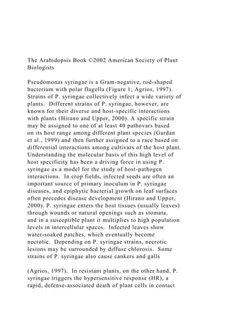
  • 4. When a suspension of 108 bacteria/ml (a high dose of bacteria) is sprayed with a surfactant onto susceptible Arabidopsis plants (e.g., ecotype Columbia), the first sign of disease is the appearance of “water-soaked” patches on leaves on day 2. The water-soaked symptom results from massive release of water and, presumably, nutrients from infected Arabidopsis cells. The water-soaked patches become necrotic and dark-colored on day 3, and the surrounding leaf tissue shows extensive chlorosis, giving the characteristic appearance of a ‘speck’ disease (Figure 2, Figure 3E and 3F). In addition, closely related, but avirulent strains (such as P. syringae pv. tomato JL1965 and P. syringae pv. maculicola M2) that became sources of some avirulence (avr) genes were also identified (Dong et al., 1991; Whalen et al., 1991; Dangl et al., 1992). The main reason for examining P. syringae strains as potential pathogens of Arabidopsis was because P. syringae had already been proven to be an excellent genetically tractable pathogen of soybean, tomato, and bean in the mid-1980s (Keen, 1990). Development of the Arabidopsis-P. syringae pathosystem would provide a system in which both the plant and the pathogen are amenable to rigorous genetic analysis. However, it is interesting to note that even after the demonstration of disease symptoms caused by P. syringae on Arabidopsis, not many people were convinced that this was a good model system for two reasons: there was no report of naturally occurring P. syringae disease in Arabidopsis in the wild, and in the laboratory inoculation required artificial methods (infiltration or use of surfactant). 1.2. Establishing the system: gene-for-gene Figure 1. A transmission electron microscope image of Pseudomonas syringae pv. tomato DC3000. Note that
  • 5. DC3000 produces polar flagella (15 nm in diameter) and a few Hrp pili (8 nm in diameter). The flagella and Hrp pili are indicated with arrows. Flagella enable bacteria to swim toward or away from specific chemical stimuli. Hrp pili are involved in type III secretion of avirulence and virulence pro- teins. Figure 2. Disease symptoms in Arabidopsis leaves caused by DC3000 infection. Leaves (indicated with arrows) were syringe-infiltrated with 5 x 10⁵ cfu/mL of Pst DC3000 and pictures were taken four days after inoculation. The whole plant is shown in (A). A close-up of a diseased leaf is shown in (B). The Arabidopsis Thaliana-Pseudomonas Syringae Interaction 3 of 35 interactions A significant milestone in the development of the Arabidopsis-P. syringae system was the demonstration that this pathosystem conforms to the gene-for-gene relationship that underlies many well-known plant- pathogen interactions in nature (such as the flax-rust fungus interaction or the soybean-P. syringae interaction) (Keen, 1990). The gene-for-gene hypothesis was advanced by H.H. Flor, based on his work on the flax-rust fungus interaction in the 1940s and 1950s (Flor, 1971). This hypothesis states that when a pathogen (in this case a P. syringae strain) has an avirulence (avr) gene, and a plant host (in this case the Arabidopsis plant) has the corresponding disease resistance (R) gene, the plant is resistant to the pathogen (Table 1). It is defined by a single plant R gene for a single pathogen avr gene, hence the
  • 6. name gene-for-gene resistance. Table 2 defines the terminology involved in gene-for-gene resistance. When the plant is resistant, the pathogen is said to be avirulent and the interaction is said to be incompatible. When the plant is susceptible, the pathogen is said to be virulent and the interaction is said to be compatible. The laboratories of Fred Ausubel and Brian Staskawicz showed that an avr gene, avrRpt2, from an avirulent P. syringae pv. tomato strain, JL1065, converted DC3000 and ES4326 into avirulent strains in the Arabidopsis ecotype Columbia (Dong et al., 1991; Whalen et al., 1991). Subsequent Arabidopsis mutagenesis and screening efforts led to the identification of mutations in the Arabidopsis RPS2 disease resistance gene (Kunkel et al., 1993; Yu et al., 1993). Thus, a demonstration of the avrRpt2-RPS2 gene- for-gene interaction was completed. Similar efforts in the laboratory of Jeff Dangl led to the identification of the avrRpm1 gene in P. syringae pv. maculicola strain M2 and the RPM1 resistance gene in Arabidopsis ecotype Columbia (Dangl et al., 1992). Interestingly, the RPM1 resistance gene also recognizes avrB, which was isolated initially from P. syringae pv. glycinea in the soybean-P. syringae interaction (Bisgrove et al., 1994). These pioneering efforts spurred subsequent research to identify additional P. syringae avr genes and the corresponding Arabidopsis resistance gene loci, the eventual cloning of the first Arabidopsis resistance gene, RPS2 (Bent et al., 1994; Mindrinos et al., 1994), and identification of non-R gene plant components involved in gene-for-gene resistance (see section 3.1.5). Early concerns about the lack of natural infection and artificial inoculation methods have still not been addressed, but the Arabidopsis-P. syringae system has flourished as a widely recognized model plant-pathogen
  • 7. system. This fact presents us a lesson on what is important in developing a new model system. A model system cannot address every single aspect of a natural system. For example, the Arabidopsis-P. syringae system is probably not an appropriate system to model a bacterial invasion process seen in P. syringae infection of beans in the field because P. syringae infection of Arabidopsis requires an artificial infection method. However, it has been a great system in which to study broadly observed phenomena, such as gene-for-gene interactions. Thus, we have to define appropriate questions to ask in a model system; it is not reasonable to dismiss a model system simply because it cannot address every single aspect of natural systems. Success in the Arabidopsis-P. syringae system encouraged development of other Arabidopsis pathogen systems (see chapters by Dangl, Somerville, Innes, Buell, and Crute). At the same time, comparisons with other plant-pathogen systems, especially the Arabidopsis-Peronospora parasitica system, have helped advancement of the Arabidopsis-P. syringae system. 2. EARLY INTERACTIONS IN THE ARABIDOPSIS INTERCELLULAR SPACE (APOPLAST) The Arabidopsis Book 4 of 35 Figure 3. Disease symptom development in a susceptible Arabidopsis plant 1, 2, 3 and 4 days after inoculation. Leaves were vacuum infiltrated with 1 x 10⁶ bacteria/ml of DC3000. A picture was taken before inoculation (A) immediately after vacuum infil-
  • 8. tration (B) and every day for 4 days (C to F). To the right of each picture is a plot of the level of bacteria present within the leaves The Arabidopsis Thaliana-Pseudomonas Syringae Interaction 5 of 35 at that particular time. Note, water-soaking symptoms, appeared at 48 to 60 hours. Significant chlorosis and necrosis occurred at 72 to 96 hours after inoculation. Note that bacteria multiplied to a near maximum level before chlorosis or massive cell death appeared. The Arabidopsis Book 6 of 35 As mentioned above, P. syringae often first flourishes on the surface of plants as an epiphyte in the wild before it enters the intercellular space to initiate pathogenesis (Hirano and Upper, 2000). The ability of P. syringae to grow epiphytically is ecologically important for pathogen survival and spread in the field and is a topic of intensive study. In the laboratory, however, where the Arabidopsis- P. syringae system is studied, we often bypass the epiphytic growth phase and place P. syringae directly in the intercellular space by syringe injection, or we artificially facilitate the entry of P. syringae into the intercellular space by spraying leaves with high doses of bacterial suspension in the presence of a surfactant (e.g., Silwet L-77). In addition, the commonly used strain DC3000 is a very poor epiphyte in the field (Kyle Willis, University of Wisconsin,
  • 9. Madison, personal communication) and therefore, the Arabidopsis-P. syringae interaction is not a good model for the study of epiphytic interaction. In this chapter, which is focused on the Arabidopsis-P. syringae pathogenic interaction, we therefore begin our discussion with the events occurring immediately after P. syringae arrives in the intercellular space, where bacteria are in direct contact with Arabidopsis cells that are about 10,000 times larger, are enveloped with a >100 nm thick cell wall, and are full of exploitable photosynthates behind the wall (Figure 4). The mesh size of the cell wall is in the order of 20 to 30 nm, and this is too small for a bacterium of a few µm to simply penetrate. Both P. syringae and Arabidopsis must react to this situation quickly, obviously for different reasons. Whatever happens in the first few hours of the encounter will determine whether P. syringae will be successful in becoming a virulent pathogen of Arabidopsis or the Arabidopsis plant will effectively stop further infection of P. syringae. The precise details of the early events after P. syringae enters the leaf apoplast are still not clear, but a few key steps have been revealed or can be speculated about. From the plant side, it is believed that Arabidopsis (and presumably all other plants) have developed mechanisms to detect the invasion of any microbe and respond with the first line (a basal level) of general defense and that this defense is effective enough to stop some microbes (e.g., saprophytes, which lack the ability to flourish in the living tissues). The first line of defense is not well characterized but presumably involves expression of some defense genes (Jakobek et al., 1993). It is relatively benign – it does not sacrifice the plant cell under attack. However, it is still costly to the cell, and that is why it is regulated. How Arabidopsis cells detect the presence of a microbe at this stage is not clear, but it likely involves sensing of
  • 10. constitutively expressed extracellular molecules or structures of the microbe. One example may be the bacterial flagellin protein, the structural protein of the bacterial flagellum. Felix et al. (1999) showed that a peptide whose sequence is well conserved among flagellins of eubacteria, including Pseudomonas, can elicit general defense responses in plants (Felix et al., 1999). An Arabidopsis gene involved in the perception of this peptide, FLS2, has been cloned and it encodes a leucine- rich-repeat (LRR) receptor-like protein kinase (Gomez- Gomez and Boller, 2000). Thus, the putative FLS2 receptor can potentially respond to a wide variety of bacterial pathogens, including P. syringae, and activate a general defense response. It is interesting that flagellins of Agrobacterium and Rhizobium, which do not elicit strong defense in plants, do not have this peptide sequence conserved (Felix et al., 1999). For P. syringae, the plant intercellular space is a potential niche from which to exploit the bulk of photosynthates and other nutrients hidden behind the host cell wall. The normal apoplast is believed to be limited in water and possibly nutrients (although theoretical considerations argue for sufficient nutrients in the apoplast, see Hancock and Huisman, 1981) and is a depository for some plant defense compounds. In order to flourish and attain an extremely high population density (typically 5x10⁷ bacteria/cm2) in the apoplast, P. syringae must produce appropriate virulence factors to cause Arabidopsis cells to ‘leak’ nutrients and water into the intercellular space and at the same time to suppress or evade Arabidopsis defense aimed at inhibiting bacterial proliferation. Because no massive host cell death occurs before P. syringae has achieved a near maximum population in infected leaves
  • 11. Figure 4. A scanning electron microscopic image of a cross section of an Arabidopsis (ecotype Columbia; susceptible) leaf infected with DC3000. HC: host cells. Ba: Bacteria. Arrows indicate the direction of type III secretion from bacte- ria in the apoplast into the host cell interior. Note that the host cell wall remains intact, physically separating bacteria and host cells until the very late stages of the interaction, when host cells collapse. The Arabidopsis Thaliana-Pseudomonas Syringae Interaction 7 of 35 (Figure 3), it is believed that nutrient and water release from host cells during the early to mid (probably most critical) stages of P. syringae infection is not caused by nonspecific host cell rupture. The exact arsenal of P. syringae virulence factors has not been determined, but two virulence systems have been shown to play a key role: the type III protein secretion system that delivers a battery of bacterial avirulence and virulence proteins (type III effectors, hereinafter) to the apoplast and also into the Arabidopsis cells (Alfano and Collmer, 1997; Lindgren, 1997; He, 1998; Preston, 2000) and a diffusible toxin coronatine that partially mimics plant hormone methyl jasmonate (MeJA) (Bender et al., 1999; Preston, 2000). Both of these virulence systems are induced in plant tissues, presumably because they are not needed before bacteria encounter plant cells. A detailed description of these two virulence systems will be presented in section 4, but it is important to mention here that direct injection of bacterial virulence proteins into host cells via the type III secretion system is a widespread phenomenon in bacterial pathogens of plants and animals, and is considered to be an
  • 12. evolutionarily critical invention of bacterial pathogens (He, 1998; Hueck, 1998; Galan and Collmer, 1999). Once P. syringae has injected type III effector proteins, which include Avr proteins (see below for more discussion of the relationship between Avr proteins and type III effector proteins), into Arabidopsis cells via the type III protein secretion system, there are two outcomes, depending on the genotypes of the infecting P. syringae and Arabidopsis plants. These two outcomes are most elegantly (albeit in an oversimplified manner) explained by the gene-for-gene hypothesis, i.e., if an infected Arabidopsis plant has an R gene that recognizes a P. syringae type III effector (i.e., an Avr protein in this situation), a rapid defense mechanism of the plant will be triggered. Alternatively, if the infected Arabidopsis plant has no corresponding R gene and/or the P. syringae strain has no avr gene, defense responses will be activated slowly, the infection will continue, and the plant will succumb to P. syringae and become diseased. In a given Arabidopsis-P. syringae system, it is possible that more than one specific combination of avr and R genes are operating at the same time, and such different combinations are often co-dominant (Table 3). For example, it is possible to have a strain of P. syringae carrying avr genes interacts with an Arabidopsis plant carrying two corresponding R genes. The two combinations, avr1/R1 and avr2/R2, may be co-dominant in the system. All known P. syringae avr genes (with the exception of avrD; Keen et al., 1990) that trigger gene-for-gene resistance encode type III effector proteins that are apparently delivered by bacteria to the plant cell via the type III secretion system (Mudgett and Staskawicz, 1998;
  • 13. Collmer et al., 2000; Kjemtrup et al., 2000; White et al., 2000). Why would P. syringae inject Avr proteins into Arabidopsis cells to trigger host resistance, thus inhibiting bacterial growth? The likely answer is that avr genes actually function as virulence genes when host plants do not carry the corresponding R genes. In fact, virulence functions of avrRpm1 and avrRpt2 on plants lacking the RPM1 and RPS2 resistance genes, respectively, have been demonstrated (Ritter and Dangl, 1995; Chen et al., 2000; Guttman and Greenberg, 2001). Thus, a more appropriate view of avr genes is probably that these are virulence genes first evolved to promote bacterial parasitism and that plants then counter-evolved surveillance systems to recognize virulence gene-based molecules (effectively turning virulence genes into avirulence genes). When P. syringae injects these proteins into Arabidopsis cells with the original purpose of parasitizing the Arabidopsis cells, it does not “know” that the recipient Arabidopsis cells may already be armed with one or more corresponding R genes, which would turn these virulence-intended proteins into defense elicitors-an elegant example of the adaptive co-evolution of pathogen virulence and plant resistance traits. 3. P. SYRINGAE ATTACKS AND ARABIDOPSIS COUNTER-ATTACKS Tremendous progress has been made in understanding how Arabidopsis recognizes P. syringae Avr proteins and The Arabidopsis Book 8 of 35 mounts effective defense against P. syringae. We now know that the pathogen recognition and defense signal
  • 14. transduction mechanisms underlying the Arabidopsis-P. syringae interaction share many common features with those observed in other Arabidopsis-pathogen interactions. Readers are encouraged to consult several excellent reviews on this topic (Glazebrook, et al., 1997; Dong, 1998; Innes, 1998; Bent, 2001; Thomma, et al., 2001; Glazebrook, 2001; Dangl and Jones, 2001; Staskawicz et al, 2001). In addition, several chapters of this book describe other Arabidopsis-pathogen systems or discuss pathogen recognition and disease signal transduction. We therefore will highlight only examples that particularly illustrate either the important contribution of using P. syringae as a model or the uniqueness of the Arabidopsis-P. syringae system. 3.1. Pathogen avirulence and plant resistance in incompatible Arabidopsis-P. syringae interactions 3.1.1. Gene-for-gene resistance in the Arabidopsis-P. syringae system Gene-for-gene incompatibility is prevalent among various plant-pathogen systems and one of the best characterized genetic relationships between plant hosts and pathogens. The prevalence of gene-for-gene resistance and similarities in associated responses among different plant- pathogen systems strongly suggest common underlying molecular mechanisms (Bent, 1996; Hammond-Kosack and Jones, 1997) and, therefore, gene-for-gene resistance was chosen to be the first target of study for the use of this model system. To study gene-for-gene resistance, first, a single avr gene was isolated by introducing a cosmid library made from an avirulent strain into a virulent strain. A cosmid clone containing an avr gene transformed the virulent strain to an avirulent strain. Use of such a strain carrying only a single avr gene created a situation in which
  • 15. only a single gene-for-gene interaction was operating in the plant-pathogen system. This step was crucial because different combinations of avr and R genes are usually co- dominant. For identification of corresponding R genes after genetic isolation of avr genes, r- plants were identified either by mutational analysis or by screens of various ecotypes. Genetically isolated avr-R gene combinations include avrRpt2-RPS2 (Kunkel et al., 1993; Yu et al., 1993), avrRpm1 (or nearly identical avrPpiA1)-RPM1 (Dangl et al. 1992), avrB-RPM1 (originally called RPS3, but later shown to be identical to RPM1) (Bisgrove et al., 1994), avrRps4- RPS4 (Hinsch and Staskawicz, 1996), avrPphB (formerly known as avrPph3)-RPS5 (Simonich and Innes, 1995), and avrPphB-PBS1 (Warren et al., 1999). Breakdowns in the narrowly defined version of the gene- for-gene concept are seen here. The R gene RPM1 corresponds to two avr genes, avrRpm1 and avrB. The avr gene avrPphB corresponds to two R genes, RPS5 and PBS1. AvrRpm1 and AvrB are not homologous, neither are RPS5 and PBS1. We should remember that the gene- for-gene hypothesis was forwarded by Flor, based on the study of flax-flax rust fungus interactions in the 1940s and 1950s (Flor, 1971), when knowledge about how genes function at the molecular level was almost non-existent. With the knowledge we currently have about molecular interaction mechanisms, we can easily imagine a number of possible molecular interaction models to explain these situations, and we should interpret the gene-for-gene concept more broadly. In a broader interpretation of the gene-for-gene resistance, the key concepts should be that a plant has pathogen recognition mechanism(s) composed of a repertoire of genetically definable recognition specificities and that pathogen recognition by these mechanism(s) leads to a successful deployment of
  • 16. defense responses in the plant. 3.1.2. R genes in the Arabidopsis-P. syringae system All of the above-mentioned Arabidopsis R genes, RPS2, RPM1, RPS4, RPS5, and PBS1, have been isolated by a map-based cloning approach (Bent et al., 1994; Mindrinos et al., 1994; Grant et al., 1995; Gassmann et al., 1999; Warren et al., 1998; Swiderski and Innes, 2001). All but PBS1 belong to the nucleotide binding site-leucine rich repeat (NBS-LRR) class of R genes. The protein encoded by an R gene of the NBS-LRR class has one NBS structure that is located N-terminal to large imperfect LRRs, which are located close to the C-terminus (Bent, 1996; van der Biezen and Jones, 1998b). NBS-LRR is the dominating class among the R genes so far isolated from dicots and monocots (Ellis et al., 2000). Although each class member usually has a high specificity to a particular pathogen of a particular genotype (i.e., with the corresponding avr gene), R gene members of this class collectively can confer resistance against all major types of plant pests, namely bacteria, oomycetes, fungi, viruses, nematodes, and aphids. This fact provides strong molecular support to the notion of common underlying mechanisms responsible for many gene-for-gene resistance phenomena. RPS2, RPM1, and RPS5 proteins have coiled-coil (cc) structures (such as a leucine zipper) at their N-termini, and The Arabidopsis Thaliana-Pseudomonas Syringae Interaction 9 of 35 are classified in the cc subclass of NBS-LRR (cc-NBS- LRR). RPS4 has a TIR (for Toll and Interleukin-1 Receptor)
  • 17. homology at its N-terminus, which is conserved among the cytoplasmic domains of Drosophila Toll protein, mammalian interleukin-1 receptors, and other NBS-LRR R proteins (Whitham et al., 1994). Thus, RPS4 belongs to the TIR subclass of NBS-LRR (TIR-NBS-LRR). Arabidopsis ecotype Col has ~140 NBS-LRR genes in its genome, including ~ 50 cc-NBS-LRRs and ~ 90 TIR-NBS-LRRs. Although it is generally assumed that the role of most, if not all, functional NBS-LRR genes is as R genes, it is not known how many of these NBS-LRR genes are functional R genes. It is conceivable that some of the NBS-LRR genes might function as a reservoir of sequence materials to create new recognition specificities through recombination. NBS-LRR proteins are believed to be intracellular, based on computer predictions from their primary sequences. RPS2 is not secreted or membrane-integrated in a heterologous in vitro system (Leister et al., 1996). RPM1 is peripherally associated with the plasma membrane (Boyes et al., 1998). The plasma membrane localization of RPM1 seems appropriate because the corresponding Avr proteins, AvrRpm1 and AvrB, are also localized at the plasma membrane (Nimchuk et al., 2000). It is possible that NBS-LRR proteins are localized at different subcellular compartments in the cell for optimal detection of signal molecules generated by the corresponding avr genes. For example, it will be interesting to see whether there are any NBS-LRR proteins localized in the nucleus to detect nuclear-transported pathogen signal molecules. PBS1 encodes a predicted cytoplasmic protein serine/threonine kinase. The tomato R gene PTO (Martin et al., 1993) is so far the only other example of an R gene of the cytoplasmic protein kinase class. Both PBS1 and the RPS5 NBS-LRR genes are required for resistance
  • 18. against a P. syringae strain carrying avrPphB (Warren et al., 1999). This combination of protein kinase and NBS-LRR genes is reminiscent of the tomato R genes PTO (a protein kinase gene) and PRF (NBS-LRR), both of which are required for resistance against a P. syringae strain carrying avrPto (Salmeron et al., 1996). Although PBS1 and PTO belong to a large subfamily of plant protein serine/threonine kinase genes, within the subfamily they are not very closely related. It is likely that the substrate specificities of these kinases are significantly different (Warren et al., 1998). 3.1.3. avr genes in the Arabidopsis-P. syringae system Although direct demonstrations are still lacking, all the above-mentioned avr gene products, AvrRpt2, AvrRpm1, AvrB, AvrRps4, and AvrPphB, are believed to be delivered from bacteria into the plant cell via the type III secretion system for the following reasons: (i) These avr genes require type III secretion genes (called hrp/hrc genes in P. syringae and other pathogenic bacteria) to express their avirulence functions when P. syringae strains carrying avr genes are inoculated into plants carrying the corresponding R genes (Pirhonen et al., 1996; Gopalan et al., 1996). (ii) Direct expression of these avr genes in the plant cell leads to the HR, which is dependent on the corresponding R genes (Alfano et al., 1997; Gopalan et al., 1996; Leister et al., 1996; Scofield et al., 1996; Tang et al., 1996; McNeillis et al., 1998; Stevens et al., 1998; Nimchuk et al., 2000; Chen et al., 2000; Figure 5) – when expressed in the plant cell, these Avr proteins are predicted to stay in the cytoplasm. (iii) All but AvrRps4 seem to be modified in the plant cell in a host cell-specific manner (Mudgett and Staskawicz, 1999; Nimchuk et al., 2000). Again, it should be emphasized that P. syringae stays outside of plant cells (i.e., in the intercellular space) until plant cells start to
  • 19. disintegrate at a very late stage of the interaction. Host cell-specific modifications of Avr proteins are Figure 5. The RPM1 resistance gene-dependent HR induced by the expression of the P. syringae avrB gene directly in Arabidopsis. Left panel: An Arabidopsis rps3-1 (an rpm1mutant; Columbia background) seedling expressing avrB under the 35S promoter. No HR is present. Right panel: An F1 seedling from a cross between the rps3-1/avrB plant and a wild-type Columbia plant (RPM1+). Arrow indicates dark HR necroses on the cotyledon leaf. This seedling died before true leaves emerged because of systemic develop- ment of the HR. The Arabidopsis Book 10 of 35 intriguing in the light of evolution of virulence/avirulence factors – evolving mechanisms that are dependent on host cell functions. AvrRpt2 protein is cleaved at a specific site when it is incubated with plant cell extracts or when it is directly expressed in the plant cell, whereas it is not cleaved when produced in P. syringae or E. coli (Mudgett and Staskawicz, 1999). The cleaving activity was not detected in Arabidopsis intercellular fluids, so the cleavage event is predicted to occur inside the plant cell. When a P. syringae strain carrying avrRpt2 is inoculated into the leaf, the inoculated tissue accumulates the cleaved form of AvrRpt2. Therefore, AvrRpt2 seems to be transported into the plant cell and specifically cleaved inside the cell. AvrRpm1, AvrB, and the processed form of AvrPphB (AvrPphB is rapidly cleaved when expressed in bacteria or plants) have canonical eukaryotic acylation sequences at
  • 20. their N-termini (Nimchuk et al., 2000). Mutations in the potential N-terminal myristoylation sites in AvrRpm1 and AvrB dramatically decreased their Avr activities when they were delivered by P. syringae (it is not known whether the mutations affected translocation of the proteins) or when they were expressed in the plant cell. When the proteins were expressed in the plant cell, they were myristoylated and localized to the plasma membrane in a myristoylation site-dependent manner. Because overexpression of the Avr proteins in the plant cell can overcome the requirement for the myristoylation site, myristoylation seems to be a mechanism to increase the concentration of the protein at the site of action, which is probably the intracellular side of the plasma membrane. AvrPphB protein expressed in the plant cell is cleaved and plasma membrane-localized in a myristoylation site-dependent manner. This observation demonstrates that proteins with canonical sites can be myristoylated post-translationally and supports the notion that proteins with canonical sites can be myristoylated after they are delivered from bacteria via the type III system. 3.1.4. Models for molecular mechanisms of gene-for- gene relationships We have yet to determine how Avr-based signals are recognized by the R-mediated mechanism. Below we discuss two popular hypotheses. Two observations led to the ligand-receptor model (or elicitor-receptor model) (Gabriel and Rolfe, 1990): (i) both avr and R genes are usually dominant, and (ii) in most cases, genetic single gene-single gene correspondence can be seen. In this model: (i) an avr gene generates a specific molecular signal (elicitor) directly (with the Avr
  • 21. protein as the signal) or indirectly (e.g., Avr protein is an enzyme that makes the signal molecule; Keen et al., 1990); (ii) the corresponding R gene encodes the receptor for the molecular signal; and (iii) this ligand-receptor interaction initiates signal transduction to induce downstream responses (Figure 6). Because the general concept of specific interactions between ligands and the cognate receptors has been known in biology for a long time, this model is an intuitively obvious one. Although the R protein in Figure 6 is depicted as a membrane receptor, the model based on genetic relationships does not specify the nature of the receptor, and the R protein could be an intracellular receptor. If an Avr protein itself is the specific molecular signal, this model predicts that the Avr protein and the corresponding R protein physically interact. The P. syringae AvrPto protein and the corresponding tomato Pto protein interact in the yeast two-hybrid assay, and the specificity for this interaction tightly correlates with the specificity in their avr and R functionalities (Scofield et al., 1996; Tang et al., 1996; Frederick et al., 1998). PTO belongs to a rare R gene class of protein kinase genes. The rice blast fungus Avr-Pita protein and the corresponding rice Pi-ta R protein interact in the yeast two-hybrid assay and in vitro (Jia et al., 2000). Pi-ta belongs to the NBS-LRR class, although its LRRs do not have a typical consensus sequence (Bryan et al., 2000). These observations of physical interactions between Avr and R proteins support the ligand-receptor model. Although the ligand-receptor model is an obvious one, it does not provide simple explanations to some observations. (i) Plants do not have an efficient mechanism to create new pathogen recognition specificities and select good ones, compared with the vertebrate adaptive immune system, in which somatic recombination creates a vast repertoire of recognition
  • 22. specificities and clonal selection provides a way to select good specificities. Given this limitation, how can plants Figure 6. The ligand-receptor model of R gene and avr gene interaction. A specific signal molecule is directly or indirectly generated by the avr gene in P. syringae. The signal mole- cule is recognized by the receptor encoded by the corre- sponding R gene in Arabidopsis. This moleuclar recognition leads to rapid induction of defense response. The Arabidopsis Thaliana-Pseudomonas Syringae Interaction 11 of 35 have enough recognition specificities, which are limited by the number of R genes in the genome, to effectively fend off most potential pathogens, when pathogens, which are in most cases microbes, can evolve much faster than plants? For example, Arabidopsis has only ~140 NBS- LRR genes. (ii) The same or very similar R genes can confer resistance against very different types of pathogens. The tomato Mi gene can confer resistance against both nematodes and aphids (Rossi et al., 1998). The potato Rx and Gpa2 genes confer resistance against potato virus X and nematode, respectively, and are highly homologous (Bendahmane et al., 1999; van der Vossen et al., 2000). Similarly, the Arabidopsis RPP8 and HRT genes, which are highly homologous, confer resistance against the Peronospora parasitica and turnip crinkle virus, respectively (McDowell et al., 1998; Kachroo et al., 2000). How could molecular signals derived from very different types of pathogens be recognized by the same or very similar R genes? (iii) In many cases, cloned R genes cannot function in different families of plants. The NBS- LRR based mechanism, for example, apparently evolved
  • 23. before major diversification of angiosperms. It is difficult to imagine that the downstream mechanism has become incompatible with the NBS-LRR upstream factors. (iv) Avr proteins in general appear to be virulence factors when the plant does not have the appropriate R genes. Is there any reason that the factors to be recognized by plants as signals of pathogen attack should have virulence functions in nature? To explain these phenomena, the “guard model” has been put forward recently (van der Biezen and Jones, 1998a). According to this model: virulence factors originating from pathogens have targets in the host to express their virulence functions; the function of an R protein is to guard such a target of a virulence factor; when the target is attacked by the virulence factor, the R protein somehow senses it and initiates signal transduction to induce defense responses (Figure 7). The guard model has been gaining popularity despite the lack of directly supporting evidence, because it can provide simple explanations to the above questions. (i) Assuming that the number of targets for virulence factors is limited, plants may not need to have a large number of R genes, nor do they have to generate new specificities quickly. A population genetic study of RPM1 alleles among ecotypes supported the trench warfare hypothesis in the evolution of avr and R genes, rather than the arms race hypothesis (Stahl et al., 1999). The trench warfare hypothesis images a battle between a host and its pathogen in which one wins sometimes and loses other times at the front line, but the overall situation does not change drastically. The arms race hypothesis, on the other hand, images a battle in which one acquires a new weapon and almost eliminates the other, then the other fights back with another new weapon. The RPM1 gene
  • 24. was not defeated by the pathogen for a long time, and its occurrence among the population fluctuates during this time. This observation is consistent with the trench warfare hypothesis and could be explained by assuming a relatively limited number of potential virulence targets (so that it is not easy for a pathogen to evolve a totally new virulence factor) and a balance between benefit and cost of resistance. (ii) If the same or very similar molecules are targeted by virulence factors derived from different types of pathogens (this situation is more likely to occur if the number of potential virulence targets is limited), it is conceivable that the same or very similar R proteins can guard the same or very similar virulence target molecules. (iii) A combination of a virulence factor target and the guarding R protein can co-evolve and drift, so that it is conceivable that after some evolutionary time, partners in orthologous combinations in different taxa of plants become unexchangeable. (iv) That virulence factors are the molecules to be recognized as signals of pathogen attack is a built-in assumption of the model. From the viewpoint of molecular recognition mechanisms, the guard model appears to be a small extension of the ligand-receptor model. The combination of the virulence factor target and the R protein can be considered as a receptor complex. However, the guard model adds more restrictions in this figure from the viewpoint of biological functions – the ligand must be intrinsically a virulence factor, and the receptor complex must contain the virulence factor target in addition to the R Figure 7. The guard model of R gene and avr gene interac- tion. When a plant does not have an appropriate R gene (r- background; left), a virulence factor derived from P. syringae interacts with the plant virulence target molecule. The viru-
  • 25. lence target molecule has a role in defense response induc- tion in the plant cell, and this function is inhibited by the interacting virulence factor. When a plant has the appropriate R gene (R+ background; right), the virulence target is guard- ed by the R protein. When the target is attacked by the viru- lence factor, the R protein senses the attack and rapidly induces defense response. The Arabidopsis Book 12 of 35 protein. These restrictions are the reason that the guard model can provide the above simple explanations. The result from co-immunoprecipitation of AvrRpt2 and RPS2 after expressing the proteins in Arabidopsis protoplasts was consistent with the notion of receptor complex (Leister and Katagiri, 2000). They were co- immunoprecipitated together with some other plant proteins from the plant extracts; they were not co- immunoprecipitated by themselves in vitro. However, this study does not tell whether the other complex components are necessary for gene-for-gene recognition, or whether one of the components is the AvrRpt2 virulence factor target. 3.1.5. Other genes involved in gene-for-gene resistance A few Arabidopsis genes in which mutations affect a group of R gene-mediated resistance are known. A mutation in NDR1 strongly affects resistance mediated by some cc- NBS-LRR R genes, whereas a mutation in EDS1 strongly affects resistance mediated by TIR-NBS-LRR R genes (Aarts et al., 1998). The most simple-minded model is that
  • 26. cc-NBS-LRR and TIR-NBS-LRR use different signal transduction pathways, and NDR1 and EDS1 are signal transducers in each pathway. For gene-for-gene resistance against P. syringae, resistance mediated by cc- NBS-LRRs (namely RPS2, RPM1, and RPS5 R genes) is affected by the ndr1mutation but not much by the eds1mutation, whereas resistance mediated by the RPS4 TIR-NBS-LRR is affected by eds1 and not by ndr1. This does not conflict with the model if we assume both pathways can independently induce a set of defense responses that are important for resistance against P. syringae. Alternatively, these pathways may induce two different sets of defense responses, and both sets are effective against P. syringae. However, there are many other simple models to explain the behavior of ndr1 and eds1 mutations. In addition, there is no reason to believe that NDR1 and EDS1 have comparable positions in the sequence of events. For example, these proteins might be needed to produce functional R proteins in a subclass- specific manner (e.g., modification, localization); one of them might be affecting pathogens directly but show differential effects due to differences in the sensitivity of R- mediated recognition (the other functions in a different way); in the guard model, they might be the targets of multiple virulence factors, guarded by multiple R proteins. It should be emphasized that the effects of ndr1 and eds1 mutations on R gene-mediated resistance are not always clear-cut. For example, RPS2-mediated resistance is strongly suppressed by the ndr1 mutation, but RPM1- mediated resistance is only partially suppressed (Century et al., 1995). NDR1 might be a quantitative factor for NBS- LRR R gene functions (Tao et al., 2000). A mutation in PBS2 affects resistance mediated by RPS2, RPM1, and RPS5, but does not affect resistance
  • 27. mediated by RPS4. Within this set of R genes, it appears that pbs2 suppresses cc-NBS-LRR mediated resistance, although a larger set of R genes needs to be tested to obtain a general conclusion about the R gene subclass specificity. There are also NBS-LRR genes whose functions are independent of any of NDR1 , EDS1 , PAD4, and PBS2 (Bittner-Eddy and Beynon, 2001). Some of such complications could stem from a signal transduction network (involving not only divergent pathways but also convergent pathways) and quantitative dynamics of the network. 3.2. General resistance in the Arabidopsis-P. syringae interaction Here we use the term “general resistance” as resistance that contributes to reduction in growth of virulent and avirulent pathogens to a similar extent. As you will see in this section, even the growth of a virulent pathogen is limited by general resistance of the plant. Note that the growth of an avirulent pathogen is limited by the gene- for-gene resistance in addition to general resistance. Even when the gene-for-gene resistance component is intact, a defect in general resistance component may allow an avirulent pathogen to grow well enough to cause disease symptoms. But in this case, without the gene- for-gene resistance component, the pathogen would grow even better. 3.2.1. General resistance against virulent P. syringae: a new view of compatible Arabidopsis-P. syringae interactions Establishment of a genetic model plant-pathogen system opened up new areas of experiments, some of which ended up challenging dogmas. One new area of
  • 28. experimentation is intensive plant mutant screens under well-controlled conditions: because Arabidopsis plants are The Arabidopsis Thaliana-Pseudomonas Syringae Interaction 13 of 35 small, mutant screens can be performed in growth chambers (even when you don’t have access to a large volume of growth chamber space) instead of greenhouses. Well-controlled conditions are crucial in reproducibility of subtle phenotype differences. This is especially important because plant-pathogen interactions are often significantly affected by environmental factors and the developmental stage of the plants. One subtle phenotype difference that was pursued was the difference in the degree of disease symptoms during compatible interactions (Glazebrook et al., 1996). Mutational analysis of Arabidopsis in response to virulent P. syringae strains led to identification of Arabidopsis mutants (e.g., npr1, eds, pad4; see below) that show enhanced disease susceptibility to normally virulent P. syringae strains (Glazebrook et al., 1997b). These Arabidopsis mutants are impaired in the activation of defense responses during compatible interactions. A long-held dogma in plant pathology textbooks led people to believe that incompatible and compatible interactions are qualitatively different. The idea was that plants are susceptible to certain pathogens because they simply cannot mount a resistance response to the pathogens. However, studies on these mutants unambiguously demonstrated that even when they are susceptible to a pathogen, plants defend themselves to slow down the pathogen-but the general defense is not effective enough to completely stop the pathogen.
  • 29. This change of the view of compatible interactions also raises a question about whether the distinction between compatible and incompatible interactions is necessarily correlated with differences in the underlying molecular mechanisms. One can easily imagine the following situation: each plant has a pool of R genes with different affinities that detect various P. syringae Avr proteins. The affinity will be highest if an Avr protein is perceived by a cognate R gene in a resistant plant and is responsible for induction of an effective defense. Although a susceptible plant lacks the cognate R gene to recognize an “Avr-like” protein of a virulent P. syringae strain, some R genes in the susceptible plant may still weakly “interact” with one or more of these ”Avr-like” proteins produced by the virulent P. syringae strain, so the resistance pathway is activated, but too slowly and/or too weakly to completely stop the virulent P. syringae infection. Nevertheless, loss-of- function mutations in these “weak” R genes or resistance signaling component genes would increase the susceptibility of the plant to virulent P. syringae. In short, compatible/incompatible interactions may not be determined by distinctive molecular mechanisms, but by quantitative or kinetic variations in the same molecular mechanism. We should be aware that traditional classification of biological phenomena may not be correlated with distinctive molecular mechanisms. An interesting observation is that some Arabidopsis mutations initially identified based on defects in gene-for- gene resistance against avirulent pathogens also affect general resistance against virulent pathogens or vice versa. For example, virulent Pst DC3000 grows better in the eds1 mutants, which were initially identified in screens for gene-for-gene interaction mutants (Aarts, et al., 1998). Mutations in PAD4 were identified initially by reduced
  • 30. general resistance, but pad4 affects some R gene- mediated resistance and general resistance to P. syringae (Glazebrook, et al., 1997b). When expressed in the plant lacking RPS2, the virulence function of AvrRpt2 affects both general resistance to virulent DC3000 and specifically RPM1-mediated gene-for-gene resistance (Chen, et al., 2000). These observations suggest that R-mediated resistance and general resistance against virulent pathogens are closely related. Responses to virulent strains might use both EDS1- and PAD4- dependent pathways, if EDS1 and PAD4 are indeed signal transducers. Furthermore, recognition of virulent strains might even use NBS-LRR type proteins but the response could be slower and/or weaker. In the guard model, the pathogens’ virulence factors may attack some molecular components for general resistance and resistance mediated by R proteins that guard the particular components could also be affected. 3.2.2. Role of SA in general resistance and gene-for- gene resistance to P. syringae Genes that contribute to general resistance against P. syringae and P. parasitica appear to be mainly involved in the SA-dependent signaling pathway for defense response regulation. Mutations in all such genes (e.g., EDS and PAD4; Aarts et al., 1998; Glazebrook et al., 1997b), except for NPR1 and DTH9, reduce SA accumulation during pathogen attack (Glazebrook, 2001). NPR1 seems to be a signal transducer downstream of SA (Delaney et al., 1995; Cao et al., 1994; Kinkema et al., 2000). Studies that involve plants that carry the NahG transgene (encoding salicylate hydroxylase which degrades SA) and general resistance mutants clearly indicate that the SA-dependent pathway is crucial for general resistance against many pathogens including P. syringae. Furthermore, many
  • 31. pathogen-responsive genes are regulated in a SA- dependent manner. SA is accumulated at a higher level during incompatible interactions than during compatible interactions at early stages of interaction. The SA- dependent pathway might be the link between general resistance and gene-for-gene resistance. However, the role of SA in gene-for-gene resistance is not that clear. The Arabidopsis Book 14 of 35 NahG plants can develop an HR upon infection of avirulent bacteria. They are not strongly affected in the gene-for- gene resistance mediated by at least some R genes, whereas the general resistance component is strongly impaired (Delaney et al., 1994). It should be noted that not only is SA a major signal transducer after recognition of pathogen attack, but it also can potentiate pathogen recognition sensitivity at a low level (Shirasu et al., 1997). One role of SA in gene-for-gene resistance may be to potentiate the recognition mechanism. In this way, only gene-for-gene interactions with relatively low sensitivities might be strongly affected by a lower SA level. 3.2.3. Role of JA and ethylene in general resistance to P. syringae In addition to the SA-dependent pathway, involvement of the JA/ethylene-dependent pathway in general defense has been observed. In many cases, the SA-dependent pathway and the JA-dependent pathway act antagonistically (Felton et al., 1999a, 1999b; Pieterse and van Loon, 1999; Thomma et al., 2001). Activation of the JA-pathway could suppress the SA-pathway and reduce general resistance against pathogens, such as P. syringae,
  • 32. against which plants mainly use the SA-pathway. In fact, some virulent P. syringae strains appear to use this antagonistic interaction to suppress Arabidopsis defense (see section 4.2.1). However, the JA/ethylene pathway, not the SA pathway, is important for induced systemic resistance (ISR), which causes resistance against virulent P. syringae strains (see section 3.5). Whereas in many cases JA and ethylene appear to function in concert, ethylene response seems to be important for general resistance or tolerance against virulent P. syringae (Ton et al., 1999a; 1999b; Bent et al., 1992). The interactions among SA-, JA-, and ethylene-dependent pathways do not appear to be simple. Complications could arise from different roles of these pathways in different stages of plant-pathogen interactions. Therefore, it will be important to obtain various measurements of a given interaction with high spatial and temporal resolution. 3.3. What defense responses are responsible for Arabidopsis resistance to P. syringae? We have very limited knowledge about which particular defense responses are important for resistance against P. syringae or any other pathogen. Phytoalexins are known to be involved in pathogen resistance in some pathosystems (Hammerschmidt and Dann, 1999). The Arabidopsis PAD3 gene encodes a P450 monooxygenase, which is likely to be one of the enzymes required for synthesis of the Arabidopsis phytoalexin camalexin (Zhou et al., 1999). A mutation in this gene abolishes camalexin accumulation after infection by P. syringae but does not affect other tested defense responses (Glazebrook and Ausubel, 1994). General and gene-for-gene resistance against P. syringae is not significantly affected in the pad3 mutant (Glazebrook and Ausubel, 1994), whereas general resistance against the fungus Alternaria brassicicola is
  • 33. reduced in the pad3 mutant (Thomma et al., 1999). Although camalexin exhibits toxicity to P. syringae in vitro (Rogers et al., 1996), it is likely that camalexin does not have a major contribution to resistance against P. syringae in Arabidopsis. We should, however, keep in mind that resistance to a given pathogen is likely to be composed of complex combinations of different defense responses. Some responses may act additively, some may act synergistically, and some may be functionally redundant. We may not detect an effect of loss of a redundant function. Reactive oxygen species, PR proteins, and host cell wall modification are additional candidate defense compounds/mechanisms, but none of them have so far been linked directly to P. syringae resistance in vivo. 3.4. Systemic resistance responses An initial localized infection of an avirulent P. syringae strain causes plant cell death (an HR in this case), which could further trigger a whole-plant level of relatively weak resistance against secondary infection by a broad range of pathogens, including normally virulent strains of P. syringae (Ryals et al., 1996). This phenomenon is called systemic acquired resistance, or SAR (see chapter by Dangl for details). SA accumulation in the systemic tissues is essential for SAR, but SA is unlikely to be the long-distance signal that initiates SAR in the uninfected parts of the plant (Vernooij et al., 1994). Because the SA- dependent mechanism is also crucial for local gene-for- gene and general defense, many Arabidopsis mutants isolated based on their deficiency in local defense against pathogens have defects in the SA signaling pathway and are also deficient in SAR (Glazebrook et al., 1997a). The level of SA accumulated in the infected local tissues is
  • 34. The Arabidopsis Thaliana-Pseudomonas Syringae Interaction 15 of 35 much higher than that in the systemic tissue. It seems that plants use the same SA pathway at different degrees for local defense and for defense in systemic tissues in SAR. This would be a very practical solution for plants because (i) plants would not have had to evolve two distinct signaling pathways for two purposes and (ii) full-blown activation of the SA pathway appears deleterious not only to pathogens, but also to the plant itself, as suggested by growth defects among plant mutants that have the SA pathway constitutively on. 3.5. Rhizobacteria-mediated induced systemic resistance (ISR) Before we leave the subject of P. syringae resistance, we would like to mention an interesting systemic resistance mechanism that is effective against P. syringae, but is mechanistically distinct from SAR described above. This resistance mechanism is called induced systemic resistance (ISR) and is triggered by non-pathogenic, root- colonizing rhizobacteria, such as Pseudomonas fluorescens strain WCS417r or P. putida strain WCS358r (Pieterse et al., 1996). In contrast to pathogen-induced SAR, ISR is not associated with SA accumulation or activation of PR genes. Jasmonic acid and ethylene appear to play an important role in this SA-independent pathway (Pieterse et al., 1998; Knoester, 1999). The Arabidopsis jasmonate response mutant jar1 and the ethylene response mutant etr1, which show a normal response to inducers of SAR, are unable to express ISR after root treatment with P. fluorescens WCS417r. Although ISR and SAR seem to follow distinct signaling pathways,
  • 35. they are both blocked in the npr1 mutant (Pieterse et al., 1998). Thus, the NPR1 protein is not only required for the SA-dependent expression of PR genes that are activated during SAR, but also for the jasmonate- and ethylene- dependent activation of so-far-unidentified defense responses resulting from ISR. Because the expression of none of the SAR-associated or jasmonate/ethylene- induced genes is affected during ISR, ISR is believed to be associated with an increase in Arabidopsis sensitivity to the defense hormones JA and ethylene (van Wees et al., 1999, 2000). 4. Pathogen virulence and host susceptibility in the compatible Arabidopsis-P. syringae interactions In the preceding section, we discussed how Arabidopsis recognizes and defends against P. syringae infection. What happens when Arabidopsis fails to rapidly recognize a P. syringae strain, due to lack of appropriate R genes? How do Arabidopsis plants become susceptible to virulent P. syringae strains? What is the molecular basis of P. syringae pathogenicity? To be a successful extracellular pathogen, virulent P. syringae (e.g., strain DC3000 in Arabidopsis ecotype Columbia) must evolve an array of pathogenic mechanisms to suppress or evade Arabidopsis defense responses that are apparently effective in preventing virulent infection by the vast majority of potential parasites. It must also develop mechanisms to release nutrients and water to the apoplast, where bacteria live. How P. syringae succeeds in doing this is not known. However, molecular genetic analysis of P. syringae pathogenicity has revealed two virulence systems that play an important role in P. syringae infection of plants: the type III protein secretion system and a toxin called coronatine. 4.1. Pathogen virulence
  • 36. 4.1.1. The type III protein secretion system: A key pathogenicity factor in P. syringae The type III protein secretion system was discovered first in the human pathogens Yersinia spp., but has now been found to be widespread among Gram-negative bacterial pathogens of plants and animals (He, 1998; Hueck, 1998; Galan and Collmer, 1999; Cornelis and Van Gijsegem, 2000). The most intriguing feature of this protein secretion system is the ability of this system to actively inject bacterial virulence proteins directly into host cells. The importance of the type III secretion system in P. syringae pathogenicity is underscored by the observation that hrp/hrc mutations that block type III secretion completely eliminate P. syringae infectivity in susceptible Arabidopsis plants (Roine et al., 1997). The ability of P. syringae to inject virulence proteins directly into the host cell is believed to be highly significant in pathogen evolution, because this injection mechanism enables the pathogen to gain access to a vast number of intracellular host targets that would not be available for bacterial virulence proteins delivered to the surface of host cells. As mentioned in section 3.1.3, in P. syringae and other plant bacterial pathogens, the majority of the known type III effectors are Avr proteins, which are identified based on their ability to trigger resistance responses following recognition by the R gene products in the incompatible interactions. How P. The Arabidopsis Book 16 of 35 syringae delivers Avr and other type III effector proteins from its cytoplasm to the host cell cytoplasm is not known. However, a P. syringae surface pilus (called the Hrp pilus)
  • 37. assembled by the type III secretion system has been shown to play a key role in this process, possibly by providing a bridge for protein transfer (Roine et al., 1997; Wei et al., 2000; Hu et al., 2001; Jin et al., 2001). With the DC3000 genome sequencing project near completion (http://www.tigr.org/tdb/mdb/mdbinprogress.html), several groups are addressing a pressing question: how many type III effectors are produced by this bacterium? Two strategies of systematic search for type III effectors are underway. First, all known type III effector genes in P. syringae contain a characteristic ‘hrp-box’ motif in their promoters (Shen and Keen, 1993; Innes et al., 1993; Xiao et al., 1994). Therefore, a BLAST search using the consensus ‘hrp-box’ motif should identify putative type III effector genes. Second, a type III secretion reporter system has been developed. A truncated AvrRpt2 protein lacking the N-terminal type III secretion signal but maintaining HR-eliciting activity once inside RPS2+ Arabidopsis cells (Mudgett et al., 2000; Guttman and Greenberg, 2001) can be used to generate random fusions in the DC3000 genome. An in-frame fusion of the N- terminal secretion signal of a type III effector with the truncated AvrRpt2 reporter will target the fusion protein into Arabidopsis cells to trigger an AvrRpt2/RPS2- dependent HR. With a combination of these two strategies, we shall soon know the exact number of type III effectors in DC3000. How do type III effectors function inside the host cell to promote plant susceptibility? This has been a major mystery in the field of plant-pathogen interactions. To date, no plant ‘susceptibility’ pathway has been clearly identified in any plant-bacteria interaction. In animal- pathogen interactions, increasing evidence suggests that
  • 38. the MAP kinase defense pathway (Orth et al., 1999), delivery of reactive oxygen-generating enzymes (Vazquez- Torres et al., 2000, 2001; Feng et al., 2001; Vazquez-Torres and Fang, 2001), ubiquitin-like molecules (Orth et al., 2000), and actin cytoskeleton (Galan and Zhou, 2000) in the host are the targets of type III virulence proteins, demonstrating that type III effectors are ‘smart bombs’ sent by pathogens. Unfortunately, the primary sequences of the identified P. syringae type III effector genes provide little clue to their functions in modulating plant signaling and metabolic processes. In Xanthomonas spp., however, there are some clues to the functions of several Avr proteins in compatible hosts. For example, AvrRxv/AvrBsT from X. campestris pv. vesicatoria shares sequence homology with YopJ/P of Yersinia and AvrA of Salmonella (Orth et al., 2000). YopJ is a MAP kinase pathway inhibitor that suppresses macrophage defenses and appears to have protease activity (Orth et al., 1999, 2000). Whether AvrRxv and AvrBsT target a MAP kinase pathway for their virulence functions in susceptible host plants is not known, but the putative protease activity of AvrBsT appears to be required for elicitation of plant resistance response (Orth et al., 2000). AvrBs2 shows similarity with agrocinopine synthase (opine production in tumors) of Agrobacterium tumefaciens, suggesting a possible (but not proven) role in the nutrition of the pathogen (Swords et al., 1996). AvrBs3 family members, which are widespread in pathogenic Xanthomonas spp., appear to be transcription factors, although the host genes directly transcribed by the AvrBs3 family proteins remain to be identified (Bonas et al., 1989; Yang and Gabriel, 1995; Zhu et al., 1998; Zhu et al., 1999; Yang et al., 2000). It is almost certain that a major function of P. syringae type III effector proteins is to suppress plant defense
  • 39. responses in the host. Supporting evidence is accumulating. For example, hrp mutant bacteria appear to induce the expression of several general defense- associated genes, such as phenylalanine ammonia lyase and chitinase genes (Jakobek et al., 1993), and papillae formation (Bestwick et al., 1995; Brown et al., 1995), whereas the wild-type strains suppress expression of these defense genes and formation of papillae. There is also bacterial genetic evidence that some avr gene products, including AvrRpt2, interfere with the function of other avr gene products in the elicitation of host resistance (Ritter and Dangl, 1996; Reuber and Ausubel, 1996; Jackson et al., 1999; Chen et al., 2000; Tsiamis et al., 2000). An increasingly popular idea, as discussed in the guard model (Figure 7), is that type III effectors, including Avr proteins, may target key components of general defense in susceptible plants, presumably to suppress host defense. However, the exact molecular mechanisms by which type III effectors modulate the plant resistance responses remain to be elucidated. Experiments involving heterologous expression of avr genes inside plant cells suggest that Avr proteins can be deleterious even in the absence of a known cognate R gene if expressed too strongly (Gopalan et al., 1996; McNeillis et al., 1998; Nimchuk et al., 2000). Whether these effects result from interaction with susceptibility targets in the host is unknown. However, the sensitivity of Arabidopsis to AvrB over-expressed in the susceptible plant cell depends on a single gene, which suggests that a specific plant target is involved in this phenomenon (Nimchuk et al., 2000). As extracellular pathogens, P. syringae and other bacterial pathogens must also cause host cells to release water and nutrients into the apoplast. Consequently, some type III effectors may be involved in water and nutrient release (Figure 8). Despite these
  • 40. accumulating clues, in no case has a specific plant ‘susceptibility’ target been identified for any type III effector in plant pathogenic bacteria. Identification of host The Arabidopsis Thaliana-Pseudomonas Syringae Interaction 17 of 35 susceptibility targets/pathways is therefore a major challenge in the field. 4.1.2. The coronatine toxin-A molecular mimic of methyl jasmonate In addition to the type III secretion system, strains DC3000 and ES4326 also produce a toxin (called coronatine) that plays a significant role in modulating host susceptibility (Bender et al., 1999). However, unlike mutations affecting the type III secretion system, mutations affecting coronatine production do not completely eliminate pathogen virulence in susceptible Arabidopsis plants, rather they have only a quantitative effect on pathogen virulence, most notably coronatine-deficient bacterial mutants cause substantially weaker disease symptoms (chlorosis and necrosis) and a subtle reduction of bacterial multiplication in host leaves (Bender et al., 1987; Mittal and Davis, 1995). The structure of coronatine (Figure 9) has two distinct components: the polyketide coronafacic acid (CFA) and coronamic acid (CMA), an ethylcyclopropyl amino acid derived from isoleucine. The primary symptom elicited by coronatine in plants is tissue chlorosis (Gnanamaickam et al., 1982). However, exogenously applied coronatine
  • 41. induces purpling of Arabidopsis leaves (Bent et al., 1992), presumably resulting from anthocyanin production. Coronatine also inhibits root elongation and stimulates ethylene production in plants (Ferguson and Mitchell, 1985; Kenyon and Turner, 1992; Feys et al., 1994). Coronatine bears remarkable structural and functional homologies to methyl jasmonate (MeJA). Furthermore, coronatine and MeJA induce similar biological responses in Arabidopsis seedlings (Feys et al., 1994) and other plants (Weiler et al., 1994; Greulich et al., 1995; Koda et al., 1996), leading to the suggestion that coronatine functions as a molecular mimic of MeJA. MeJA accumulates in response to wounding or insect chewing and is involved in a systemic defense response to invading insects via production of defense compounds, including proteinase inhibitors (Ryan and Pearce, 1998). Why would bacteria produce a toxin whose function mimics that of MeJA? As mentioned above, coronatine-deficient DC3000 mutants have reduced virulence in susceptible Arabidopsis, so one must hypothesize that coronatine somehow conditions host plants to be more susceptible for bacterial infection via activation of the JA signaling pathway. Emerging evidence suggests that the JA-mediated insect defense and SA-controlled pathogen resistance are sometimes antagonistic to each other so that activation of one pathway leads to inhibition of the other (Felton et al., 1999a, 1999b; Pieterse and van Loon, 1999; Thomma et al., 2001). This has been interpreted as plants prioritizing energy-consuming defense responses towards specific insults (e.g., insects vs. some microbial pathogens). If this is true, coronatine could act as a suppressor of the plant’s SA-dependent microbial defense system by triggering JA- mediated insect defense response. Consistent with this idea, a study shows that a coronatine mutant of DC3000 induces the expression of two pathogen defense-
  • 42. associated genes (phenylalanine ammonia lyase and Figure 8. A hypothetical model of the potential targets of type III effector proteins in the host cell. P. syringae is an extracellular pathogen, living and multiplying in the leaf apoplast. Some effector proteins must therefore be involved in releasing water, carbohydrates and other nutrients from the host cell. Other effector proteins are likely involved in suppressing or evading host defense responses. In the top right corner is a scanning electron microscopic image of a cross section of an Arabidopsis leaf infected with DC3000 (see Figure 4). Figure 9. The structure of coronatine. The Arabidopsis Book 18 of 35 glutathione S-transferase) more highly than wild-type DC3000 (Mittal and Davis, 1995). However, we have yet to resolve the precise mechanism by which coronatine tricks Arabidopsis and other plants to turn on the JA pathway and to presumably inhibit effective plant defense against P. syringae. Remember that ISR against P. syringae is actually dependent on the JA pathway components (see section 3.5). How can the same pathway be involved in processes leading to opposing effects on P. syringae resistance/susceptibility? 4.2. Host susceptibility 4.2.1. Arabidopsis coi1 mutant-Blocking the action of the coronatine toxin Based on the ability of coronatine to inhibit Arabidopsis
  • 43. root growth, Feys et al. (1994) isolated a coronatine- insensitive (coi1) mutant of Arabidopsis. Interestingly, the same mutant is also insensitive to MeJA, further suggesting a similar mode of action of coronatine and MeJA. Consistent with the role of coronatine in the virulence of toxin-producing P. syringae strains, the coi1 mutant plants are highly resistant to P. syringae pv. atropurpurea (Feys et al., 1994), and pv. tomato DC3000 (Kloek et al., 2001; R. Thilmony and S. Y. He, unpublished), with markedly reduced disease symptoms and bacterial multiplication. However, the coi1 mutant has increased susceptibility to insects and certain necrotrophic fungal pathogens and is male sterile (Feys et al., 1994; Thomma et al., 1998). At present, it is not known how the same COI1 protein participates in seemingly different pathways leading to pathogen defense/susceptibility and pollen development. The COI1 protein belongs to a family of conserved proteins (e.g., human Skp2 involved in cell division, yeast Grr1 involved in cell division and nutrient uptake, and Arabidopsis TIR1 involved in auxin response) that contain an F box and leucine-rich repeats (Xie et al., 1998). Proteins of this family are known to be involved in selectively recruiting substrate regulatory proteins (e.g., transcription repressors) into a complex for ubiquitination and subsequent removal by proteolysis (Latres et al., 1999; Spencer et al., 1999; Spiegelman et al., 2001). Thus, the COI1 protein could control pathogen defense responses and pollen development by targeting distinct repressors for degradation in different cell types and possibly in response to different signals. Recently, Barbara Kunkel’s lab isolated additional alleles of the coi1 mutant from a direct screening for Arabidopsis mutants with enhanced resistance (Kloek et al., 2001). They made the important finding that DC3000 resistance
  • 44. (restriction of DC3000 multiplication) in the coi1 plants is largely abolished when the coi1 plants are crossed to SA- deficient nahG plants. These results suggest that the inhibition of DC3000 multiplication in the coi1 mutant is dependent on accumulation of SA. Perhaps the coi1 plants can mount a more aggressive SA-mediated pathogen defense in the absence of the otherwise antagonistic JA defense pathway. Interestingly the coi1nahG plants still show less severe disease symptoms than wild-type plants after DC3000 infection. This observation argues that the COI1 protein is required for DC3000-induced symptom development, in addition to its involvement in cross-talk with the SA-mediated defense pathway. Compared with the bacterial cor- mutant phenotype, the Arabidopsis coi1 mutant phenotype is much more drastic, suggesting that lack of perception of coronatine is not the only defect in the coi1 mutant. Specifically, whereas DC3000 multiplies about 100-fold less in coi1 plants (compared with in wild-type Col plants) and causes no disease symptoms when 106 cfu/ml of DC3000 are infiltrated into leaves, DC3000 cor- mutants multiply similarly as the wild-type bacteria in Col plants (at most, a 10-fold reduction) and still cause some disease symptoms, albeit at a lower frequency (Kloek et al., 2001; R. Thilmony and S. Y. He, unpublished results). COI1 could be required for the action of additional DC3000 virulence factors. One possibility is that some type III effectors also target a COI1-dependent pathway to modulate host susceptibility, which, if proven, could assign a major host target for P. syringae type III effectors. Alternatively, coronatine may activate only a subset of COI1-dependent, SA-antagonistic pathways, whereas the coi1 mutation may eliminate all COI1-mediated, SA-antagonistic pathways.
  • 45. As discussed in section 3.2, a variety of other Arabidopsis mutants also influence susceptibility to virulent P. syringae infection via their effects on general defense in the plant. Some mutants have increased susceptibility owing to a block in the accumulation or perception of SA (e.g., npr1, eds, ndr1, pad4, eds5, sid2); others have enhanced resistance owing to constitutive expression of SA and defense genes (e.g., cpr, dnd, acd, lesion mimic mutants, MAP kinase mutants; see chapter by Dangl). A key distinction between the Arabidopsis coi1 mutant and these other Arabidopsis mutants is that we know that the COI1 pathway is the target of a known P. syringae virulence factor (coronatine). As we continue to unravel the host components targeted by the DC3000 virulence factors, it would not be surprising to learn that some of the known Arabidopsis defense pathway components are additional targets of DC3000 virulence factors during compatible interactions. This prediction is The Arabidopsis Thaliana-Pseudomonas Syringae Interaction 19 of 35 in line with the adaptive co-evolution theory that both plant and pathogen evolve to overcome each other’s lethal weapons, and with the guard model of R gene/avr gene interaction. 5. ARABIDOPSIS AND P. SYRINGAE GENOMICS In addition to the genetic tractability of both host and pathogen genomes, the Arabidopsis-P. syringae pathosystem now offers another advantage: rapidly accumulating genomics resources. Completion of Arabidopsis genome sequencing in 2000 (The Arabidopsis
  • 46. Genome Initiative, 2000) and expected completion of the P. syringae pv. tomato DC3000 genome sequencing in 2002 (http://www.tigr.org/tdb/mdb/mdbinprogress.html) are making the Arabidopsis-P. syringae system an attractive genomics-amenable system. Powerful structural, computational, and functional genomics approaches can be used to examine this interaction in ways that cannot yet be done with many other pathosystems. For example, genome-wide monitoring of changes in gene, protein, and metabolite expression will give us a global picture of the intricate cross-talk among interconnecting signaling and metabolic pathways during the Arabidopsis-P. syringae interaction. Knowledge of host and pathogen genomes will also facilitate systematic mutational analysis of host and pathogen determinants involved in incompatible and compatible interactions. 5.1. Host genomics Analysis of the Arabidopsis genome has identified hundreds of genes that show significant sequence similarities with known disease resistance genes, disease signal transduction components, and downstream defense response genes, confirming the belief that Arabidopsis (and likely other plants) devotes a substantial portion of its genome to combating pathogens. Some of these genes likely encode additional recognition components of distinct specificities, checkpoints of sub- branches of disease signaling pathways, and redundant defense substances. They represent a rich resource for discovery of new genes controlling Arabidopsis-P. syringae interactions. Global gene expression profiling is already an established technology for Arabidopsis (Zhu and Wang,
  • 47. 2000). It has been used in the study of host gene regulation in interaction with P. syringae. One of the discoveries made by Maleck et al. (2000) is that many of the genes regulated by DC3000/avrRpt2 in the systemic leaves clustered with genes regulated by Peronospora parasitica and also by the SA analog BTH (within the first 4 h), suggesting that the DC3000/avrRpt2-induced SAR is transcriptionally related to P. parasitica-induced SAR and to an early phase of BTH-induced SAR (Maleck et al., 2000). The inclusion of the DC3000/avrRpt2 treatment in this study thus helped to sort out the biologically relevant transcripts that are strongly associated with SAR. The identified novel SAR-associated genes are candidates for further study using functional genomics approaches. 5.2. P. syringae genomics At the writing of this edition, the DC3000 genome (approximately 6.2 mb) has not been annotated completely. However, even the incompletely annotated DC3000 genome is giving us an opportunity to perform many specific analyses. For example, a Blast search using the ‘hrp box’ motif, which is present in the promoters of all known P. syringae avr genes, has revealed about 70 putative open reading frames (J. Zwiesler-Vollick, A. Plovanich-Jones, and S. Y. He, unpublished). Some of these genes could encode bona fide type III effectors and are therefore candidates for further verification by experimentation. The DC3000 genome also contains genes involved in cell wall degradation, extracellular polysaccharides, and production of phytohormones (e.g., IAA), which have been shown to be important in the virulence of other bacterial pathogens (Alfano and Collmer, 1996). These potential virulence factors (in addition to the type III secretion system and coronatine toxin) could therefore contribute to DC3000 aggressiveness in
  • 48. Arabidopsis tissues. In summary, we are entering an exciting phase of research on the Arabidopsis-P. syringae interaction using a variety of functional genomics tools. We anticipate that many more structural, computational, and functional genomics studies of both Arabidopsis and P. syringae will be completed in the near future, which will provide a comprehensive picture of interconnecting host and pathogen pathways that define incompatible and compatible interactions. We also anticipate the construction of a comprehensive plant-P. syringae functional genomics interaction database soon, so expect a much expanded version of this section in the next edition! The Arabidopsis Book 20 of 35 6. CONCLUDING REMARKS Since the discovery of P. syringae as a pathogen of Arabidopsis in the late 1980s, tremendous progress has been made in our understanding of pathogen virulence and avirulence determinants, the mechanism of host recognition of pathogen avirulence factors, resistance signal transduction pathways, and some aspects of host susceptibility. This progress is made possible because of the vision of a few pioneering scientists in the late 1980s. The genetic tractability of both Arabidopsis and P. syringae has played a critical role in the success of developing the Arabidopsis-P. syringae pathosystem into a remarkable model for research on plant-pathogen interactions. Despite the exciting progress, a number of fundamental
  • 49. questions remain to be answered. For incompatible Arabidopsis-P. syringae interactions, we still have no idea of the actual mechanisms/compounds that stop P. syringae infection in the apoplast of a resistant plant. Although the gene-for-gene interactions have been molecularly defined and provide a clear explanation for host resistance to avirulent strains of a compatible P. syringae pathovar (i.e., a pathovar in which some strains can cause disease in Arabidopsis), we do not know the molecular basis of the more prevalent nonhost resistance of Arabidopsis that is extremely effective against the vast majority of pathogens that are capable of causing disease in some other plants, but never Arabidopsis. For example, why don’t any of the P. syringae pv. phaseolicola strains, which infect bean, infect Arabidopsis? Is this because Arabidopsis contains all the resistance genes that could recognize all P. syringae pv. phaseolicola strains? Or is this because the virulence factors of P. syringae pv. phaseolicola that are adapted to the bean ‘susceptibility’ targets are not optimized for Arabidopsis? Recently, an Arabidopsis mutant, nho1 (for nonhost resistance 1), has been isolated that allows P. syringae pv. phaseolicola, P. syringae pv. tomato DC3000 hrp mutants, and the saprophyte P. fluorescens to grow significantly (Lu et al., 2001). In addition, the nho1 mutation also compromised resistance mediated by RPS2, RPS4, RPS5, and RPM1, providing evidence that nonhost resistance is controlled, at least in part, by general resistance that functions in gene-for-gene resistance. Further characterization of this mutant will shed light on the molecular basis of Arabidopsis nonhost resistance against P. syringae. For compatible Arabidopsis-P. syringae interactions, the main challenge is to identify the susceptibility targets of P. syringae virulence factors and to learn how P. syringae has evolved to circumvent Arabidopsis defense and at the same time to cause Arabidopsis cells to release water and
  • 50. nutrients into the apoplast. It is important to point out that we are at the beginning of an era when we can apply powerful scientific approaches toward understanding biological systems at a global level, in addition to understanding narrowly reduced, specific parts of the systems. This capability will drastically change the way biological research can be done, the way we think about biology, and the level of understanding we have in biology. Starting with a model biological system that is powerful in both genetics and genomics makes sense for collective efforts toward global levels of understanding. For the study of plant-pathogen interactions, the Arabidopsis-P. syringae system is such an ideal model system. We anticipate a new wave of system- based discoveries, which will reveal the dynamic, interconnecting, and flexible nature of the Arabidopsis and P. syringae signaling networks during interaction. In the end, we hope that study of this model interaction will contribute to our appreciation of how plants and bacterial pathogens have evolved to survive each others’ attacks and counterattacks, which will in turn help us to develop sustained control measures by guided interception of bacterial virulence and/or by selective activation of plant defense. ACKNOWLEDGMENTS We would like to thank Jane Glazebrook and members of our laboratories for critical reading of the manuscript, and Karen Bird for editing. Work in Sheng Yang He’s laborato- ry is supported by funds from the U. S. Department of Energy, National Science Foundation, and Department of Agriculture. REFERENCES
  • 51. Aarts, N., Metz, M., Holub, E., Staskawicz, B. J., and Daniels, M.J. (1998) Different requirements for EDS1 and NDR1 by disease resistance genes define at least two R gene-mediated signaling pathways in Arabidopsis. Proc. Natl. Acad. Sci. USA 95, 10306-10311. Agrios, G.N. (1997) Plant Pathology. (San Diego: Academic Press). Alfano, J.R., and Collmer, A. (1996) Bacterial pathogens in plants: Life up against the wall. Plant Cell 8, 1683-1698. Alfano, J.R., and Collmer, A. (1997) The type III (Hrp) secretion pathway of plant pathogenic bacteria: Trafficking harpins, Avr proteins, and death. J. Bacteriol. 179, 5655-5662. Alfano, J.R., Kim, H.-S., Delaney, T.P., and Collmer, A. (1997) The Arabidopsis Thaliana-Pseudomonas Syringae Interaction 21 of 35 Evidence that the Pseudomonas syringae pv. syringae hrp- linked hrmA gene encodes an Avr-like protein that acts in an hrp-dependent manner within tobacco cells. Mol. Plant- Microbe Interact. 10, 580-588. Arabidopsis Genome Initiative, The (2000) Analysis of the genome sequence of the flowering plant Arabidopsis thaliana. Nature 408, 796-813. Bendahmane, A., Kanyuka, K., and Baulcombe, D.C. (1999) The
  • 52. Rx gene from potato controls separate virus resistance and cell death responses. Plant Cell 11, 781-792. Bender, C.L., Stone, H.E., Sims, J.J., and Cooksey, D.A. (1987) Reduced pathogen fitness of Pseudomonas syringae pv. tomato Tn5 mutants defective in coronatine production. Physiol. Mol. Plant Pathol. 30, 273-283. Bender, C.L., Alarcon-Chaidez, F., and Gross, D.C. (1999) Pseudomonas syringae phytotoxins: mode of action, regulation, and biosynthesis by peptide and polyketide synthetases. Microbiol. Mol. Biol. Rev. 63, 266-292. Bent, A.F., Innes, R.W., Ecker, J.R., and Staskawicz, B.J. (1992) Disease development in ethylene-insensitive Arabidopsis thaliana infected with virulent and avirulent Pseudomonas and Xanthomonas pathogens. Mol. Plant-Microbe Interact. 5, 372- 378. Bent, A.F., Kunkel, B.N., Dahlbeck, D., Brown, K.L., Schmidt, R., Giraudat, J., Leung, J., and Staskawicz, B.J. (1994) RPS2 of Arabidopsis thaliana: A leucine-rich repeat class of plant disease resistance genes. Science 265, 1856-1860. Bent, A.F. (1996) Plant disease resistance genes: Function meets structure. Plant Cell 8, 1757-1771. Bent, A.F. (2001) Plant mitogen-activated protein kinase cascades: Negative regulatory roles turn out positive. Proc. Natl. Acad. Sci. USA 98, 784-786. Bergey, D.R., Howe, G.A., and Ryan, C.A. (1996) Polypeptide signaling for plant defensive genes exhibits analogies to defense signaling in animals. Proc. Natl. Acad. Sci. USA 93,
  • 53. 12053-12058. Bestwick, C.S., Bennett, M.H., and Mansfield, J.W. (1995) Hrp mutant of Pseudomonas syringae pv phaseolicola induces cell wall alterations but not membrane damage leading to the hypersensitive reaction in lettuce. Plant Physiol. 108, 503-516. Bisgrove, S.R., Simonich, M.T., Smith, N.M., Sattler, A., and Innes, R.W. (1994) A disease resistance gene in Arabidopsis with specificity for two different pathogen avirulence genes. Plant Cell 6, 927-933. Bittner-Eddy, P.D., and Beynon, J.L. (2001) The Arabidopsis downy mildew resistance gene, RPP13-Nd, functions independently of NDR1 and EDS1 and does not require the accumulation of salicylic acid. Mol. Plant-Microbe Interact. 14, 416-421. Bonas, U., Stall, R.E., and Staskawicz, B. (1989) Genetic and structural characterization of the avirulence gene avrBs3 from Xanthomonas campestris pv. vesicatoria. Mol. Gen. Genet. 218, 127-136. Boyes, D.C., Nam, J., and Dangl, J.L. (1998) The Arabidopsis thaliana RPM1 disease resistance gene product is a peripheral plasma membrane protein that is degraded coincident with the hypersensitive response. Proc. Natl. Acad. Sci. USA 95, 15849- 15854. Brown, I., Mansfield, J., and Bonas, U. (1995) hrp genes in Xanthomonas campestris pv. vesicatoria determine ability to suppress papilla deposition in pepper mesophyll cells. Mol. Plant-Microbe Interact. 8, 825-836. Bryan, G.T., Wu, K.S., Farrall, L., Jia, Y., Hershey, H.P.,
  • 54. McAdams, S.A., Faulk, K.N., Donaldson, G.K., Tarchini, R., and Valent, B. (2000) A single amino acid difference distinguishes resistant and susceptible alleles of the rice blast resistance gene Pi-ta. Plant Cell 12, 2033-2046. Cao, H., Bowling, S.A., Gordon, S., and Dong, X. (1994) Characterization of an Arabidopsis mutant that is nonresponsive to inducers of systemic acquired resistance. Plant Cell 6, 1583-1592. Century, K.S., Holub, E.B., and Staskawicz, B.J. (1995) NDR1, a locus of Arabidopsis thaliana that is required for disease resistance to both a bacterial and a fungal pathogen. Proc. Natl. Acad. Sci. USA 92, 6597-6601. Chen, Z., Kloek, A.P., Boch, J., Katagiri, F., and Kunkel, B.N. (2000) The Pseudomonas syringae avrRpt2 gene product promotes pathogen virulence from inside plant cells. Mol. Plant- Microbe Interact. 13, 1312-1321. Collmer, A., Badel, J.L., Charkowski, A.O., Deng, W.L., Fouts, D.E., Ramos, A.R., Rehm, A.H., Anderson, D.M., Schneewind, O., van Dijk, K., and Alfano, J.R. (2000) Pseudomonas syringae Hrp type III secretion system and effector proteins. Proc. Natl. Acad. Sci. U S A 97, 8770-8777. Cornelis, G.R., and Van Gijsegem, F. (2000) Assembly and function of type III secretory systems. Annu. Rev. Microbiol. 54, 735-774. Crute, I., Beynon, J., Dangl, J., Huolub, E., Mauch-Mani, B., Slusarenko, A., Staskawicz, B.J., and Ausubel, F. (1994) Microbial pathogenesis of Arabidopsis. In Arabidopsis E.M.
  • 55. Meyerowitz and C.R. Somerville (eds). (Cold Spring Harbor: Cold Spring Harbor Laboratory Press), pp. 705-747. Dangl, J.L., Ritter, C., Gibbon, M.J., Mur, L.A.J., Wood, J.R., Goss, S., Mansfield, J., Taylor, J.D., and Vivian, A. (1992) Functional homologs of the Arabidopsis RPM1 disease resistance gene in bean and pea. Plant Cell 4, 1359-1369. Dangl, J.L., Dietrich, R.A., and Richberg, M.H. (1996) Death don’t have no mercy: cell death programs in plant-microbe interactions. Plant Cell 8, 1793-1807. Dangl, J.L., and Jones, J.D.G. (2001) Plant pathogens and integrated defense responses to infection. Nature 411, 826-833. Delaney, T.P., Uknes, S., Vernooij, B., Friedrich, L., Weymann, K., Negrotto, D., Gaffney, T., Gut-Rella, M., Kessmann, H., Ward, E., and Ryals, J. (1994) A central role of salicylic acid in plant disease resistance. Science 266, 1247-1250. Delaney, T.P., Friedrich, L., and Ryals, J.A. (1995) Arabidopsis signal transduction mutant defective in chemically and biologically induced disease resistance. Proc. Natl. Acad. Sci. USA 92, 6602-6606. Dong, X. (1998) SA, JA, ethylene, and disease resistance in plants. Curr. Opin. Plant Biol. 1, 316-323. Dong, X., Mindrinos, M., Davis, K.R., and Ausubel, F.M. (1991) Induction of Arabidopsis defense genes by virulent and avirulent Pseudomonas syringae strains and by a cloned avirulence gene. Plant Cell 3, 61-72.
  • 56. Ellis, J., Dodds, P., and Pryor, T. (2000) Structure, function and evolution of plant disease resistance genes. Curr. Opin. Plant Biol. 3, 278-284. The Arabidopsis Book 22 of 35 Felix, G., Duran, J.D., Volko, S., and Boller, T. (1999) Plants have a sensitive perception system for the most conserved domain of bacterial flagellin. Plant J. 18, 265-276. Felton, G.W., Bi, J.L., Mathews, M.C., Murphy, J.B., Korth, K., Wesley, S.V., Lamb, C., and Dixon, R.A. (1999a) Cross-talk between the signal pathways for pathogen-induced systemic acquired resistance and grazing-induced insect resistance. Novartis Found. Symp. 223, 166-171. Felton, G.W., Korth, K.L., Bi, J.L., Wesley, S.V., Huhman, D.V., Mathews, M.C., Murphy, J.B., Lamb, C., and Dixon, R.A. (1999b) Inverse relationship between systemic resistance of plants to microorganisms and to insect herbivory. Curr. Biol. 9, 317-320. Feng, Y., Wente, S.R., and Majerus, P.W. (2001) Overexpression of the inositol phosphatase SopB in human 293 cells stimulates cellular chloride influx and inhibits nuclear mRNA export. Proc. Natl. Acad. Sci. USA 98, 875-879. Ferguson, I.B., and Mitchell, R.E. (1985) Stimulation of ethylene production in bean leaf discs by the pseudomonad phytotoxin
  • 57. coronatine. Plant Physiol. 77, 969-973. Feys, B.J.F., Benedetti, C.E., Penfold, C.N., and Turner, J.G. (1994) Arabidopsis mutants selected for resistance to the phytotoxin coronatine are male sterile, insensitive to methyl jasmonate, and resistant to a bacterial pathogen. Plant Cell 6, 751-759. Flor, H.H. (1971) Current status of gene-for-gene concept. Ann. Rev. Phytopathol. 9, 275-296. Frederick, R.D., Thilmony, R.L., Sessa, G., and Martin, G.B. (1998) Recognition specificity for the bacterial avirulence protein AvrPto is determined by Thr-204 in the activation loop of the tomato Pto kinase. Mol. Cell 2, 241-245. Gabriel, D.W., and Rolfe, B.G. (1990) Working models of specific recognition in plant-microbe interactions. Ann. Rev. Phytopathol. 28, 365-391. Galan, J.E., and Collmer, A. (1999) Type III secretion machines: Bacterial devices for protein delivery into host cells. Science 284, 1322-1328. Galan, J.E., and Zhou, D. (2000) Striking a balance: modulation of the actin cytoskeleton by Salmonella. Proc. Natl. Acad. Sci. USA 97, 8754-8761. Gardan, L., Shafik, H., Belouin, S., Broch, R., Grimont, F., and Grimont, P.A. (1999) DNA relatedness among the pathovars of Pseudomonas syringae and description of Pseudomonas tremae sp. nov. and Pseudomonas cannabina sp. nov. (ex Sutic
  • 58. and Dowson 1959). Int. J. Syst. Bacteriol. 49, 469-478. Gassmann, W., Hinsch, M.E., and Staskawicz, B.J. (1999) The Arabidopsis RPS4 bacterial-resistance gene is a member of the TIR-NBS-LRR family of disease-resistance genes. Plant J. 20, 265-277. Glazebrook, J. (2001) Genes controlling expression of defense responses in Arabidopsis - 2001 status. Curr. Opin. Plant Biol. 4, 301-308. Glazebrook, J., and Ausubel, F.M. (1994) Isolation of phytoalexin- deficient mutants of Arabidopsis thaliana and characterization of their interactions with bacterial pathogens. Proc. Natl. Acad. Sci. USA 91, 8955-8959. Glazebrook, J., Rogers, E.E., and Ausubel, F.M. (1996) Isolation of Arabidopsis mutants with enhanced disease susceptibility by direct screening. Genetics 143, 973-982. Glazebrook, J., Rogers, E.E., and Ausubel, F.M. (1997a) Use of Arabidopsis for genetic dissection of plant defense responses. Annu. Rev. Genet. 31, 547-569. Glazebrook, J., Zook, M., Mert, F., Kagan, I., Rogers, E.E., Crute, I.R., Holub, E.B., Hammerschmidt, R., and Ausubel, F.M. (1997b) Phytoalexin-deficient mutants of Arabidopsis reveal that PAD4 encodes a regulatory factor and that four PAD genes contribute to downy mildew resistance. Genetics 146, 381-192. Gnanamaickam, S.S., Starratt, A.N., and Ward, E.W.B. (1982) Coronatine production in vitro and in vivo and its relation to symptom development in bacterial blight of soybean. Can. J.
  • 59. Microbiol. 60, 645-650. Gomez-Gomez, L., and Boller, T. (2000) FLS2: an LRR receptor- like kinase involved in the perception of the bacterial elicitor flagellin in Arabidopsis. Mol. Cell 5, 1003-1011. Gopalan, S., Bauer, D.W., Alfano, J.R., Loniello, A.O., He, S.Y., and Collmer, A. (1996) Expression of the Pseudomonas syringae avirulence protein AvrB in plant cells alleviates its dependence on the hypersensitive response and pathogenicity (Hrp) secretion system in eliciting genotype-specific hypersensitive cell death. Plant Cell 8, 1095-1105. Grant, M.R., Godiard, L., Straube, E., Ashfield, T., Lewald, J., Sattler, A., Innes, R.W., and Dangl, J.L. (1995) Structure of the Arabidopsis RPM1 gene which enables dual-specificity disease resistance. Science 269, 843-846. Greenberg, J.T. (1996) Programmed cell death: A way of life for plants. Proc. Natl. Acad. Sci., USA 93, 12094-12097. Greulich, F., Yoshihara, T., and Ichihara, A. (1995) Coronatine, a bacterial phytotoxin, acts as a stereospecific analog of jasmonate type signals in tomato cells and potato tissues. J. Plant Physiol. 147, 359-366. Guttman, D.S., and Greenberg, J.T. (2001) Functional analysis of the type III effectors AvrRpt2 and AvrRpm1 of Pseudomonas syringae with the use of a single-copy genomic integration system. Mol. Plant-Microbe Interact. 14, 145-155.
  • 60. Hammerschmidt, R., and Dann, E. K. (1999) The role of phytoalexins in plant protection. Novartis Found. Symp. 223, 175-187. Hammond-Kosack, K.E., and Jones, J.D.G. (1997) Plant disease resistance genes. Annu. Rev. Plant Physiol. Plant Mol. Biol. 48, 575-607. Hancock, J.G., and Huisman, O.C. 1981. Nutrient movement in host-pathogen systems. Annu. Rev. Phytopathol. 19, 309-331. He, S.Y. (1998) Type III secretion systems in animal and plant pathogenic bacteria. Annu. Rev. Phytopathol. 36, 363-392. Hinsch, M., and Staskawicz, B. (1996) Identification of a new Arabidopsis disease resistance locus, RPS4, and cloning of the corresponding avirulence gene, avrRps4, from Pseudomonas syringae pv. pisi. Mol. Plant-Microbe Interact. 9, 55-61. Hirano, S.S., and Upper, C.D. (2000) Bacteria in the leaf ecosystem with emphasis on Pseudomonas syringae-a pathogen, ice nucleus, and epiphyte. Microbiol. Mol. Biol. Rev. 64, 624-653. Hu, W., Yuan, J., Jin, Q.L., Hart, P., and He, S.Y. (2001) Immunogold labeling of Hrp pili of Pseudomonas syringae pv. tomato assembled in minimal medium and in planta. Mol. Plant- Microbe Interact. 14, 234-241. Hueck, C.J. (1998) Type III protein secretion systems in bacterial The Arabidopsis Thaliana-Pseudomonas Syringae Interaction 23 of 35
  • 61. pathogens of animals and plants. Microbiol. Mol. Biol. Rev. 62, 379-433. Innes, R.W. (1998) Genetic dissection of R gene signal transduction pathways. Curr. Opin. Plant Biol. 1, 299-304. Innes, R., Bent, A., Kunkel, B., Bisgrove, S., and Staskawicz, B. (1993) Molecular analysis of avirulence gene avrRpt2 and identification of a putative regulatory sequence common to all known Pseudomonas syringae avirulence genes. J. Bacteriol. 175, 4859-4869. Jackson, R.W., Athanassopoulos, E., Tsiamis, G., Mansfield, J.W., Sesma, A., Arnold, D.L., Gibbon, M.J., Murillo, J., Taylor, J.D., and Vivian, A. (1999) Identification of a pathogenicity island, which contains genes for virulence and avirulence, on a large native plasmid in the bean pathogen Pseudomonas syringae pathovar phaseolicola. Proc. Natl. Acad. Sci. USA 96, 10875-10880. Jakobek, J.L., Smith, J.A., and Lindgren, P.B. (1993) Suppression of bean defense responses by Pseudomonas syringae. Plant Cell 5, 57-63. Jia, Y., McAdams, S.A., Bryan, G.T., Hershey, H.P., and Valent, B. (2000) Direct interaction of resistance gene and avirulence gene products confers rice blast resistance. EMBO J. 19, 4004- 4014. Jin, Q., Hu, W., Brown, I., McGhee, G., Hart, P., Jones, A.L., and He, S.Y. (2001) Visualization of secreted Hrp and Avr proteins along the Hrp pilus during type III secretion in Erwinia