
Recommended
More Related Content
What's hot
What's hot (20)
Similar to Splenic injuries ppt by manjusb
Similar to Splenic injuries ppt by manjusb (20)
Recently uploaded
Recently uploaded (20)
Splenic injuries ppt by manjusb
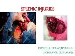
- 1. SPLENIC INJURIES PRESENTER: DR MANJUNATHA S B MODERATOR: DR BHARATH J
- 2. Contents Introduction Epidemiology Anatomy & Physiology Evaluation Management Surgical procedures Guidelines Conclusion References
- 3. History : • Hippocrates in the fourth century bc was one of the first to write on the spleen. He taught broadly on the need for balance and equilibrium between the patient and his environment. • The great ancient Roman physician, surgeon and philosopher Claudius Galen (129-216 AD) described the spleen as “Plenum mysterii organum” or “the organ full of mystery” as he struggled to elucidate its function. • In modern English, "to vent one's spleen" means to vent one's anger, e.g. by shouting (BAD TEMPER)
- 4. • In 1893, Reigner published the first documented successful splenectomy in the German literature. • Operative mortality rates remained high until the 1950s • Non operative care during this period was predominantly fatal. • With the widespread availability of computed tomography surgeons began to focus on those needing surgery and those who could be observed safely.
- 5. Anatomy: • Ovoid/wedge, usually purplish, pulpy mass • Largest lymphoid organ, part of lymphoreticular system • About size and shape of one’s fist • Most vulnerable abdominal organ for traumatic injury • Located in left upper quadrant or LHC • Protected by lower thoracic cage • Completely encircled and covered with peritoneum except at hilum
- 6. 1×3×5×7×9×11 rule • Size 1 inch thickness 3 inch wide 5 inch long • Weight 7 ounce= 200gm. • Related ribs 9-11 (along long axis of 10th rib)
- 7. ANTERIOR: Stomach POSTERIOR: Left diaphragm Lung Costodiaphragmatic recess 9-11 ribs INFERIOR: Left colic flexure MEDIAL: Left kidney and pancreas
- 9. Blood supply
- 11. • LIGAMENTS: • Gastrosplenic Short gastric vessels and left gastro-omental vessels • Splenorenal (lienorenal) splenic vessels and tail of pancreas • Splenocolic in contact with lower pole of spleen; at danger during spleenectomy
- 13. • BORDERS: • Superior(notched) border • Inferior border • Anterior border • ENDS: • Posterior end • Anterior end • SURFACES: • Diaphragmatic • Visceral
- 14. Anatomical variations: Accessory Spleens: splenenculi • MC congenital anomaly • 20 -30% incidence • More incidence in haematological diseases • Found near hilum >80%, and vascular pedicle
- 15. Locations in decreasing order: • Hilum 80% • Gastrocolic ligament • Tail of pancreas • Greater omentum • Greater curvature of stomach • Splenocolic ligament • Mesentry • Lt spermatic cord/ broad lig
- 16. Heterotaxy Syndromes: • Disturbance in the usual right left distribution of thoracic and abdominal organs. • Occurs from early embryological developmental disturbance. • 2 types • Right isomerism – Presents as CHD • Left isomerism – Intestinal malrotation / Mid gut volvulus
- 17. Asplenia syndrome: • Situs ambiguus with asplenia or Ivemark syndrome is characterized by an absent spleen and duplication of right- sided structures. • Affecting males twice as commonly as females.
- 18. Polysplenia syndrome: • Multiple small spleens. Also referred to as situs ambiguus with left isomerism. • Equally in males and females.
- 19. Physiology: • Filter of Reticulo endothelial system. • Humoral immunity, producing Ig M, Opsonins, tufstin and properidin. • Activation of complement system. • Source of extramedullary hematopoiesis.
- 20. Splenic injury: • Splenic injury most commonly occurs following blunt trauma due to motor vehicle collisions • Penetrating splenic trauma is less common than blunt injury • Iatrogenic traumatic injuries to the spleen can result from surgical or endoscopic manipulation colon, stomach, pancreas, Kidney. • Primary mechanism is capsular tear, laceration from retraction devices, or tension on the spleen during manipulation of the colon
- 21. • Importance of history- victims located on the left side of car • Type & nature of weapon is important in penetrating injuries • Caliber of the gun
- 22. • Blunt Trauma: Rapid deceleration(MVA) Direct blows to the abdomen(domestic violence, or leisure and play activities such as bicycling) • Penetrating Trauma • Combination of above explosive type injuries warfare and civilian bombing • Iatrogenic: Post procedure. • Spontaneous Rupture: Malaria, IMN
- 23. Types of Injury: • Splenic Hematoma Subcapsular Intraparenchymal • Lacerated wound • Clean incised wound • Hilar/vascualr injuries
- 24. Associated Injuries: • Fracture Left lower ribs (30 %) • Left sided hemothorax • Left lung and diaphragm injury • Left lobe liver injury • Tail of pancreas injury • Left kidney • Left colonic injury • Small bowel injury
- 25. Presentation • Pain abdomen • Shock • Pain in lower part of chest on left side • Abdominal distension • Wound in left upper quadrent • Symptoms of associated injuries
- 26. Examination : • Tachycardia • Feeble pulse • Hypotension • Tachypnea • Anemia
- 27. Examination: Per Abdomen • External bruise may be seen in LHC • Abdominal distension • Contusions • Decreased movement of LHC region • Tenderness in LHC or all over abdomen
- 28. Signs: • Kehr’s sign: Hans Kher, Germany • Clot collected under left diaphragm irritates it and the phrenic nerve( C3, C4) causing referred pain in left shoulder 15 minutes after foot end elevation • Because the supraclavicular nerves have the same cervical nerves origin as the phrenic nerve, C3 and C4
- 29. Signs: • Seat belt sign : Abdominal wall contusion or hematoma.
- 30. SIGNS: • BALLANCE SIGN: Charles Alfred Ballance,UK • Persistent dullness to percussion in the left flank due to coagulated blood • shifting dullness to percussion in the right flank due to fluid blood
- 31. Evaluation: • Clinical signs. • Hematology • Radiology • Prepare the team for surgery • Reserve ICU bed
- 32. Investigations: USG • FAST. • Look for position of spleen, hematoma, lacerration, hilar structures, vascualarity, hemoperitoneum. • Associated injuries to stomach, colon, Lt Kidney, Pancreas, Lt lobe of Liver, lower segments of Lung,
- 34. USG: Hematoma and Rupture
- 35. CECT Abdomen: • Hemoperitoneum – Localized fluid collections around the spleen (especially those with an elevated HU measurement) are highly suggestive of hemoperitoneum. • Briskly bleeding splenic lacerations may establish blood density fluid throughout the abdomen. • Hypodensity – Hypodense regions represent areas of parenchymal disruption, intraparenchymal hematoma or subcapsular hematoma. • Contrast blush or extravasation – Contrast blush describes hyperdense areas within the splenic parenchyma that represent traumatic disruption or pseudoaneurysm of the splenic vasculature. Active extravasation of contrast implies ongoing bleeding and the need for urgent intervention
- 36. AAST Grading:
- 38. AAST 1
- 39. AAST 2
- 40. AAST 3
- 41. AAST 4
- 42. AAST 5
- 43. Management Non-operative management of splenic injury (NOMSI) • Conservative • Interventional radiology: Splenic artery angio embolization Operative management • Splenorraphy: procedure to preserve spleen done in past, now replaced by NOMSI • Splenectomy
- 45. The standard criteria for NOM are: • Hemodynamic stability/ readily stabilizable • Lack of rebound and guarding • Blood transfusions ≤ 4 units • No lack of consciousness; • Age <55 yrs.
- 46. NOMSI: • Lower hospital cost • Earlier discharge • Avoiding nontherapeutic celiotomies (and their associated cost and morbidity), • Fewer intra-abdominal complications, and • Reduced transfusion rates
- 47. • The only absolute indication for emergency laparotomy is hemodynamic instability • Complex/severe splenic injuries, age, preexistent splenic diseases, number of units of transfused blood, brain injuries are no longer considered absolute contraindications for NOM
- 48. Splenic artery angioembolization: • Adjunct to NOM of high grade injuries 1. Proximal splenic artery embolization: Distal to dorsal pancreatic artery. 2. Distal selective embolization: High failure rate. Indicated in pts with evidence of active extravasation of dye or pseudo aneurysm
- 49. Unsuccessful NOM: Occurs in • Hemodynamic instability (systolic BP < 90 mmHg despite adequate resuscitation) • Age > 55 years old • > 4 units of transfused blood to maintain a Hblevel over > 10 g/dl • Persistent leucocytosis • The onset or aggravating sings of peritoneal irritation • Worsening imaging signs of splenic injury (repeated US exams)-post-traumatic splenic defect • Intra-abdominal compartment syndrome (intravesical pressure > 20 cm H2O).
- 50. Splenorrhaphy: • Parenchyma saving surgery of spleen • The technique is dictated by the magnitude of the splenic injury 1.superficial hemostatic strategies like fibrin glue,gel foam,argon beem coagulation,diathermy,topical thrombin 2.non absorbable suture repair 3.absorbable mesh wrap(poly galactin) 4.resectional debridement
- 55. Splenectomy: Partial • Grade IV to V splenic injury may require anatomic resection, including ligation of the lobar artery.
- 56. Total Splenectomy: Open Position: Supine GA Inciscion: midline, Left subcostal. In large spleens and trauma of other organs full midline laparotomy inciscions.
- 57. • Incision deepened to access the peritoneal cavity. • Pack the 4 quadrant of the peritoneal cavity • Suck out all free blood and clot • Remove packs starting from least area of bleeding. • Use your fingers to temporarily secure hemostasis at the hilum(to prevent clamping of the tail of pancreas) • Place the left hand on the spleen and draw it down to divide the lieno renal ligament lying posteriorly • Deliver the spleen into the abdominal incision • Then a non-crushing clamp is applied at the hilum safeguarding the pancreas • Examine the spleen for grade of injury • Ligate and divide; the short gastric arteries, left gastroepiploic arteries. Slightly away from the stomach with non absorbable suture
- 58. Approaches:
- 59. Ligation of the Splenic Pedicle:Anterior Approach • Clamp, incise, and ligate the left part of the gastrocolic ligament and the gastroepiploic artery and vein. • Locate the splenic artery at the superior border of the body of the pancreas. Carefully ligate the artery in continuity and doubly, with ligatures being placed as distally as possible
- 61. Ligation of the Splenic Pedicle: Posterior Approach • Hold the spleen medially. • Divide the splenorenal, splenophrenic, and splenocolic ligaments • Lift the spleen outside the peritoneal cavity, • being particularly careful with the tail of the pancreas. • Dissect rapidly and mobilize the bleeding spleen immediately.
- 62. • Bleeding can be controlled by manually compressing the splenic artery and vein and the tail of the pancreas between the thumb and index finger or with a non crushing clamp • Ligate the arterial and venous branches close to the hilum using 2–0 and 3–0 ligatures. Doubly ligate the splenic artery • Ligate the short gastric vessels. • Remove the spleen and secure any bleeding points. • Close the abdominal wall.
- 65. Complications: INTRAOPERATIVE • Haemorrhage • Pancreatic injury • Bowel injury(stomach & colon) • Diaphragmatic injury
- 66. Complications EARLY POST OP • Hematoma/seroma • Wound infection • Subphrenic abscess • Atelectasis • Pneumonia • Pleural effusion • Portal vein thrombosis • DVT • Paralytic ileus
- 68. OPSI • A rapidly fatal infection following removal of spleen • Incidence: 0.23-0.42% per year • Most Occur 1st 2 years after splenectomy • Lifetime risk <1-5%, • Common organisms • 1.S.pneumonia-50-90% • 2.H.influenza • 3.N.meningitis • Mortality rate : 50-80%
- 69. • Mechanism-organism with polysaccharide capsules need Opsonization with IGg3 or C3B which attaches to special macrophages found in the spleen • Post splenectomy patients lack of macrophages • Starts with flu like symptoms, Meningitis or sepsis • Rapidly progressive 12-48 hrs
- 70. • Other organisms include: streptococcus species, salmonella, Capnocytophaga canimorous, Babesiosis. High risk: • children<5yrs old/>50 yrs, • Splenectomy for Haemoglobiniopathies [Thalasemeia, sickle cell a], Myelodysplasia, malignancies.
- 71. Prevention: Immunoprophylaxis: • Vaccines aganst Strepto Pneumoniae [PPV23], H.Infl [H influenza type B], Meningococcus • Elective: At least 2 wks prior • Emergency: PPV 23 immediate post op & Other two 2 wks after surgery. • [All 3 delayed for at least 2wks; because transient immune suppression post op] Antibody titre: • No correlation between ab titre & clinical immunity • Only in about 50% cases protective levels abs formed against pneumococci • Revaccination: CDC [united statescommunicable disese control & prevention] to be revaccinated ppv 232-6 yrs after splencetomy.
- 72. Antibiotic prophylaxis: • Children: Until 5 yrs of age or at least 5 yrs after surgery • Penicillin, Amox, amoxyclav • Adults: scanty evidence. Provided with antibiotics to be taken at the sign of infection.
- 73. Prevention
- 74. EAST Guidelines: • Level 1 Patients who have diffuse peritonitis or who are hemodynamically unstable after blunt abdominal trauma should be taken urgently for laparotomy.
- 75. EAST Guidelines: • Level 2 1. A routine laparotomy is not indicated in the hemodynamically stable patient without peritonitis presenting with an isolated splenic injury. 2. The severity of splenic injury (as suggested by CT grade or degree of hemoperitoneum), neurologic status, age >55 and/or the presence of associated injuries are not contraindications to a trial of nonoperative management in a hemodynamically stable patient. 3. In the hemodynamically normal blunt abdominal trauma patient without peritonitis, an abdominal CT scan with intravenous contrast should be performed to identify and assess the severity of injury to the spleen.
- 76. EAST Guidelines: 4. Angiography should be considered for patients with AAST grade of greater than III injuries, presence of a contrast blush, moderate hemoperitoneum, or evidence of ongoing splenic bleeding. 5. Non operative management of splenic injuries should only be considered in an environment that provides capabilities for monitoring, serial clinical evaluations, and an operating room available for urgent laparotomy.
- 77. EAST Guidelines:Level 3 1. After blunt splenic injury, clinical factors such as a persistent systemic inflammatory response, increasing/persistent abdominal pain, or an otherwise unexplained drop in Hb should dictate the frequency of and need for follow-up imaging for a patient with blunt splenic injury. 2. Contrast blush on CT scan alone is not an absolute indication for an operation or angiographic intervention. Factors such as patient age, grade of injury, and presence of hypotension need to be considered in the clinical management of these patients. 3. Angiography may be used either as an adjunct to nonoperative management for patients who are thought to be at high risk for delayed bleeding or as an investigative tool to identify vascular abnormalities such as pseudoaneurysms that pose a risk for delayed hemorrhage. 4. Pharmacologic prophylaxis to prevent venous thromboembolism can be used for patients with isolated blunt splenic injuries without increasing the failure rate ofnonoperative management, although the optimal timing of safe initiation has not been determined.
- 78. Unanswered questions:??? 1. Frequency of Hb measurements 2. Frequency of abdominal examinations 3. Intensity and duration of monitoring 4. Is there a transfusion trigger after which operative or angiographic intervention should be considered? 5. Time to reinitiating oral intake 6. The duration and intensity of restricted activity (both in hospital and after discharge) 7. Optimum length of stay for both the intensive care unit (ICU) and hospital 8. Necessity of repeated imaging 9. Timing of initiating chemical deep venous thrombosis (DVT) prophylaxis after a splenic injury 10. Should patients with severe injuries/or embolized injuries receive postsplenectomy vaccines? 11. Is there an immunologic deficiency after splenic embolization?
- 79. Conclusion: • Spleen is important organ, try to conserve it. • Clinical examination has vital role in diagnosing and treating splenic injuries. • Activate the team as soon as splenic injury is suspected. • CECT is the investigation of choice. • Hemodynamically unstable patient : Directly to OR • Keep adequate blood ready before opening the abdomen. • Splenic artery embolisation has got definitive role.
- 80. • Enlarged spleens are more susceptible to injury. • Hemodynamic instability is the only absolute contraindication for NOMSI • Left LL Pneumonia is the MCC following splenectomy. • OPSI is the devastating sequelae of asplenia. • Prophylaxis against OPSI is must.
- 81. References: 1. Bailey & Love Short practice of Surgery,26th ed. 2. Sabiston Textbook of Surgery, 20th ed. 3. Schwartz Principles of Surgery, 9th ed. 4. Fischers Mastery of Surgery, Vol2, 6th ed. 5. Kirk’s General Surgical Operations, 6th ed. 6. Shakelford’s Surgery of Alimentary Tract, 7th ed. 7. EAST Guidelines. 8. Amith Ashish Surgery for PGMEE, 10th ed. 9. Velmahos GC, Chan LS, Kamel E, Murray JA, Yassa N, Kahaku D, Berne TV, Demetriades D. Nonoperative management of splenic injuries; have we gone too far ? Arch Surg. 2000; 135: 674-681. 10. Cocanour CS, Moore FA, Ware DN, Marvin RG, Clark M, Duke JH. Delayed complications of nonoperative management of blunt adult splenic trauma. Arch Surg. 1998;133: 619-625.
- 82. ...Thank you...