SlideShare a Scribd company logo
1 of 119
ANATOMICAL, BIOLOGICALAND HISTOLOGICAL
CONSIDERATION OF MAXILLARY ANATOMICAL
LANDMARKS
PRESENTED BY
Dr . KOMALPREET KAUR
( PG IST YEAR)
CONTENTS
1. Introduction
2. Osteology
3. Myology
4. Anatomy of supporting structures
I. Oral mucous membrane
II. Primary stress bearing areas
III. Secondary stress bearing areas
IV. Relief areas
3. Anatomy of limiting structure
4. REFERENCE
INTRODUCTION
 A prosthesis must function in harmony with the tissues that support
them and surround them.
 Dentist must understand the macroscopic and microscopic anatomy
of supporting and limiting structures of the denture
 The denture must extend as far as possible without interfering the
health of tissues
Maxillary denture is supported by two pairs of bone
 The maxillae
 And palatine bones
OSTEOLOGY
 The osseous structure not only support the dentures,
but have a direct bearing on
 Impression – making procedures ,
 the position of teeth ,
 and the contours of finished denture base.
 There are two maxillae, each consisting of a central
body and four processes
PALATINE PROCESS OF MAXILLA
 The palatine processes of maxillary bones arises as horizontal
plates from the body of maxilla.
 Two horizontal plates joins in the mid line forming a suture ,
mid palatal suture
 The horizontal palatine process of the maxillary bones appear to
resist resorption for longer period of time.
PALATINE BONE
 The horizontal plates of palatine bones articulate with
the posterior rough border of the horizontal process of
maxillae.
 Posterior border of the horizontal plates of the palatine
bones unite at the midline to form a sharp spine called as
POSTERIOR NASAL SPINE .
 It serves as attachment for the aponeurosis of soft palate.
The POSTERIOR PALTAL SEAL should follow the posterior
border of the hard palate:
ALVEOLAR BONE
 DEFINITION :The portion of maxilla and mandible that
forms and supports the tooth sockets.
 Based on functional adaptation : 2 types
1. Alveolar bone proper
2. Supporting alveolar bone
 Based on radiographic experience.
1. Type I
2. Type II
ALVEOLAR BONE PROPER
 The bony wall of tooth socket is alveolar bone proper.
 Compact bone
 Bundle bone
RADIOGRAPHIC APPEARANCE
 It appears as dense white line called as
Lamina dura
 SUPPORTING ALVEOLAR BONE
 It consists of two parallel plates of cortical bone( buccolingual
or labio lingual)
 Between cortical bone there is spongy bone.
 The labial and buccal cortical plates are relatively thin,
especially over the cuspids and central incisor.
 ALVEOLAR CREST
 Formed when inner and outer cortical plates meet.
o TYPE I : regular interradicular and interdental trabeculae ,
horizontal in ladder like arrangement
o common in mandible.
 TYPE II : irregularly arranged , numerous , delicate ,interdental and
interradicular trabeculae .
 Common in maxilla
PTERYGOID HAMULUS
 Although does not provide support for maxillary denture,
its position is in osseous limits of the maxillary denture
base
 It is thin , curved process at the terminal end of medial
pterygoid plate of sphenoid bone.
 It has two important relations , related to its function
1. Tensor veli palatine tendon passes immediately behind it.
2. Superior attachment of the pterygomandibular raphe
ZYGOMATIC PROCESS
 Distal to buccal frenum lies the root of zygoma , which
is located opposite the first molar region
 With increase in resorption of ridge , it becomes more
noticeable
 Relief is indicated.
MYOLOGY
 MUSCLES OF FACIAL EXPRESSION
 These muscles lies superficially under the skin
 the action of these muscles is responsible for the facial
postures associated with smiling , laughing or frowning .
ORIGINS
 The origins of several of the muscles of facial expression
are near enough to the denture-bearing areas that their
actions must be considered as definitely influencing the
denture borders.
 their influence is in proportion to the contour and quantity
of residual ridge present in a vertical direction.
 The higher the residual ridge the less influence will be
exerted.
INSERTION
 Bundles of muscles insert partly into the skin , partly into
the mucous membrane of the lips and the immediate
vicinity.
 Area situated laterally and slightly above the corner of
mouth is the concentration of many fibers of this muscle
group , this area is called MODIOLOUS
 The labial flanges of the maxillary denture frequently
need to be reduced lateromedially in the area of the
modiolus .
 Can be studied under following groups
 Orbital: occipito frontalis, orbicularis oculi,
corrugator supercilli
 Nasal: nasalis , procerus , depressor septi nasii
 Oral: orbicularis oris, levator anguli ori, depressor
anguli oris,risorus, mentalis , buccinator , levator
labii superiorus , depressor labii inferiorus ,
zygomaticus major , zygomaticus minor
ORBICULARIS ORIS
ORIGIN
Tissue surrounding lips
INSERTION
Underneath skin at corner of mouth.
FUNCTIONS
 Closes the mouth
 Purses and protrudes the lip.
 We use this muscle when we whistle or blow
BUCCINATOR
ORIGIN
Alveolar process of maxilla and mandible.
Pterygoid hamulus and pterygomandibular raphe
INSERTION
In the fibres of orbicularis oris
FUNCTIONS:
 Prevents the build up of food between cheeks and
teeth.
 Aids in forcefull expulsion of air from mouth.
LEVATOR LABI SUPERIORUS
ORIGIN
 Infra -orbital margin
INSERTION
 Skin and muscle of upper lip
FUNCTIONS
 Elevates the upper lip
ZYGMATICUS MINOR
 It is one of our smiling muscles.
FUNCTIONS
 Pull the corner of mouth upwards and outwards .
ZYGOMATICUS MAJOR
 ORIGIN
Zygomatic bone
 INSERTION
Underneath skin at corner of mouth
 FUNCTIONS
Draws the corner of mouth upwards
and outwards.
LEVATOR ANGULI ORIS
 Deep to zygomaticus major there is levator anguli
oris muscle
 ORIGIN
Anterior surface of maxilla
Below infra orbital foramen
 INSERTION
modiolous
 FUNCTIONS
Lifts the corner of mouth.
RISORIOUS
Gets its name from latin “risus” which means laughter.
ORIGIN
Fascia surrounding parotid salivary gland
INSERTION
Angle of mouth
FUNCTIONS
 Retracts the angle of mouth
DEPRESSOR ANGULI ORIS
ORIGIN
Anterolateral surface of body.
INSERTION
Angle of mouth.
FUNCTION
 Antagonizes levator anguli oris
 pulls the corner of mouth downwards.
DEPRESSOR LABII INFERIORIS
ORIGIN
Mandible between the anterior midline and the mental
foramen.
INSERTION
Skin of lower lip
FUNCTIONS
 Pulls the lower lip downwards and
outwards
MENTALIS
ORIGIN
Incisive fossa of mandible.
INSERTION
Skin of chin.
FUNCTIONS
 Lifts and protrude lower lip
SIGNIFICANCE
 If these are not properly supported , either by natural or
artificial dentition , none of the facial expression appear
normal.
 The nasolabial sulcus , the philtrum , the commissure of lips,
and mentolabial sulcus will not have their normal contour.
 Incorrectly contoured denture base or incorrectly positioned
teeth will destroy the normal tonicity of the muscles.
 Lack of support leads to sagging,
 stretching : retards the normal contracture of muscles and
results in loss of tonicity
MUSCLES OF SOFT PALATE
 Levator veli palatine
 Tensor veli palatine
 Glossopalatinus
 palatoglossus
 palatopharyngeus
LEVATOR VELI PALATINI
Origin
 petrous part of temporal bone
 inferior aspect of auditory tube
Insertion
 upper surface of palatal aponeurosis
action
 elevates the soft palate
TENSOR VELI PALATINI
Origin
 Lateral side of auditory tube
 Scaphoid fossa of sphenoid bone
Insertion
 Palatine aponeurosis
Action
 Tightens the soft palate
 Opens the auditory tube
PALATOPHARYNGEUS
 The wall of soft palte is formed principally by this
muscle.
Origin
 Superior surface of palatine aponeurosis
Insertion
 Pharyngeal wall
Action
 Depress the soft palate
 Elevates pharynx
PALATOGLOSSUS
Origin
 Inferior surface of palatine aponeurosis
Insertion
 Lateral margin of tongue.
Action
 Depress palate
 Moves palatoglossus arch toward midline
 Elevates back of tongue.
MUSCULUS UVULAE
Origin
 Posterior nasal spine of hard palate
Insertion
 Connective tissue uvulae
Action
 Elevates and retract uvula
 Thickens central of region of soft palate.
WHAT IS THE ANATOMICAL LANDMARK?
 Anatomical Landmark is a recognizable anatomic
structure used as a point of reference. [GPT-9]
 They acts as positive guides to the limits of the
impression
Impression surface of a denture is comprised of
 stress bearing areas/supporting areas
 Relief areas
 Peripheral or limiting areas
ANATOMY OF SUPPORTING STRUCTURES
 Foundation of denture is made up of bone of the hard
palate and residual ridge ,covered by mucous
membrane.
 The denture base rests on mucous membrane ,which
serves as a cushion between the base and supporting
bone.
MUCOUS MEMBRANE
 The mucous membrane is composed of
 Mucosa
 Sub –mucosa(it makes the bulk of mucous membrane)
MUCOSA
 Mucosa is formed by stratified squamous epithelium, which
often is keratinized
 subjacent narrow layer of connective tissue is known as
lamina propria.
 It exhibits regional modifications to suit the functional needs
of specific area
It can be classified as( based on function)
1. Masticatory mucosa
2. Lining / reflecting mucosa
3. Specialized mucosa
MASTICATORY MUCOSA:
 it is characterized by well defined keratinized layer on its
outermost surface.
 In edentulous patient the mucosa covering the hard
palate , and the crest of residual alveolar ridge ,including
the residual attached gingiva
LINING OR REFLECTING MUCOSA:
o Formed by non keratinized epithelium.
o And thin layer of connective tissue, the lamina propria.
o It covers lips , cheeks, labial & buccal mucosa, vestibular
fornix, slopes of residual ridge, ventral surface of tongue,
floor of mouth and soft palate
SPECIALIZED MUCOSA
o It is keratinized.
o Covers the dorsal surface of tongue , taste buds.
SUBMUCOSA
 Submucosa is formed of connective tissue that varies in
character from dense to loose areolar tissue and also
varies considerably in thickness.
 It may contain glandular, fat , or muscle cells and
transmits the blood and nerve supply to mucosa.
 The thickness and consistency of sub mucosa are largely
responsible for the support that the mucous membrane
affords a denture.
 In healthy mouth, submucosa is attached to the periosteum of
underlying bone and will usually successfully bears the
pressures of the denture.
 When submucosa is thin , the soft tissues will be non resilient
, and the mucous membrane will get traumatized easily.
 When it loosely attached to periosteum , or it is inflamed or
edematous , the tissue is easily displaceable , and the stability
and support of the denture are adversely affected.
STRESS BEARING AREAS
 These are load bearing areas
 Denture should be made such that most of the load is
concentrated on these areas
PRIMARY STRESS BEARING AREAS
 The horizontal portion of hard palate lateral to the midline
 Maxillary tuberosities
SECONDARY STRESS BEARING AREAS
 Residual alveolar ridge
 Rugae
*bouchers 13 edition
HARD PALATE
 The ultimate support for a
maxillary denture is
 Bone of two maxillae and
the palatine bone
HISTOLOGY
 Epithelium is keratinized throughout
 Mucous membrane is tightly fixed to the underlying
periosteum and therefore immovable
 Lamina propria ( a layer of dense CT) is thick in anterior
part and thin in posterior region)
 Submucosa: it is different in various regions
Anterolateral portion
With abundant adipose tissue
Posterolateral portion
With abundant gland tissue
Although this tissue is displaceable it contribute to support
 Primary support : horizontal portion of hard palate lateral to
mid line
MAXILLARY TUBEROSITY
 The maxillary tuberosity is the most posterior part of the
alveolar ridge
 lies distal to the position of the last molar
o The posterior part of the tuberosity rarely resorbs and it covered
with dense connective tissue
o SIGNIFICANCE
 denture base should cover the maxillary tuberosity and fill the
hamular notches .
 The last posterior tooth should not be placed on tuberosity.
CLINICAL SIGNIFICANCE
 The enlarged tuberosities limits the space and will
interfere the occlusal plane and distal extension of lower
denture.
 Surgical reduction should be done
RUGAE
 They are folds of irregular
mucous membrane extends
bilaterally from midline in the
region of upper 6 anteriors
and sometime bicuspid
 In the area of rugae palate is
set to an angle with residual
ridge .
 Resist the anterior
displacement of the denture
RESIDUAL RIDGE
 DEFINITION
 The portion of the alveolar ridge and its soft tissue covering
which remains following the removal of teeth- GPT
 The mucous membrane covering
the crest of ridge in a healthy
mouth is firmly attached to the
periosteum of bone
 Submucosa is characterized by
dense collagenous fibres that are
contiguous with the lamina propria
and provides adequate resiliency to
support of denture .
 provides good support , however
bone is subject to resorption, which
limits its potential for support ,
unlike palate which is resistant to
resorption
 SLOPES OD RESIDUAL ALVEOLAR RIDGE
 As the mucous membrane extends from the crest along
the slope of residual ridge, It loses its firm attachment to
underlying bone.
 Epithelium is non keratinized or slightly keratinized
 Submucosa contains loose connective tissue and elastic
fibres.
 This loosely attached tissue cannot withstand the forces
of mastication.
RELIEF AREAS
Mid palatine raphe
 It is the junction between two palatine processes of maxilla
 submucosa is extremely thin
 Mucosa is in close contact with the bone
CLINICAL SIGNIFICANCE
 Little or no stress can be placed in this region during the
making of the final impression or in the completed denture.
Otherwise, the denture will tend to rock over the centre of the
palate when vertical forces are applied to the teeth.
INCISIVE PAPILLA
 It is a small tubercle overlies incisive foramen
 Situated on a line immediately behind and between central
incisors.
 Nasopalatine nerve and vessels pass through it at right angle
to the bony foramen .
 As resorption progresses, it comes to lie nearer the crest of
ridge
 Anterior border and center of incisive papilla are
likely to change after extraction of incisor teeth
 the posterior border is relatively stable .
 Papilla becomes round after extraction of incisor
teeth due to changes in the anterior border.
 Gives positional relation to central incisor which are
8-10 mm anterior to the CENTRE incisive papilla.
 Beside this, canine–papilla line is a useful guide to
orient the upper canine teeth in complete dentures
 a perpendicular is drawn posterior to centre of incisive
papilla to saggital plane passes through canine.
CLINICAL SIGNFICANCE
 pressure should not be applied on this when final
impression is made
 Pressure obliteration of vessels/compression
nerves deprive of nutrition necrosis
and paraesthesia of anterior palate
FOVEA PALATINI
 Several ducts of mucous glands coalesce to form fovea
 This acts an arbitrary guide to locate the posterior border
of denture.
 Denture can extend 1-2 mm beyond this
 Secretion from this spreads as a thin film on denture
which aids in retention
 It should be relieved in a patient with thick ropy saliva.
TORUS PALATINUS
 This is a hard bony enlargement that occurs in midline of
the roof of the mouth
 Found in about 20% of population.
 FEMALE : MALE RATION 2:1
CLINICAL SIGNIFICANCE
 Relief is indicated for less extensive tori, and surgical
removal for more extensive tori.
 Removed when it has undercuts , if impedes the normal
movement of tongue , or if act as fulcrum.
ANATOMY OF LIMITING STRUCTURES
 Limiting structures: To determine and confine the extent
of denture:
1. Labial frenum
2. Labial vestibule
3. Buccal frenum
4. Buccal vestibule
5. Hamular notch
6. Vibrating line
LABIAL FRENUM
 The upper medial labial frenum, or frenulum, is a fold of
mucous membrane that overlies loose connective tissue .
 It contains no muscle fibers and insert in a vertical direction
 It anchors the upper lip to the gingiva.
 The frenum can vary in size
 Can be single or multiple
 In many edentulous patients, resorption of the alveolar bone
brings the crest of the alveolar ridge closer to the frenum.
CLINICAL CONSIDERATION
• Lip should be streched horizontally outwards for proper
recording of frenum
The labial notch must be wide and deep enough to allow the frenum
to pass through the lips without the manipulation of the lip
When it is abnormally large, it extends to the interdental papilla between the two
central incisors. An enlarged upper median labial frenum is frequently found in
association with a diastema .
LABIAL VESTIBULE
 Definition
 The portion of the oral cavity that is bounded on one side by
teeth , gingiva, and alveolar ridge(in edentulous mouth ,
residual ridge) and on other by lips anterior to buccal frenum.
GPT8
 Main , muscle of lip which forms the outer surface of
vestibule is orbicularis oris
 Its tone depends on the support given by labial denture flange
and position of teeth
HISTOLOGY
 The mucous membrane lining the vestibule is thin with
an epithelium that is non keratinized (lining mucosa)
 Submucosal layer is thick and contains large amount of
loose areolar tissue and elastic fibres.
FORNIX  The fornix of the vestibule is
the site where the mucous
membrane lining of the lips
and cheeks reflects and joins
the unattached gingiva, or
alveolar mucosa.
 The depth of the vestibule in
the upper and lower jaws is
determined by the site of the
fornix.
if while taking an upper impression, the lips are pulled
vertically instead of horizontally
artificially increase the depth of the vestibule
the denture will extend into the subcutaneous space
irritation of the mucosa / alteration of the facial appearance.
 Following extraction of teeth, alveolar bone undergoes
resorption and therefore the depth of the vestibule
become shallower.
 Surgical creation of a new fornix is required.
BUCCAL FRENUM
 The buccal frenum is a band of tissue that overlies the levator
anguli oris muscle.
 It is sometime single fold of mucous membrane , sometime
double and sometime broad and fan shaped
 Buccal frenum is part of continuous band of tissue going from
maxilla through the modiolous in the corner of the mouth to
the buccal frenum on the mandible .
 It has attachment of following muscles
• Levator anguli oris: affects the position of frenum
• Orbicularis oris: pulls the frenum forwards
• Buccinator: pulls frenum in backward
CLINICAL SIGNIFICANCE
 For the proper action of these muscles , proper clearance
should be provided in the denture
 Inadequate provision can cause dislodgement
 Cheek is elevated and then pulled outward, downward,
and inward and moved backward and forward to simulate
movement of frenum
BUCCAL VESTIBULE
 The buccal vestibule lies opposite the tuberosity
 extends from the buccal frenum to hamular notch
 the size of buccal vestibule varies with the
1. contraction of buccinator muscle ,
2. the position of mandible, and the
3. amount of bone lost from of the maxilla
 The size and the shape of the distal end of buccal flange
must be adjusted to accommodate ramus ,coronoid
process and masseter muscle in function
 When mandible is wide opened and moved laterally, the
width and height of this area is reduced.
 When masseter muscle contracts under heavy closing
prrssures it also reduces this space
CORONO-MAXILLARY SPACE
 The corono-maxillary space is that anatomic region that lies
medial to coronoid process ,lateral to maxillary tuberosity
and bounded anteriorly by base of zygomatic process and
posteriorly by pterygo-maxillary / hamular notch.
 Two types of individual anatomical variations can affect the
size of this space:
 With vertical variations, the space increases or does not vary
when the mouth is opened; therefore, the prosthesis border
must be thicker to obtain adequate retention.
 With lateral variations, the corono maxillary space is
reduced when the mouth is opened; therefore, the prosthesis
border must be thin.
The use of a mouth mirror provides adequate
information about the height of this space
 a diagnostic technique—a space impression tool—
has been proposed to clinically delineate the type of
space present and to help visualize dimensions.
 This technique consists of the use of a modified
tongue blade
 that, in conjunction with low-fusing impression
compound
 If the coronomaxillary space broadens or maintains
its size when the mouth is opened
 it is advisable not to have the patient open the
mouth wide, to protrude to move laterally during
border molding or impression procedures.
 A gentle molding of the region by pulling the cheek
out, down, and in will yield more successful results.
 For the patients, where opening the mandible can
result in narrowing of the space
 Border molding procedures in this region should
include opening and closing together with
protrusion and lateral movements of the jaw.
HAMULAR NOTCH
 Lying between maxillary tuberosity and pterygoid hamulus is a groove
called the hamular notch.
 distolateral border of denture should rest in hamular notch
 it is the soft area of loose areolar tissue , so can be displaced to achieve the
posterior palatal seal
 it is located by T-shaped burnisher or mouth mirror
CLINICAL SIGNIFICANCE
 Over extension can cause soreness due to pressure on
hamular process and interference of pterygomandibular
raphe
 If under extended or rest on tuberosity it will lost
retentive properties as the tissue there is non resilient and
border seal could not be achieved
POSTERIOR PALATAL SEAL
 It is defined as “ the soft tissues along the junction of the
hard and soft palates on which pressure within the
physiologic limits of the tissues can be applied by a
denture to aid in the retention of the denture.” GPT
RELEVANCE
 At the posterior aspect of the denture there is no cheek tissue
to seal the denture border , therefore provide optimum
retention.
 Horizontal forces and lateral torqueing of maxillary denture
can only be resisted by adequate border seal
 Terminating the denture borders at soft tissue will allow the
mucosa to move with the denture base during function and
thereby maintaining denture seal .
FUNCTIONS
 Primary purpose is retention of maxillary denture
 Maintains contact with the anterior portion of the soft palate
 the seal prevents passage of air between the denture and the
tissues ..
 Reduces the patient awareness to this area and also
subsequent reduction in area in gag reflex.
 Reduces the food accumulation beneath the posterior aspect
of denture.
 Reduces the patient discomfort
 Compensate for the volumetric shrinkage
 Creates a partial vacuum that only gets activated when
horizontal and tipping forces are directed against the
denture base
 The range of the soft palate movement and the degree of
displaceability of seal area differ in every individual
 House classification: (based on the angle that soft palate
makes with the hard palate and the soft palate muscle
activity that is essential to establish velo phayngeal)
closure
 Class I
 Class ii
 Class iii
 •In class I, the soft palate is horizontal as it extends
 It extends posteriorly, requiring minimal muscular
activity for velopharyngeal closure
 Allows more than 5 mm of seal area
 Class II type of soft palatal contour lie somewhere
between
class I and class III classes
 allows 1 to 5 mm of seal area
 In class III, the soft palate is more acute in relation to
the hard palate
 necessitating marked elevation of the musculature for
velopharyngeal closure
 permits a narrow seal of less than 1 mm
 The posterior palatal seal is
divided into two.
 The postpalatal seal
extends medially from one
tuberosity to the other.
 Laterally, the
pterygomaxillary seal
extends
 through hamular notch
continuing for 3 to 4 mm
anterolaterally
approximating the
mucogingival junction
SIGNIFICANCE
 Notch is covered by pterygomandibular fold, which
extends from the posterior aspect of the tuberosity
posterior – inferiorly to insert into retromolar pad.
 The fold of tissue can influence the posterior border seal
if the mouth is in a wide open position during the final
impression procedure..
VIBRATING LINES
 Anterior vibrating line is an imaginary line located at
the junction of the attached tissues overlying the hard
palate and movable tissues of the immediately adjacent
soft palate.
 This should not be confused with the junction of soft and
hard palate.
 It is always present on soft palate.
 How to locate anterior vibrating line?
 Ask the patient to perform valsalva maneuver , which
requires both nostrils be held firmly while patient blows
gently from nose.
 And can be visualized by instructing the patient to say
“ah” with short vigorous burst.
 Posterior vibrating line is an imaginary line at the
junction of aponeurosis of tensor veli palatine and
muscular portion of soft palate.
 It marks the most distal extension of denture base.
 How to locate posterior vibrating line?
 It can visualized by instructing the patient to say “ah” in
short burst in normal , un – exaggerated fashion.
Patient position during impression making of palatal
seal area
 this region should be recorded in function.
Therefore, an impression should be made when
 the patient is seated in upright position with head
flexed 30 degree forward, below FH plane to allow
the soft palate to reach its functionally depressed
position.
 The patients tongue should be placed under
tension against either the handle of the impression
tray or the dentist's finger .
REFERENCE
 BOUCHER’ S Prosthodontic treatment of edentulous patients (9th ,11th
and 12th edition 13th edition
 SHELDON WINKLER essentials of complete denture prosthodontics(2nd
edition)
 CHARLES M HEARTWELL syllabus of complete denture (4th edition)
 ORBAN’S oral histology and embryology(14th edition)
 ARTHUR O RAHN textbook of complete denture(6th edition)
 Coronomaxillary space and its significance in complete denture
retention-Case reportsJuly 2012General Dentistry 60(4):e263-
7SourcePubMed
 doi: 10.1007/s13191-012-0169-yThe Incisive Papilla: A
Significant Landmark in ProsthodonticsE. G. R. Solomon1 and K.
S. Arunachalam2
 https://www.ijoprd.com/doi/IJOPRD/pdf/10.5005/jp-journals-
10019-1020 posterior palatal seal a literture review
Maxillary landmarks

More Related Content

What's hot

Anatomical landmarks of maxilla and mandible [autosaved]
Anatomical landmarks of maxilla and mandible [autosaved]Anatomical landmarks of maxilla and mandible [autosaved]
Anatomical landmarks of maxilla and mandible [autosaved]Pooja Langote
 
compensating curve.ppt
compensating curve.pptcompensating curve.ppt
compensating curve.pptShrimant Raman
 
Intraoral Anatomical Landmarks
Intraoral Anatomical LandmarksIntraoral Anatomical Landmarks
Intraoral Anatomical LandmarksMohammed Fouad
 
Pulpal reactions to caries and dental procedures
Pulpal reactions to caries and dental proceduresPulpal reactions to caries and dental procedures
Pulpal reactions to caries and dental proceduresMohammed Alshehri
 
Complete denture impressions
Complete denture impressionsComplete denture impressions
Complete denture impressionsguest7e8451
 
hard and soft palate in prosthodontics
 hard and soft palate in prosthodontics hard and soft palate in prosthodontics
hard and soft palate in prosthodonticsmadhushreepatil5
 
Vertical jaw relation in Complete Dentures- Kelly
Vertical jaw relation in Complete Dentures- KellyVertical jaw relation in Complete Dentures- Kelly
Vertical jaw relation in Complete Dentures- KellyKelly Norton
 
Residual ridge resorption
Residual ridge resorptionResidual ridge resorption
Residual ridge resorptionNone None
 
Anatomical Landmarks Mandibular prosthodontics
Anatomical Landmarks Mandibular prosthodonticsAnatomical Landmarks Mandibular prosthodontics
Anatomical Landmarks Mandibular prosthodonticsSUBHRAKANTI PANDIT
 
04.principles of tooth preparation
04.principles of tooth preparation04.principles of tooth preparation
04.principles of tooth preparationDr.Jaffar Raza BDS
 
Soft liners and tissue conditioners
Soft liners and tissue conditionersSoft liners and tissue conditioners
Soft liners and tissue conditionersDHANANJAYSHETH1
 
Anatomical Landmarks of Mandible
Anatomical Landmarks of MandibleAnatomical Landmarks of Mandible
Anatomical Landmarks of MandibleSabnoor Aujla
 
TMJ and Its Applied Aspects in Prosthodontics
TMJ and Its Applied Aspects in ProsthodonticsTMJ and Its Applied Aspects in Prosthodontics
TMJ and Its Applied Aspects in ProsthodonticsTanmay Popat
 
Glass ionomer cement
Glass ionomer cementGlass ionomer cement
Glass ionomer cementAnkit Patel
 

What's hot (20)

Anatomical landmarks of maxilla and mandible [autosaved]
Anatomical landmarks of maxilla and mandible [autosaved]Anatomical landmarks of maxilla and mandible [autosaved]
Anatomical landmarks of maxilla and mandible [autosaved]
 
compensating curve.ppt
compensating curve.pptcompensating curve.ppt
compensating curve.ppt
 
Intraoral Anatomical Landmarks
Intraoral Anatomical LandmarksIntraoral Anatomical Landmarks
Intraoral Anatomical Landmarks
 
Inferior alveolar nerve
Inferior alveolar nerveInferior alveolar nerve
Inferior alveolar nerve
 
Pulpal reactions to caries and dental procedures
Pulpal reactions to caries and dental proceduresPulpal reactions to caries and dental procedures
Pulpal reactions to caries and dental procedures
 
Complete denture impressions
Complete denture impressionsComplete denture impressions
Complete denture impressions
 
hard and soft palate in prosthodontics
 hard and soft palate in prosthodontics hard and soft palate in prosthodontics
hard and soft palate in prosthodontics
 
Vertical jaw relation in Complete Dentures- Kelly
Vertical jaw relation in Complete Dentures- KellyVertical jaw relation in Complete Dentures- Kelly
Vertical jaw relation in Complete Dentures- Kelly
 
Residual ridge resorption
Residual ridge resorptionResidual ridge resorption
Residual ridge resorption
 
Anatomical Landmarks Mandibular prosthodontics
Anatomical Landmarks Mandibular prosthodonticsAnatomical Landmarks Mandibular prosthodontics
Anatomical Landmarks Mandibular prosthodontics
 
Anatomical landmarks of denture bearing area of.pptx final
Anatomical landmarks of denture bearing area of.pptx finalAnatomical landmarks of denture bearing area of.pptx final
Anatomical landmarks of denture bearing area of.pptx final
 
Deglutition
DeglutitionDeglutition
Deglutition
 
04.principles of tooth preparation
04.principles of tooth preparation04.principles of tooth preparation
04.principles of tooth preparation
 
Soft liners and tissue conditioners
Soft liners and tissue conditionersSoft liners and tissue conditioners
Soft liners and tissue conditioners
 
Anatomical Landmarks of Mandible
Anatomical Landmarks of MandibleAnatomical Landmarks of Mandible
Anatomical Landmarks of Mandible
 
TMJ and Its Applied Aspects in Prosthodontics
TMJ and Its Applied Aspects in ProsthodonticsTMJ and Its Applied Aspects in Prosthodontics
TMJ and Its Applied Aspects in Prosthodontics
 
2.anatomy of the denture foundation areas
2.anatomy  of the denture foundation areas2.anatomy  of the denture foundation areas
2.anatomy of the denture foundation areas
 
Landmarks
LandmarksLandmarks
Landmarks
 
Alveolar bone
Alveolar boneAlveolar bone
Alveolar bone
 
Glass ionomer cement
Glass ionomer cementGlass ionomer cement
Glass ionomer cement
 

Similar to Maxillary landmarks

Anatomical landmarks for edentulous patients and facial landmarks
Anatomical landmarks for edentulous patients and facial landmarksAnatomical landmarks for edentulous patients and facial landmarks
Anatomical landmarks for edentulous patients and facial landmarksMohammed Aziz
 
Osteology and mucose membrane of maxi & mandible
Osteology and mucose membrane of maxi & mandibleOsteology and mucose membrane of maxi & mandible
Osteology and mucose membrane of maxi & mandiblepranav verma
 
Maxillary and mandbular anatomical landmarks
Maxillary and mandbular anatomical landmarksMaxillary and mandbular anatomical landmarks
Maxillary and mandbular anatomical landmarksRajvi Nahar
 
anatomyandclinicalsignificanceofdenturebearingareas-160731195726-converted.pptx
anatomyandclinicalsignificanceofdenturebearingareas-160731195726-converted.pptxanatomyandclinicalsignificanceofdenturebearingareas-160731195726-converted.pptx
anatomyandclinicalsignificanceofdenturebearingareas-160731195726-converted.pptxSALAWUSAMUELADINOYI
 
significance of maxillary denture bearing area
significance of maxillary denture bearing area significance of maxillary denture bearing area
significance of maxillary denture bearing area Narayan Sukla
 
ANATOMICAL-LANDMARKS-OF-CD.pptx
ANATOMICAL-LANDMARKS-OF-CD.pptxANATOMICAL-LANDMARKS-OF-CD.pptx
ANATOMICAL-LANDMARKS-OF-CD.pptxAmanBobal1
 
Anatomical landmarks
Anatomical landmarksAnatomical landmarks
Anatomical landmarksBarkhaBhatia5
 
oral anatomy
oral anatomyoral anatomy
oral anatomyshammasm
 
Complete denture and denture material Seminar
Complete denture and denture material SeminarComplete denture and denture material Seminar
Complete denture and denture material Seminaralinoori55
 
MANDIBULAR ANATOMICAL LANDMARK APOORWA - Copy - Copy.pptx
MANDIBULAR ANATOMICAL LANDMARK APOORWA - Copy - Copy.pptxMANDIBULAR ANATOMICAL LANDMARK APOORWA - Copy - Copy.pptx
MANDIBULAR ANATOMICAL LANDMARK APOORWA - Copy - Copy.pptxDrApoorwaAwasthi
 
1. BIOLOGICAL CONSIDERATIONS IN MANDIBULAR IMPRESSION.pptx
1. BIOLOGICAL CONSIDERATIONS IN MANDIBULAR IMPRESSION.pptx1. BIOLOGICAL CONSIDERATIONS IN MANDIBULAR IMPRESSION.pptx
1. BIOLOGICAL CONSIDERATIONS IN MANDIBULAR IMPRESSION.pptxmanjulikatyagi
 
Muscle of mastication and muscle of facial expression
Muscle of mastication and muscle of facial expressionMuscle of mastication and muscle of facial expression
Muscle of mastication and muscle of facial expressionNitesh Singh
 
Myology related to complete denture.pdf
Myology related to 
 complete denture.pdfMyology related to 
 complete denture.pdf
Myology related to complete denture.pdfKirkuk University
 
Physiology of tooth form
Physiology of tooth formPhysiology of tooth form
Physiology of tooth formDeepa Bhaskaran
 
2. hand out
2. hand out2. hand out
2. hand outshammasm
 
Mandibular anatomical landmarks
Mandibular anatomical landmarksMandibular anatomical landmarks
Mandibular anatomical landmarksNishu Priya
 
Anatomy complete denture
Anatomy complete denture Anatomy complete denture
Anatomy complete denture sara mahmoud
 
Mastication, orientatory lines and dental xray
Mastication, orientatory lines and dental xrayMastication, orientatory lines and dental xray
Mastication, orientatory lines and dental xrayKarolinaSczkowska2
 

Similar to Maxillary landmarks (20)

Anatomical landmarks for edentulous patients and facial landmarks
Anatomical landmarks for edentulous patients and facial landmarksAnatomical landmarks for edentulous patients and facial landmarks
Anatomical landmarks for edentulous patients and facial landmarks
 
Osteology and mucose membrane of maxi & mandible
Osteology and mucose membrane of maxi & mandibleOsteology and mucose membrane of maxi & mandible
Osteology and mucose membrane of maxi & mandible
 
Maxillary and mandbular anatomical landmarks
Maxillary and mandbular anatomical landmarksMaxillary and mandbular anatomical landmarks
Maxillary and mandbular anatomical landmarks
 
anatomyandclinicalsignificanceofdenturebearingareas-160731195726-converted.pptx
anatomyandclinicalsignificanceofdenturebearingareas-160731195726-converted.pptxanatomyandclinicalsignificanceofdenturebearingareas-160731195726-converted.pptx
anatomyandclinicalsignificanceofdenturebearingareas-160731195726-converted.pptx
 
significance of maxillary denture bearing area
significance of maxillary denture bearing area significance of maxillary denture bearing area
significance of maxillary denture bearing area
 
ANATOMICAL-LANDMARKS-OF-CD.pptx
ANATOMICAL-LANDMARKS-OF-CD.pptxANATOMICAL-LANDMARKS-OF-CD.pptx
ANATOMICAL-LANDMARKS-OF-CD.pptx
 
Anatomical landmarks
Anatomical landmarksAnatomical landmarks
Anatomical landmarks
 
oral anatomy
oral anatomyoral anatomy
oral anatomy
 
Complete denture and denture material Seminar
Complete denture and denture material SeminarComplete denture and denture material Seminar
Complete denture and denture material Seminar
 
MANDIBULAR ANATOMICAL LANDMARK APOORWA - Copy - Copy.pptx
MANDIBULAR ANATOMICAL LANDMARK APOORWA - Copy - Copy.pptxMANDIBULAR ANATOMICAL LANDMARK APOORWA - Copy - Copy.pptx
MANDIBULAR ANATOMICAL LANDMARK APOORWA - Copy - Copy.pptx
 
1. BIOLOGICAL CONSIDERATIONS IN MANDIBULAR IMPRESSION.pptx
1. BIOLOGICAL CONSIDERATIONS IN MANDIBULAR IMPRESSION.pptx1. BIOLOGICAL CONSIDERATIONS IN MANDIBULAR IMPRESSION.pptx
1. BIOLOGICAL CONSIDERATIONS IN MANDIBULAR IMPRESSION.pptx
 
2.anatomy of the denture foundation areas
2.anatomy  of the denture foundation areas2.anatomy  of the denture foundation areas
2.anatomy of the denture foundation areas
 
Muscle of mastication and muscle of facial expression
Muscle of mastication and muscle of facial expressionMuscle of mastication and muscle of facial expression
Muscle of mastication and muscle of facial expression
 
Myology related to complete denture.pdf
Myology related to 
 complete denture.pdfMyology related to 
 complete denture.pdf
Myology related to complete denture.pdf
 
Physiology of tooth form
Physiology of tooth formPhysiology of tooth form
Physiology of tooth form
 
2. hand out
2. hand out2. hand out
2. hand out
 
Mandibular anatomical landmarks
Mandibular anatomical landmarksMandibular anatomical landmarks
Mandibular anatomical landmarks
 
Anatomy complete denture
Anatomy complete denture Anatomy complete denture
Anatomy complete denture
 
Mastication, orientatory lines and dental xray
Mastication, orientatory lines and dental xrayMastication, orientatory lines and dental xray
Mastication, orientatory lines and dental xray
 
osteology.pptx
osteology.pptxosteology.pptx
osteology.pptx
 

Recently uploaded

(Rocky) Jaipur Call Girl - 09521753030 Escorts Service 50% Off with Cash ON D...
(Rocky) Jaipur Call Girl - 09521753030 Escorts Service 50% Off with Cash ON D...(Rocky) Jaipur Call Girl - 09521753030 Escorts Service 50% Off with Cash ON D...
(Rocky) Jaipur Call Girl - 09521753030 Escorts Service 50% Off with Cash ON D...indiancallgirl4rent
 
Call Girls Ludhiana Just Call 9907093804 Top Class Call Girl Service Available
Call Girls Ludhiana Just Call 9907093804 Top Class Call Girl Service AvailableCall Girls Ludhiana Just Call 9907093804 Top Class Call Girl Service Available
Call Girls Ludhiana Just Call 9907093804 Top Class Call Girl Service AvailableDipal Arora
 
All Time Service Available Call Girls Marine Drive 📳 9820252231 For 18+ VIP C...
All Time Service Available Call Girls Marine Drive 📳 9820252231 For 18+ VIP C...All Time Service Available Call Girls Marine Drive 📳 9820252231 For 18+ VIP C...
All Time Service Available Call Girls Marine Drive 📳 9820252231 For 18+ VIP C...Arohi Goyal
 
Lucknow Call girls - 8800925952 - 24x7 service with hotel room
Lucknow Call girls - 8800925952 - 24x7 service with hotel roomLucknow Call girls - 8800925952 - 24x7 service with hotel room
Lucknow Call girls - 8800925952 - 24x7 service with hotel roomdiscovermytutordmt
 
♛VVIP Hyderabad Call Girls Chintalkunta🖕7001035870🖕Riya Kappor Top Call Girl ...
♛VVIP Hyderabad Call Girls Chintalkunta🖕7001035870🖕Riya Kappor Top Call Girl ...♛VVIP Hyderabad Call Girls Chintalkunta🖕7001035870🖕Riya Kappor Top Call Girl ...
♛VVIP Hyderabad Call Girls Chintalkunta🖕7001035870🖕Riya Kappor Top Call Girl ...astropune
 
Book Paid Powai Call Girls Mumbai 𖠋 9930245274 𖠋Low Budget Full Independent H...
Book Paid Powai Call Girls Mumbai 𖠋 9930245274 𖠋Low Budget Full Independent H...Book Paid Powai Call Girls Mumbai 𖠋 9930245274 𖠋Low Budget Full Independent H...
Book Paid Powai Call Girls Mumbai 𖠋 9930245274 𖠋Low Budget Full Independent H...Call Girls in Nagpur High Profile
 
💎VVIP Kolkata Call Girls Parganas🩱7001035870🩱Independent Girl ( Ac Rooms Avai...
💎VVIP Kolkata Call Girls Parganas🩱7001035870🩱Independent Girl ( Ac Rooms Avai...💎VVIP Kolkata Call Girls Parganas🩱7001035870🩱Independent Girl ( Ac Rooms Avai...
💎VVIP Kolkata Call Girls Parganas🩱7001035870🩱Independent Girl ( Ac Rooms Avai...Taniya Sharma
 
Call Girls Jabalpur Just Call 9907093804 Top Class Call Girl Service Available
Call Girls Jabalpur Just Call 9907093804 Top Class Call Girl Service AvailableCall Girls Jabalpur Just Call 9907093804 Top Class Call Girl Service Available
Call Girls Jabalpur Just Call 9907093804 Top Class Call Girl Service AvailableDipal Arora
 
Top Rated Bangalore Call Girls Richmond Circle ⟟ 8250192130 ⟟ Call Me For Gen...
Top Rated Bangalore Call Girls Richmond Circle ⟟ 8250192130 ⟟ Call Me For Gen...Top Rated Bangalore Call Girls Richmond Circle ⟟ 8250192130 ⟟ Call Me For Gen...
Top Rated Bangalore Call Girls Richmond Circle ⟟ 8250192130 ⟟ Call Me For Gen...narwatsonia7
 
Call Girls Faridabad Just Call 9907093804 Top Class Call Girl Service Available
Call Girls Faridabad Just Call 9907093804 Top Class Call Girl Service AvailableCall Girls Faridabad Just Call 9907093804 Top Class Call Girl Service Available
Call Girls Faridabad Just Call 9907093804 Top Class Call Girl Service AvailableDipal Arora
 
Call Girls Dehradun Just Call 9907093804 Top Class Call Girl Service Available
Call Girls Dehradun Just Call 9907093804 Top Class Call Girl Service AvailableCall Girls Dehradun Just Call 9907093804 Top Class Call Girl Service Available
Call Girls Dehradun Just Call 9907093804 Top Class Call Girl Service AvailableDipal Arora
 
Call Girls Service Jaipur Grishma WhatsApp ❤8445551418 VIP Call Girls Jaipur
Call Girls Service Jaipur Grishma WhatsApp ❤8445551418 VIP Call Girls JaipurCall Girls Service Jaipur Grishma WhatsApp ❤8445551418 VIP Call Girls Jaipur
Call Girls Service Jaipur Grishma WhatsApp ❤8445551418 VIP Call Girls Jaipurparulsinha
 
Russian Escorts Girls Nehru Place ZINATHI 🔝9711199012 ☪ 24/7 Call Girls Delhi
Russian Escorts Girls  Nehru Place ZINATHI 🔝9711199012 ☪ 24/7 Call Girls DelhiRussian Escorts Girls  Nehru Place ZINATHI 🔝9711199012 ☪ 24/7 Call Girls Delhi
Russian Escorts Girls Nehru Place ZINATHI 🔝9711199012 ☪ 24/7 Call Girls DelhiAlinaDevecerski
 
Call Girls Cuttack Just Call 9907093804 Top Class Call Girl Service Available
Call Girls Cuttack Just Call 9907093804 Top Class Call Girl Service AvailableCall Girls Cuttack Just Call 9907093804 Top Class Call Girl Service Available
Call Girls Cuttack Just Call 9907093804 Top Class Call Girl Service AvailableDipal Arora
 
Call Girls Bhubaneswar Just Call 9907093804 Top Class Call Girl Service Avail...
Call Girls Bhubaneswar Just Call 9907093804 Top Class Call Girl Service Avail...Call Girls Bhubaneswar Just Call 9907093804 Top Class Call Girl Service Avail...
Call Girls Bhubaneswar Just Call 9907093804 Top Class Call Girl Service Avail...Dipal Arora
 
Call Girls Bangalore Just Call 9907093804 Top Class Call Girl Service Available
Call Girls Bangalore Just Call 9907093804 Top Class Call Girl Service AvailableCall Girls Bangalore Just Call 9907093804 Top Class Call Girl Service Available
Call Girls Bangalore Just Call 9907093804 Top Class Call Girl Service AvailableDipal Arora
 
Best Rate (Hyderabad) Call Girls Jahanuma ⟟ 8250192130 ⟟ High Class Call Girl...
Best Rate (Hyderabad) Call Girls Jahanuma ⟟ 8250192130 ⟟ High Class Call Girl...Best Rate (Hyderabad) Call Girls Jahanuma ⟟ 8250192130 ⟟ High Class Call Girl...
Best Rate (Hyderabad) Call Girls Jahanuma ⟟ 8250192130 ⟟ High Class Call Girl...astropune
 
Low Rate Call Girls Kochi Anika 8250192130 Independent Escort Service Kochi
Low Rate Call Girls Kochi Anika 8250192130 Independent Escort Service KochiLow Rate Call Girls Kochi Anika 8250192130 Independent Escort Service Kochi
Low Rate Call Girls Kochi Anika 8250192130 Independent Escort Service KochiSuhani Kapoor
 
Call Girls Horamavu WhatsApp Number 7001035870 Meeting With Bangalore Escorts
Call Girls Horamavu WhatsApp Number 7001035870 Meeting With Bangalore EscortsCall Girls Horamavu WhatsApp Number 7001035870 Meeting With Bangalore Escorts
Call Girls Horamavu WhatsApp Number 7001035870 Meeting With Bangalore Escortsvidya singh
 
Call Girls Bareilly Just Call 9907093804 Top Class Call Girl Service Available
Call Girls Bareilly Just Call 9907093804 Top Class Call Girl Service AvailableCall Girls Bareilly Just Call 9907093804 Top Class Call Girl Service Available
Call Girls Bareilly Just Call 9907093804 Top Class Call Girl Service AvailableDipal Arora
 

Recently uploaded (20)

(Rocky) Jaipur Call Girl - 09521753030 Escorts Service 50% Off with Cash ON D...
(Rocky) Jaipur Call Girl - 09521753030 Escorts Service 50% Off with Cash ON D...(Rocky) Jaipur Call Girl - 09521753030 Escorts Service 50% Off with Cash ON D...
(Rocky) Jaipur Call Girl - 09521753030 Escorts Service 50% Off with Cash ON D...
 
Call Girls Ludhiana Just Call 9907093804 Top Class Call Girl Service Available
Call Girls Ludhiana Just Call 9907093804 Top Class Call Girl Service AvailableCall Girls Ludhiana Just Call 9907093804 Top Class Call Girl Service Available
Call Girls Ludhiana Just Call 9907093804 Top Class Call Girl Service Available
 
All Time Service Available Call Girls Marine Drive 📳 9820252231 For 18+ VIP C...
All Time Service Available Call Girls Marine Drive 📳 9820252231 For 18+ VIP C...All Time Service Available Call Girls Marine Drive 📳 9820252231 For 18+ VIP C...
All Time Service Available Call Girls Marine Drive 📳 9820252231 For 18+ VIP C...
 
Lucknow Call girls - 8800925952 - 24x7 service with hotel room
Lucknow Call girls - 8800925952 - 24x7 service with hotel roomLucknow Call girls - 8800925952 - 24x7 service with hotel room
Lucknow Call girls - 8800925952 - 24x7 service with hotel room
 
♛VVIP Hyderabad Call Girls Chintalkunta🖕7001035870🖕Riya Kappor Top Call Girl ...
♛VVIP Hyderabad Call Girls Chintalkunta🖕7001035870🖕Riya Kappor Top Call Girl ...♛VVIP Hyderabad Call Girls Chintalkunta🖕7001035870🖕Riya Kappor Top Call Girl ...
♛VVIP Hyderabad Call Girls Chintalkunta🖕7001035870🖕Riya Kappor Top Call Girl ...
 
Book Paid Powai Call Girls Mumbai 𖠋 9930245274 𖠋Low Budget Full Independent H...
Book Paid Powai Call Girls Mumbai 𖠋 9930245274 𖠋Low Budget Full Independent H...Book Paid Powai Call Girls Mumbai 𖠋 9930245274 𖠋Low Budget Full Independent H...
Book Paid Powai Call Girls Mumbai 𖠋 9930245274 𖠋Low Budget Full Independent H...
 
💎VVIP Kolkata Call Girls Parganas🩱7001035870🩱Independent Girl ( Ac Rooms Avai...
💎VVIP Kolkata Call Girls Parganas🩱7001035870🩱Independent Girl ( Ac Rooms Avai...💎VVIP Kolkata Call Girls Parganas🩱7001035870🩱Independent Girl ( Ac Rooms Avai...
💎VVIP Kolkata Call Girls Parganas🩱7001035870🩱Independent Girl ( Ac Rooms Avai...
 
Call Girls Jabalpur Just Call 9907093804 Top Class Call Girl Service Available
Call Girls Jabalpur Just Call 9907093804 Top Class Call Girl Service AvailableCall Girls Jabalpur Just Call 9907093804 Top Class Call Girl Service Available
Call Girls Jabalpur Just Call 9907093804 Top Class Call Girl Service Available
 
Top Rated Bangalore Call Girls Richmond Circle ⟟ 8250192130 ⟟ Call Me For Gen...
Top Rated Bangalore Call Girls Richmond Circle ⟟ 8250192130 ⟟ Call Me For Gen...Top Rated Bangalore Call Girls Richmond Circle ⟟ 8250192130 ⟟ Call Me For Gen...
Top Rated Bangalore Call Girls Richmond Circle ⟟ 8250192130 ⟟ Call Me For Gen...
 
Call Girls Faridabad Just Call 9907093804 Top Class Call Girl Service Available
Call Girls Faridabad Just Call 9907093804 Top Class Call Girl Service AvailableCall Girls Faridabad Just Call 9907093804 Top Class Call Girl Service Available
Call Girls Faridabad Just Call 9907093804 Top Class Call Girl Service Available
 
Call Girls Dehradun Just Call 9907093804 Top Class Call Girl Service Available
Call Girls Dehradun Just Call 9907093804 Top Class Call Girl Service AvailableCall Girls Dehradun Just Call 9907093804 Top Class Call Girl Service Available
Call Girls Dehradun Just Call 9907093804 Top Class Call Girl Service Available
 
Call Girls Service Jaipur Grishma WhatsApp ❤8445551418 VIP Call Girls Jaipur
Call Girls Service Jaipur Grishma WhatsApp ❤8445551418 VIP Call Girls JaipurCall Girls Service Jaipur Grishma WhatsApp ❤8445551418 VIP Call Girls Jaipur
Call Girls Service Jaipur Grishma WhatsApp ❤8445551418 VIP Call Girls Jaipur
 
Russian Escorts Girls Nehru Place ZINATHI 🔝9711199012 ☪ 24/7 Call Girls Delhi
Russian Escorts Girls  Nehru Place ZINATHI 🔝9711199012 ☪ 24/7 Call Girls DelhiRussian Escorts Girls  Nehru Place ZINATHI 🔝9711199012 ☪ 24/7 Call Girls Delhi
Russian Escorts Girls Nehru Place ZINATHI 🔝9711199012 ☪ 24/7 Call Girls Delhi
 
Call Girls Cuttack Just Call 9907093804 Top Class Call Girl Service Available
Call Girls Cuttack Just Call 9907093804 Top Class Call Girl Service AvailableCall Girls Cuttack Just Call 9907093804 Top Class Call Girl Service Available
Call Girls Cuttack Just Call 9907093804 Top Class Call Girl Service Available
 
Call Girls Bhubaneswar Just Call 9907093804 Top Class Call Girl Service Avail...
Call Girls Bhubaneswar Just Call 9907093804 Top Class Call Girl Service Avail...Call Girls Bhubaneswar Just Call 9907093804 Top Class Call Girl Service Avail...
Call Girls Bhubaneswar Just Call 9907093804 Top Class Call Girl Service Avail...
 
Call Girls Bangalore Just Call 9907093804 Top Class Call Girl Service Available
Call Girls Bangalore Just Call 9907093804 Top Class Call Girl Service AvailableCall Girls Bangalore Just Call 9907093804 Top Class Call Girl Service Available
Call Girls Bangalore Just Call 9907093804 Top Class Call Girl Service Available
 
Best Rate (Hyderabad) Call Girls Jahanuma ⟟ 8250192130 ⟟ High Class Call Girl...
Best Rate (Hyderabad) Call Girls Jahanuma ⟟ 8250192130 ⟟ High Class Call Girl...Best Rate (Hyderabad) Call Girls Jahanuma ⟟ 8250192130 ⟟ High Class Call Girl...
Best Rate (Hyderabad) Call Girls Jahanuma ⟟ 8250192130 ⟟ High Class Call Girl...
 
Low Rate Call Girls Kochi Anika 8250192130 Independent Escort Service Kochi
Low Rate Call Girls Kochi Anika 8250192130 Independent Escort Service KochiLow Rate Call Girls Kochi Anika 8250192130 Independent Escort Service Kochi
Low Rate Call Girls Kochi Anika 8250192130 Independent Escort Service Kochi
 
Call Girls Horamavu WhatsApp Number 7001035870 Meeting With Bangalore Escorts
Call Girls Horamavu WhatsApp Number 7001035870 Meeting With Bangalore EscortsCall Girls Horamavu WhatsApp Number 7001035870 Meeting With Bangalore Escorts
Call Girls Horamavu WhatsApp Number 7001035870 Meeting With Bangalore Escorts
 
Call Girls Bareilly Just Call 9907093804 Top Class Call Girl Service Available
Call Girls Bareilly Just Call 9907093804 Top Class Call Girl Service AvailableCall Girls Bareilly Just Call 9907093804 Top Class Call Girl Service Available
Call Girls Bareilly Just Call 9907093804 Top Class Call Girl Service Available
 

Maxillary landmarks

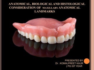
  • 1. ANATOMICAL, BIOLOGICALAND HISTOLOGICAL CONSIDERATION OF MAXILLARY ANATOMICAL LANDMARKS PRESENTED BY Dr . KOMALPREET KAUR ( PG IST YEAR)
  • 2. CONTENTS 1. Introduction 2. Osteology 3. Myology 4. Anatomy of supporting structures I. Oral mucous membrane II. Primary stress bearing areas III. Secondary stress bearing areas IV. Relief areas 3. Anatomy of limiting structure 4. REFERENCE
  • 3. INTRODUCTION  A prosthesis must function in harmony with the tissues that support them and surround them.  Dentist must understand the macroscopic and microscopic anatomy of supporting and limiting structures of the denture  The denture must extend as far as possible without interfering the health of tissues
  • 4. Maxillary denture is supported by two pairs of bone  The maxillae  And palatine bones
  • 5. OSTEOLOGY  The osseous structure not only support the dentures, but have a direct bearing on  Impression – making procedures ,  the position of teeth ,  and the contours of finished denture base.
  • 6.  There are two maxillae, each consisting of a central body and four processes
  • 7. PALATINE PROCESS OF MAXILLA  The palatine processes of maxillary bones arises as horizontal plates from the body of maxilla.  Two horizontal plates joins in the mid line forming a suture , mid palatal suture  The horizontal palatine process of the maxillary bones appear to resist resorption for longer period of time.
  • 9.  The horizontal plates of palatine bones articulate with the posterior rough border of the horizontal process of maxillae.  Posterior border of the horizontal plates of the palatine bones unite at the midline to form a sharp spine called as POSTERIOR NASAL SPINE .  It serves as attachment for the aponeurosis of soft palate.
  • 10. The POSTERIOR PALTAL SEAL should follow the posterior border of the hard palate:
  • 11. ALVEOLAR BONE  DEFINITION :The portion of maxilla and mandible that forms and supports the tooth sockets.  Based on functional adaptation : 2 types 1. Alveolar bone proper 2. Supporting alveolar bone  Based on radiographic experience. 1. Type I 2. Type II
  • 12. ALVEOLAR BONE PROPER  The bony wall of tooth socket is alveolar bone proper.  Compact bone  Bundle bone RADIOGRAPHIC APPEARANCE  It appears as dense white line called as Lamina dura
  • 13.  SUPPORTING ALVEOLAR BONE  It consists of two parallel plates of cortical bone( buccolingual or labio lingual)  Between cortical bone there is spongy bone.  The labial and buccal cortical plates are relatively thin, especially over the cuspids and central incisor.  ALVEOLAR CREST  Formed when inner and outer cortical plates meet.
  • 14. o TYPE I : regular interradicular and interdental trabeculae , horizontal in ladder like arrangement o common in mandible.
  • 15.  TYPE II : irregularly arranged , numerous , delicate ,interdental and interradicular trabeculae .  Common in maxilla
  • 16. PTERYGOID HAMULUS  Although does not provide support for maxillary denture, its position is in osseous limits of the maxillary denture base  It is thin , curved process at the terminal end of medial pterygoid plate of sphenoid bone.
  • 17.  It has two important relations , related to its function 1. Tensor veli palatine tendon passes immediately behind it. 2. Superior attachment of the pterygomandibular raphe
  • 18. ZYGOMATIC PROCESS  Distal to buccal frenum lies the root of zygoma , which is located opposite the first molar region  With increase in resorption of ridge , it becomes more noticeable  Relief is indicated.
  • 19. MYOLOGY  MUSCLES OF FACIAL EXPRESSION  These muscles lies superficially under the skin  the action of these muscles is responsible for the facial postures associated with smiling , laughing or frowning .
  • 20. ORIGINS  The origins of several of the muscles of facial expression are near enough to the denture-bearing areas that their actions must be considered as definitely influencing the denture borders.  their influence is in proportion to the contour and quantity of residual ridge present in a vertical direction.  The higher the residual ridge the less influence will be exerted.
  • 21. INSERTION  Bundles of muscles insert partly into the skin , partly into the mucous membrane of the lips and the immediate vicinity.  Area situated laterally and slightly above the corner of mouth is the concentration of many fibers of this muscle group , this area is called MODIOLOUS  The labial flanges of the maxillary denture frequently need to be reduced lateromedially in the area of the modiolus .
  • 22.  Can be studied under following groups  Orbital: occipito frontalis, orbicularis oculi, corrugator supercilli  Nasal: nasalis , procerus , depressor septi nasii  Oral: orbicularis oris, levator anguli ori, depressor anguli oris,risorus, mentalis , buccinator , levator labii superiorus , depressor labii inferiorus , zygomaticus major , zygomaticus minor
  • 23. ORBICULARIS ORIS ORIGIN Tissue surrounding lips INSERTION Underneath skin at corner of mouth. FUNCTIONS  Closes the mouth  Purses and protrudes the lip.
  • 24.  We use this muscle when we whistle or blow
  • 25. BUCCINATOR ORIGIN Alveolar process of maxilla and mandible. Pterygoid hamulus and pterygomandibular raphe INSERTION In the fibres of orbicularis oris FUNCTIONS:  Prevents the build up of food between cheeks and teeth.  Aids in forcefull expulsion of air from mouth.
  • 26.
  • 27. LEVATOR LABI SUPERIORUS ORIGIN  Infra -orbital margin INSERTION  Skin and muscle of upper lip FUNCTIONS  Elevates the upper lip
  • 28. ZYGMATICUS MINOR  It is one of our smiling muscles. FUNCTIONS  Pull the corner of mouth upwards and outwards .
  • 29. ZYGOMATICUS MAJOR  ORIGIN Zygomatic bone  INSERTION Underneath skin at corner of mouth  FUNCTIONS Draws the corner of mouth upwards and outwards.
  • 30. LEVATOR ANGULI ORIS  Deep to zygomaticus major there is levator anguli oris muscle  ORIGIN Anterior surface of maxilla Below infra orbital foramen  INSERTION modiolous  FUNCTIONS Lifts the corner of mouth.
  • 31. RISORIOUS Gets its name from latin “risus” which means laughter. ORIGIN Fascia surrounding parotid salivary gland INSERTION Angle of mouth FUNCTIONS  Retracts the angle of mouth
  • 32. DEPRESSOR ANGULI ORIS ORIGIN Anterolateral surface of body. INSERTION Angle of mouth. FUNCTION  Antagonizes levator anguli oris  pulls the corner of mouth downwards.
  • 33. DEPRESSOR LABII INFERIORIS ORIGIN Mandible between the anterior midline and the mental foramen. INSERTION Skin of lower lip FUNCTIONS  Pulls the lower lip downwards and outwards
  • 34. MENTALIS ORIGIN Incisive fossa of mandible. INSERTION Skin of chin. FUNCTIONS  Lifts and protrude lower lip
  • 35. SIGNIFICANCE  If these are not properly supported , either by natural or artificial dentition , none of the facial expression appear normal.  The nasolabial sulcus , the philtrum , the commissure of lips, and mentolabial sulcus will not have their normal contour.  Incorrectly contoured denture base or incorrectly positioned teeth will destroy the normal tonicity of the muscles.  Lack of support leads to sagging,  stretching : retards the normal contracture of muscles and results in loss of tonicity
  • 36. MUSCLES OF SOFT PALATE  Levator veli palatine  Tensor veli palatine  Glossopalatinus  palatoglossus  palatopharyngeus
  • 37.
  • 38. LEVATOR VELI PALATINI Origin  petrous part of temporal bone  inferior aspect of auditory tube Insertion  upper surface of palatal aponeurosis action  elevates the soft palate
  • 39. TENSOR VELI PALATINI Origin  Lateral side of auditory tube  Scaphoid fossa of sphenoid bone Insertion  Palatine aponeurosis Action  Tightens the soft palate  Opens the auditory tube
  • 40. PALATOPHARYNGEUS  The wall of soft palte is formed principally by this muscle. Origin  Superior surface of palatine aponeurosis Insertion  Pharyngeal wall Action  Depress the soft palate  Elevates pharynx
  • 41. PALATOGLOSSUS Origin  Inferior surface of palatine aponeurosis Insertion  Lateral margin of tongue. Action  Depress palate  Moves palatoglossus arch toward midline  Elevates back of tongue.
  • 42. MUSCULUS UVULAE Origin  Posterior nasal spine of hard palate Insertion  Connective tissue uvulae Action  Elevates and retract uvula  Thickens central of region of soft palate.
  • 43. WHAT IS THE ANATOMICAL LANDMARK?  Anatomical Landmark is a recognizable anatomic structure used as a point of reference. [GPT-9]  They acts as positive guides to the limits of the impression
  • 44. Impression surface of a denture is comprised of  stress bearing areas/supporting areas  Relief areas  Peripheral or limiting areas
  • 45. ANATOMY OF SUPPORTING STRUCTURES  Foundation of denture is made up of bone of the hard palate and residual ridge ,covered by mucous membrane.  The denture base rests on mucous membrane ,which serves as a cushion between the base and supporting bone.
  • 46. MUCOUS MEMBRANE  The mucous membrane is composed of  Mucosa  Sub –mucosa(it makes the bulk of mucous membrane)
  • 47.
  • 48. MUCOSA  Mucosa is formed by stratified squamous epithelium, which often is keratinized  subjacent narrow layer of connective tissue is known as lamina propria.  It exhibits regional modifications to suit the functional needs of specific area It can be classified as( based on function) 1. Masticatory mucosa 2. Lining / reflecting mucosa 3. Specialized mucosa
  • 49. MASTICATORY MUCOSA:  it is characterized by well defined keratinized layer on its outermost surface.  In edentulous patient the mucosa covering the hard palate , and the crest of residual alveolar ridge ,including the residual attached gingiva
  • 50. LINING OR REFLECTING MUCOSA: o Formed by non keratinized epithelium. o And thin layer of connective tissue, the lamina propria. o It covers lips , cheeks, labial & buccal mucosa, vestibular fornix, slopes of residual ridge, ventral surface of tongue, floor of mouth and soft palate
  • 51. SPECIALIZED MUCOSA o It is keratinized. o Covers the dorsal surface of tongue , taste buds.
  • 52. SUBMUCOSA  Submucosa is formed of connective tissue that varies in character from dense to loose areolar tissue and also varies considerably in thickness.  It may contain glandular, fat , or muscle cells and transmits the blood and nerve supply to mucosa.  The thickness and consistency of sub mucosa are largely responsible for the support that the mucous membrane affords a denture.
  • 53.  In healthy mouth, submucosa is attached to the periosteum of underlying bone and will usually successfully bears the pressures of the denture.  When submucosa is thin , the soft tissues will be non resilient , and the mucous membrane will get traumatized easily.  When it loosely attached to periosteum , or it is inflamed or edematous , the tissue is easily displaceable , and the stability and support of the denture are adversely affected.
  • 54. STRESS BEARING AREAS  These are load bearing areas  Denture should be made such that most of the load is concentrated on these areas PRIMARY STRESS BEARING AREAS  The horizontal portion of hard palate lateral to the midline  Maxillary tuberosities SECONDARY STRESS BEARING AREAS  Residual alveolar ridge  Rugae *bouchers 13 edition
  • 55. HARD PALATE  The ultimate support for a maxillary denture is  Bone of two maxillae and the palatine bone
  • 56. HISTOLOGY  Epithelium is keratinized throughout  Mucous membrane is tightly fixed to the underlying periosteum and therefore immovable  Lamina propria ( a layer of dense CT) is thick in anterior part and thin in posterior region)  Submucosa: it is different in various regions
  • 57. Anterolateral portion With abundant adipose tissue Posterolateral portion With abundant gland tissue Although this tissue is displaceable it contribute to support
  • 58.  Primary support : horizontal portion of hard palate lateral to mid line
  • 59. MAXILLARY TUBEROSITY  The maxillary tuberosity is the most posterior part of the alveolar ridge  lies distal to the position of the last molar
  • 60. o The posterior part of the tuberosity rarely resorbs and it covered with dense connective tissue o SIGNIFICANCE  denture base should cover the maxillary tuberosity and fill the hamular notches .  The last posterior tooth should not be placed on tuberosity.
  • 61. CLINICAL SIGNIFICANCE  The enlarged tuberosities limits the space and will interfere the occlusal plane and distal extension of lower denture.  Surgical reduction should be done
  • 62. RUGAE  They are folds of irregular mucous membrane extends bilaterally from midline in the region of upper 6 anteriors and sometime bicuspid  In the area of rugae palate is set to an angle with residual ridge .  Resist the anterior displacement of the denture
  • 63. RESIDUAL RIDGE  DEFINITION  The portion of the alveolar ridge and its soft tissue covering which remains following the removal of teeth- GPT
  • 64.  The mucous membrane covering the crest of ridge in a healthy mouth is firmly attached to the periosteum of bone  Submucosa is characterized by dense collagenous fibres that are contiguous with the lamina propria and provides adequate resiliency to support of denture .  provides good support , however bone is subject to resorption, which limits its potential for support , unlike palate which is resistant to resorption
  • 65.  SLOPES OD RESIDUAL ALVEOLAR RIDGE  As the mucous membrane extends from the crest along the slope of residual ridge, It loses its firm attachment to underlying bone.  Epithelium is non keratinized or slightly keratinized  Submucosa contains loose connective tissue and elastic fibres.  This loosely attached tissue cannot withstand the forces of mastication.
  • 66. RELIEF AREAS Mid palatine raphe  It is the junction between two palatine processes of maxilla
  • 67.  submucosa is extremely thin  Mucosa is in close contact with the bone
  • 68. CLINICAL SIGNIFICANCE  Little or no stress can be placed in this region during the making of the final impression or in the completed denture. Otherwise, the denture will tend to rock over the centre of the palate when vertical forces are applied to the teeth.
  • 69. INCISIVE PAPILLA  It is a small tubercle overlies incisive foramen  Situated on a line immediately behind and between central incisors.  Nasopalatine nerve and vessels pass through it at right angle to the bony foramen .  As resorption progresses, it comes to lie nearer the crest of ridge
  • 70.  Anterior border and center of incisive papilla are likely to change after extraction of incisor teeth  the posterior border is relatively stable .  Papilla becomes round after extraction of incisor teeth due to changes in the anterior border.
  • 71.  Gives positional relation to central incisor which are 8-10 mm anterior to the CENTRE incisive papilla.
  • 72.  Beside this, canine–papilla line is a useful guide to orient the upper canine teeth in complete dentures  a perpendicular is drawn posterior to centre of incisive papilla to saggital plane passes through canine.
  • 73. CLINICAL SIGNFICANCE  pressure should not be applied on this when final impression is made  Pressure obliteration of vessels/compression nerves deprive of nutrition necrosis and paraesthesia of anterior palate
  • 74. FOVEA PALATINI  Several ducts of mucous glands coalesce to form fovea  This acts an arbitrary guide to locate the posterior border of denture.  Denture can extend 1-2 mm beyond this  Secretion from this spreads as a thin film on denture which aids in retention  It should be relieved in a patient with thick ropy saliva.
  • 75.
  • 76. TORUS PALATINUS  This is a hard bony enlargement that occurs in midline of the roof of the mouth  Found in about 20% of population.  FEMALE : MALE RATION 2:1
  • 77. CLINICAL SIGNIFICANCE  Relief is indicated for less extensive tori, and surgical removal for more extensive tori.  Removed when it has undercuts , if impedes the normal movement of tongue , or if act as fulcrum.
  • 78. ANATOMY OF LIMITING STRUCTURES  Limiting structures: To determine and confine the extent of denture: 1. Labial frenum 2. Labial vestibule 3. Buccal frenum 4. Buccal vestibule 5. Hamular notch 6. Vibrating line
  • 79. LABIAL FRENUM  The upper medial labial frenum, or frenulum, is a fold of mucous membrane that overlies loose connective tissue .  It contains no muscle fibers and insert in a vertical direction  It anchors the upper lip to the gingiva.  The frenum can vary in size  Can be single or multiple  In many edentulous patients, resorption of the alveolar bone brings the crest of the alveolar ridge closer to the frenum.
  • 80. CLINICAL CONSIDERATION • Lip should be streched horizontally outwards for proper recording of frenum
  • 81. The labial notch must be wide and deep enough to allow the frenum to pass through the lips without the manipulation of the lip
  • 82. When it is abnormally large, it extends to the interdental papilla between the two central incisors. An enlarged upper median labial frenum is frequently found in association with a diastema .
  • 83. LABIAL VESTIBULE  Definition  The portion of the oral cavity that is bounded on one side by teeth , gingiva, and alveolar ridge(in edentulous mouth , residual ridge) and on other by lips anterior to buccal frenum. GPT8  Main , muscle of lip which forms the outer surface of vestibule is orbicularis oris  Its tone depends on the support given by labial denture flange and position of teeth
  • 84. HISTOLOGY  The mucous membrane lining the vestibule is thin with an epithelium that is non keratinized (lining mucosa)  Submucosal layer is thick and contains large amount of loose areolar tissue and elastic fibres.
  • 85. FORNIX  The fornix of the vestibule is the site where the mucous membrane lining of the lips and cheeks reflects and joins the unattached gingiva, or alveolar mucosa.  The depth of the vestibule in the upper and lower jaws is determined by the site of the fornix.
  • 86. if while taking an upper impression, the lips are pulled vertically instead of horizontally artificially increase the depth of the vestibule the denture will extend into the subcutaneous space irritation of the mucosa / alteration of the facial appearance.
  • 87.  Following extraction of teeth, alveolar bone undergoes resorption and therefore the depth of the vestibule become shallower.  Surgical creation of a new fornix is required.
  • 88. BUCCAL FRENUM  The buccal frenum is a band of tissue that overlies the levator anguli oris muscle.  It is sometime single fold of mucous membrane , sometime double and sometime broad and fan shaped  Buccal frenum is part of continuous band of tissue going from maxilla through the modiolous in the corner of the mouth to the buccal frenum on the mandible .
  • 89.  It has attachment of following muscles • Levator anguli oris: affects the position of frenum • Orbicularis oris: pulls the frenum forwards • Buccinator: pulls frenum in backward
  • 90. CLINICAL SIGNIFICANCE  For the proper action of these muscles , proper clearance should be provided in the denture  Inadequate provision can cause dislodgement
  • 91.  Cheek is elevated and then pulled outward, downward, and inward and moved backward and forward to simulate movement of frenum
  • 92. BUCCAL VESTIBULE  The buccal vestibule lies opposite the tuberosity  extends from the buccal frenum to hamular notch  the size of buccal vestibule varies with the 1. contraction of buccinator muscle , 2. the position of mandible, and the 3. amount of bone lost from of the maxilla
  • 93.  The size and the shape of the distal end of buccal flange must be adjusted to accommodate ramus ,coronoid process and masseter muscle in function  When mandible is wide opened and moved laterally, the width and height of this area is reduced.  When masseter muscle contracts under heavy closing prrssures it also reduces this space
  • 94. CORONO-MAXILLARY SPACE  The corono-maxillary space is that anatomic region that lies medial to coronoid process ,lateral to maxillary tuberosity and bounded anteriorly by base of zygomatic process and posteriorly by pterygo-maxillary / hamular notch.
  • 95.  Two types of individual anatomical variations can affect the size of this space:  With vertical variations, the space increases or does not vary when the mouth is opened; therefore, the prosthesis border must be thicker to obtain adequate retention.  With lateral variations, the corono maxillary space is reduced when the mouth is opened; therefore, the prosthesis border must be thin.
  • 96.
  • 97. The use of a mouth mirror provides adequate information about the height of this space
  • 98.  a diagnostic technique—a space impression tool— has been proposed to clinically delineate the type of space present and to help visualize dimensions.  This technique consists of the use of a modified tongue blade  that, in conjunction with low-fusing impression compound
  • 99.  If the coronomaxillary space broadens or maintains its size when the mouth is opened  it is advisable not to have the patient open the mouth wide, to protrude to move laterally during border molding or impression procedures.  A gentle molding of the region by pulling the cheek out, down, and in will yield more successful results.
  • 100.  For the patients, where opening the mandible can result in narrowing of the space  Border molding procedures in this region should include opening and closing together with protrusion and lateral movements of the jaw.
  • 101. HAMULAR NOTCH  Lying between maxillary tuberosity and pterygoid hamulus is a groove called the hamular notch.  distolateral border of denture should rest in hamular notch  it is the soft area of loose areolar tissue , so can be displaced to achieve the posterior palatal seal  it is located by T-shaped burnisher or mouth mirror
  • 102. CLINICAL SIGNIFICANCE  Over extension can cause soreness due to pressure on hamular process and interference of pterygomandibular raphe  If under extended or rest on tuberosity it will lost retentive properties as the tissue there is non resilient and border seal could not be achieved
  • 103. POSTERIOR PALATAL SEAL  It is defined as “ the soft tissues along the junction of the hard and soft palates on which pressure within the physiologic limits of the tissues can be applied by a denture to aid in the retention of the denture.” GPT
  • 104. RELEVANCE  At the posterior aspect of the denture there is no cheek tissue to seal the denture border , therefore provide optimum retention.  Horizontal forces and lateral torqueing of maxillary denture can only be resisted by adequate border seal  Terminating the denture borders at soft tissue will allow the mucosa to move with the denture base during function and thereby maintaining denture seal .
  • 105. FUNCTIONS  Primary purpose is retention of maxillary denture  Maintains contact with the anterior portion of the soft palate  the seal prevents passage of air between the denture and the tissues ..  Reduces the patient awareness to this area and also subsequent reduction in area in gag reflex.  Reduces the food accumulation beneath the posterior aspect of denture.
  • 106.  Reduces the patient discomfort  Compensate for the volumetric shrinkage  Creates a partial vacuum that only gets activated when horizontal and tipping forces are directed against the denture base
  • 107.  The range of the soft palate movement and the degree of displaceability of seal area differ in every individual  House classification: (based on the angle that soft palate makes with the hard palate and the soft palate muscle activity that is essential to establish velo phayngeal) closure  Class I  Class ii  Class iii
  • 108.  •In class I, the soft palate is horizontal as it extends  It extends posteriorly, requiring minimal muscular activity for velopharyngeal closure  Allows more than 5 mm of seal area
  • 109.  Class II type of soft palatal contour lie somewhere between class I and class III classes  allows 1 to 5 mm of seal area
  • 110.  In class III, the soft palate is more acute in relation to the hard palate  necessitating marked elevation of the musculature for velopharyngeal closure  permits a narrow seal of less than 1 mm
  • 111.  The posterior palatal seal is divided into two.  The postpalatal seal extends medially from one tuberosity to the other.  Laterally, the pterygomaxillary seal extends  through hamular notch continuing for 3 to 4 mm anterolaterally approximating the mucogingival junction
  • 112. SIGNIFICANCE  Notch is covered by pterygomandibular fold, which extends from the posterior aspect of the tuberosity posterior – inferiorly to insert into retromolar pad.  The fold of tissue can influence the posterior border seal if the mouth is in a wide open position during the final impression procedure..
  • 113. VIBRATING LINES  Anterior vibrating line is an imaginary line located at the junction of the attached tissues overlying the hard palate and movable tissues of the immediately adjacent soft palate.  This should not be confused with the junction of soft and hard palate.  It is always present on soft palate.
  • 114.  How to locate anterior vibrating line?  Ask the patient to perform valsalva maneuver , which requires both nostrils be held firmly while patient blows gently from nose.  And can be visualized by instructing the patient to say “ah” with short vigorous burst.
  • 115.
  • 116.  Posterior vibrating line is an imaginary line at the junction of aponeurosis of tensor veli palatine and muscular portion of soft palate.  It marks the most distal extension of denture base.  How to locate posterior vibrating line?  It can visualized by instructing the patient to say “ah” in short burst in normal , un – exaggerated fashion.
  • 117. Patient position during impression making of palatal seal area  this region should be recorded in function. Therefore, an impression should be made when  the patient is seated in upright position with head flexed 30 degree forward, below FH plane to allow the soft palate to reach its functionally depressed position.  The patients tongue should be placed under tension against either the handle of the impression tray or the dentist's finger .
  • 118. REFERENCE  BOUCHER’ S Prosthodontic treatment of edentulous patients (9th ,11th and 12th edition 13th edition  SHELDON WINKLER essentials of complete denture prosthodontics(2nd edition)  CHARLES M HEARTWELL syllabus of complete denture (4th edition)  ORBAN’S oral histology and embryology(14th edition)  ARTHUR O RAHN textbook of complete denture(6th edition)  Coronomaxillary space and its significance in complete denture retention-Case reportsJuly 2012General Dentistry 60(4):e263- 7SourcePubMed  doi: 10.1007/s13191-012-0169-yThe Incisive Papilla: A Significant Landmark in ProsthodonticsE. G. R. Solomon1 and K. S. Arunachalam2  https://www.ijoprd.com/doi/IJOPRD/pdf/10.5005/jp-journals- 10019-1020 posterior palatal seal a literture review