SYNOVIA FLUIDS.pptx
•Download as PPTX, PDF•
0 likes•166 views

BIOCHEMISTRY . BODY FLUIDS

Recommended
More Related Content
What's hot
What's hot (20)
Similar to SYNOVIA FLUIDS.pptx
Similar to SYNOVIA FLUIDS.pptx (20)
More from International Medicine School - Management and Science University
More from International Medicine School - Management and Science University (20)
Recently uploaded
Recently uploaded (20)
SYNOVIA FLUIDS.pptx
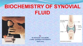
- 1. By Dr KHALED ALGARIRI CAMS- QASSIM UNIVERSITY April 2023
- 2. Synovial Fluid Synovial syn(like) + ovia (egg) “Joint Fluid INTRODUCTION
- 3. Synovial Fluid Viscous fluid found in the cavities of movable joints. Synovial fluid is necessary for normal joint function. Synovial fluid moves into the cartilage when a joint is resting, and moves out the joint space when the joint is active, particularly when the joint is engaged in a weight-bearing activity such as exercise
- 5. Secretion of Synovial Fluid Synovial tissue is composed of connective tissue that lacks a basement membrane. There are two main types of synovial lining cells: Type A cells are macrophage-like and have primarily a phagocytic function which is important to remove microbes and the debris that results from normal wear and tear in the joint. Type B cells are fibroblast-like and produce hyaluronate, which accounts for the increased viscosity of synovial fluid. • .
- 6. Composition of Synovial Fluid Synovial fluid is made of hyaluronic acid and lubricin, proteinases, and collagenases. Normal synovial fluid contains 3-4 mg/ml hyaluronate (hyaluronic acid), a polymer of disaccharides composed of D-glucuronic acid and D-N-acetylglucosamine joined by alternating beta-1,4 and beta-1,3 glycosidic bonds. Synovial fluid also contains lubricin secreted by synovial cells. It is chiefly responsible for so-called boundary-layer lubrication, which reduces friction between opposing surfaces of cartilage. It may also have a role in synovial cell growth.
- 7. Functions of Synovial A- Supplying oxygen and nutrients of articular cartilage by acting as a transport medium for nutritional substances, such as glucose. Articular cartilage has no blood, nerve, or lymphatic supply. Supplying oxygen and nutrients to and removing carbon dioxide and metabolic wastes from the chondrocytes within articular cartilage. Synovial fluid is believed to have two main functions:
- 9. Functions of Synovial B- To aid in the mechanical function of joints by lubrication of the articulating surfaces. Lubrication reduces frictional resistance between bearing surfaces by keeping them apart. During movement, the synovial fluid held in the cartilage is squeezed out mechanically to maintain a layer of fluid on the cartilage surface. The lubricating properties of synovial fluid on articular cartilage are due to the presence of hyaluronate or mucin and a glycoprotein in it.
- 10. Synovial Fluid Analysis A synovial fluid analysis is used to help diagnose the cause of joint pain and inflammation. Inflammation is the body's response to injury or infection. It can cause pain, swelling, redness, and loss of function in the affected area. Causes of joint problems include: 1-Osteoarthritis, the most common form of arthritis. It is a chronic, progressive disease that causes joint cartilage to break down. It can be painful and lead to loss of mobility and function. 2-Gout, a type of arthritis that causes inflammation in one or more joints, usually in the big toe.
- 11. 3-Rheumatoid arthritis, a condition in which the body's immune system attacks healthy cells in your joints 4-Joint effusion, a condition that happens when too much fluid builds up around a joint. It often affects the knee. When it affects the knee, it may be referred to as knee effusion or fluid on the knee. Synovial Fluid Analysis
- 12. 5-Infection in a joint such as septic arthritis 6-Bleeding disorder, such as hemophilia. Hemophilia is an inherited disorder that can cause excessive bleeding. Sometimes the excess blood ends up in the synovial fluid Synovial Fluid Analysis
- 13. Physical Characteristics of Synovial Fluid Normal synovial fluid is clear, pale yellow, viscid, and does not clot. Volume: The amount of fluid contained in joints is usually small. The knee joint normally contains up to 4 mL of fluid. An increase in synovial fluid enough to aspirates is due to some disease.
- 14. Physical Characteristics of Synovial Fluid Viscosity: Synovial fluid is very viscous due to its high concentration of polymerized hyaluronic acid. A string test can be used to evaluate the level of synovial fluid viscosity. After removing the needle or cap from the syringe, synovial fluid is expressed into a test tube one drop at a time. Normal synovial fluid will form a “string” approximately 5 cm long before breaking. In addition, the fluid may cling to the side of the test tube rather than running down to the bottom. Low viscosity of synovial indicates the presence of an inflammatory process.
- 15. Color and clarity Normal synovial fluid is colorless and clear. Other appearances may indicate various disease states. Loss of clarity : crystals, increased cellularity, infective, cartilage debris E.g. Yellow/clear (non inflammatory effusions), yellow/cloudy (inflammatory processes), white/ cloudy (crystals); and red brown (hemorrhage).
- 16. Physical Characteristics of Synovial Fluid Clot formation: Because of lack of fibrinogen and other clotting factors synovial fluid doesn’t clot Inflammation allow the plasma clotting factors to escape in to joint fluid which then clot
- 18. Microscopic Examinations Cell count and differential staining Cell count should be finished without any delay to avoid clumping of leukocytes. • Degeneration of Leukocytes happens if stored more than 1 hour after sample collection • If the sample is very thick , to facilitate cell counting sample can be incubated at 37°C with hyaluronidase enzyme. • RBCs are usually very low in number. Traumatic tap may result in high number
- 19. Microscopic Examinations Types of cells seen 1-Lymphocytes 2- Synovial tissue cells 3-Polymorpho nuclear cells 4-Macrophages 5-Fat bodies 6-Bacteria
- 20. BIOCHEMICAL EXAMINATIONS These high–molecular-weight proteins include fibrinogen, beta 2 macroglobulin, and alpha 2 macroglobulin, and can be absent or present in very low amounts. Most commonly used serum protein procedures can be used to measure synovial fluid protein. The normal range for synovial fluid protein is 1–3 g/dL. • Protein: Synovial fluid contains all proteins found in plasma, except various high– molecular weight proteins.
- 21. Increased synovial fluid protein levels are seen in ankylosing spondylitis, arthritis, arthropathies that accompany Crohn disease, gout, psoriasis, Reiter syndrome, and ulcerative colitis. Reiter syndrome ulcerative colitis psoriasis
- 23. BIOCHEMICAL EXAMINATIONS Glucose: Synovial fluid glucose levels should be interpreted using serum glucose levels. A fasting specimen should be used or at least one 6–8 hours ally, synovial fluid glucose levels are less than 10 mg/dL lower than serum levels. Joint disorders that are classified as infectious demonstrate large decreases in synovial fluid glucose and can be as much as 20–100 mg/dL less than serum levels. Other groups of joint disorders demonstrate a less of a decrease in synovial fluid glucose, 0–20 mg/dL
- 24. BIOCHEMICAL EXAMINATIONS • Uric acid: Synovial fluid uric acid normally ranges from 6 to 8 mg/dL. • The presence of uric acid in synovial fluid is helpful in diagnosis gout. • Usually, crystal identification is used for this determination, but synovial fluid uric acid levels may be performed in laboratories that do not have a light polarizing microscope.
- 25. • Lactic acid: Lactic acid is rarely measured in synovial fluid but can be helpful in diagnosing septic arthritis. Lactate levels in the synovial fluid have the highest diagnostic potential for predicting septic arthritis. • Normally, synovial fluid lactate is less than 25 mg/dL but can be as high as 1000 mg/dL in septic arthritis.
- 26. BIOCHEMICAL EXAMINATIONS Enzymes: Alkaline phosphatase, acid phosphatase, lactic dehydrogenase, and other enzymes are present in detectable quantities. Synovial fluid to serum ratios of these and other enzymes vary with the presence of articular disease. Enzymes enter the synovial fluid directly from the plasma or may be produced locally by the synovial membrane or released by synovial fluid macrophages.
- 27. BIOCHEMICAL EXAMINATION Lactate dehydrogenase: Lactate dehydrogenase (LD) can be elevated in synovial fluid, while serum levels remain normal. Synovial fluid LD levels are usually increased in RA, infectious arthritis, and gout.
- 28. BIOCHEMICAL EXAMINATION Complement levels: Normal synovial fluid complement levels in humans are approximately 10% of the serum values. In the inflamed joint synovial fluid complement levels will vary. The long-term patterns of variation have some prognostic value in human rheumatoid arthritis patients. Rheumatoid factor (RF) is an antibody to immunoglobulins. RF is present in the serum of most patients with RA, whereas just more than half of these patients will demonstrate RF in synovial fluid.
Editor's Notes
- An increase in synovial fluid enough to aspirates is due to some disease.
- String test showing normal synovial fluid viscosity. Non- inflammatory conditions : Normal viscosity Inflammatory : low viscosity due to reduced content of hyaluronate & reduced polymerization (low molecular wt.) Inflammatory : fluid falling as free droplets Will be variable in case of septic and hemorrhagic
- Reiter's syndrome, also known as reactive arthritis, is the classic triad of conjunctivitis, urethritis, and arthritis occurring after an infection, particularly those in the urogenital orgastrointestinal tract.
- Lactate levels in the synovial fluid have the highest diagnostic potential for predicting septic arthritis.
- Vascular permeability and synovial membrane permeability are altered by inflammation, which accounts for protein content changes in diseased synovial fluid. Immunoglobulins, immune complexes, and complement are produced by cells accumulating in the inflamed synovial membrane and periarticular lymph nodes and find their way to the synovial fluid.