
Recommended
More Related Content
Similar to eye (general physiology & Anatomy).pptx
Similar to eye (general physiology & Anatomy).pptx (20)
More from kashischaudhary
More from kashischaudhary (15)
Recently uploaded
Recently uploaded (20)
eye (general physiology & Anatomy).pptx
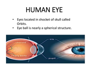
- 1. HUMAN EYE • Eyes located in shocket of skull called Orbits. • Eye ball is nearly a spherical structure.
- 3. Eye muscles
- 4. Layer of eye
- 5. • The wall of the eye ball its composed of three layers. • The external layer -a dense connective tissue called the sclera. • The anterior portion of eye sclera layer is called the cornea. • The middle layer, choroid, contains many blood vessels and looks bluish in colour. • The choroid layer is thin over the posterior two- third of the eye ball, but its become thick in the anterior part of cilliary body.
- 7. • The ciliary body itself continues forward to from a pigmented and opaque structure called the iris which is the visible coloured portion of the eye. • The eye ball contains a trsanparent crystalline lens which is held in place by ligaments attached to the ciliary body. • In front of the lens, the aperture surrounded by the iris is called the pupil. • The diameter of the pupil is regulated by the iris is called the pupil
- 8. 3 layers of neural cells in retina • The inner layer is the retina and its contains three layer of nerual cell-from inside to outside - ganglion cell, bipolor cells and photoreceptor cell.
- 10. 2 Type Photoreceptor cells 1. Rods 2. Cones
- 11. • These cells contain the Light-sensitive proteins called the photo pigments. • The day light(photopic) vision and colour vision are function of cones and the twilight (Scotopic) vision is the function of the rods. • The rods contain a pruplish-red protein called the rhodosin or visual purple, which contains a derivative of vitamine A.
- 12. • In the human eye, there are three types of cones which possess their own characteristic photo pigments that respond to red, green and blue light. • The sensations of the different colours are produced by various combinations of these cones and their photo pigments. • When these cones are stimulated equally, a sensation of which light is produced.
- 13. Optic nerve
- 15. • The optic nerves leave the eye and the retina blood vessels enter it at a point medial to and slightly above the posterior pole of the eye ball. • At the posterior pole of the eye lateral to the blind spot, there is a yellowish pigmented spot called macula lutea with a central pit called the fovea. • The fovea is a thinned-out portion of the point where ghe retina where the only the cones are densely packed. It is the point where the visual acuity(resolution) is the greatest. • Photoreceptor cell are not present in that region and hence it is called the blind spot.
- 17. Optic and other eye nerve
- 18. Fluid filled chamber of eye • The space between the cornea and the lens is called the aquous chamber and contains a thin watery fluid called aquous humor. • The space between the lens and retina is called the vitreous chamber and is filled with a transparent gel called vitreous humor.
- 21. Mechanisms of vision • The light rays in visible wavelength focussed on the retina through the cornea & lens generate potentials (impulses) in rods & cones. • The photosensitive compounds(Photopigments) in the human eye is composed of opsin (a protin)and retainal (an aldehyde of vitamin A). • Light induces dissociation of the retinal from opsin resulting in change in the structure of the opsin. This cause membrane permeability changes.
- 23. • As a result, potential differences are generated in the photoreceptor cell. • This produces a signal that generates action potentials in the in the ganglion cells through the bipolar cells. • This action potentials (impulse) are transmitted by the optic nerves to the visual cortex area of the brain. Where the neural impulses are analysed and the image formed on the retina is recognised based on earlier memory and experience.