2. DNA binding activities through dimerization comes from X-
ray crystallography, pulldown assays, chemical cross-linking,
gel filtration, analytical ultracentrifugation, and mutagenesis
studies and has been inferred from its kinetic profile for DNA
binding (22, 25–27, 42, 44, 45, 47, 69, 72, 81). Consistent with
a dimer autorepression mechanism, TBP mutations that impair
dimerization cause transcriptional derepression in vivo (24,
45). Their effect is dominant to wild-type TBP but is partially
suppressed by wild-type TBP overexpression (45). TBP over-
expression might drive unstable dimer mutants into somewhat
more stable heterodimers with wild-type TBP.
Despite several observations that support a physiological
role for TBP dimerization (45), the topic remains controversial
(20). Ultimately, we wish to understand how a variety of TBP
inhibitory mechanisms are coordinated to regulate TBP. A
necessary step in this effort is the mapping of surfaces on TBP
that are targeted for inhibition in vivo and the identification of
factors responsible for that inhibition. In an effort to more
completely examine the potential physiological significance of
TBP dimerization, we mutated 24 amino acids that comprise
the crystallographic dimer interface. Using in vitro pulldown
and electrophoretic mobility assays, we characterized their
abilities to dimerize, bind TAND, and bind TATA DNA. The
binding data were in good agreement with the crystallographic
and NMR structures of these or related complexes.
Next, we sought to determine whether mutations along the
crystallographic dimer interface affect a variety of phenotypes
in Saccharomyces cerevisiae that we previously linked to defects
in dimerization (24, 45). These include increased TBP turn-
over, transcriptional derepression, partial dominant inhibition
of cell growth (toxicity), and synthetic toxicity in a ⌬TAND
strain. With the collection of mutations throughout the dimer
interface, we observed a correlation between these phenotypes
and relative dimer stability measured in vitro. Mutations that
knock out interfaces between TBP and other inhibitors did not
give similar phenotypes. Taken together, the data provide fur-
ther support for the notion that TBP dimerizes in vivo and is a
physiologically important negative regulator of gene expres-
sion.
MATERIALS AND METHODS
Plasmids. All TBP mutations were created by oligonucleotide-directed mu-
tagenesis. The mutations and the integrity of the entire open reading frame were
confirmed in both the bacterial and yeast expression vectors by DNA sequencing.
Escherichia coli plasmids expressing the His-TBP mutants were designated
pET16b-yTBP(xxx), where xxx indicates the mutation. The plasmid pGEX-
TFIID-C, which encodes human GST-TBP(core), has been described previously
(41). Plasmid pGEX-yTBP(181C) has been described previously (45). The plas-
mid pGEX-scTAF1(10-88) was constructed by PCR amplification of the TAF1
gene and was inserted in frame with the glutathione S-transferase (GST)-coding
sequence of pGEX-3X. Derivatives of this plasmid, pGEX-scTAF1(10-88,
D66K) and pGEX-scTAF1(10-88, F23K, D66K), were constructed by oligonu-
cleotide-directed mutagenesis. The integrity of the entire TAND coding region
was verified by DNA sequencing. Plasmids expressing the HA3-TBP mutants in
S. cerevisiae were designated pCALF-T(xxx)(PGK), where CALF refers to the
CEN/ARS origin, LEU2 marker, Flu3 (or HA3) tagged; T indicates TBP; xxx is
the mutation; and PGK is the promoter controlling HA3-TBP expression. The
plasmid is derived from pDP15-flu3-yTBP (73), in which the SPT15 promoter was
replaced by the PGK1 promoter, as described previously (45). The plasmid
pCALF-T(K145E)(GAL) contains the GAL10 promoter in place of the PGK1
promoter as described previously (24). pADH1-lacZ contains the core (lacking
the upstream activation sequence) ADH1 promoter downstream of four glucose-
repressible Gal4 binding sites (15). The plasmids TAF1/Ura, TAF1/Trp, and
TAF1(⌬TAND)/Trp have been described previously as pYN1/TAF145, pYN2/
TAF145, and pYN2/taf145(⌬10-73), respectively (52).
Strains. YTW22 [MAT␣ ura3-52 trp1-⌬1 his3-⌬200 leu2-⌬1 lys2-801amber
ade2-
101ocher
⌬spt15::TRP1(pCW16-TBP-WT)] is a TBP plasmid shuffle strain (74).
YPH252 (MAT␣ ura3-52 trp1-⌬1 his3-⌬200 leu2-⌬1 lys2-801amber
ade2-101ocher
)
is wild type for TBP (SPT15) and has been described previously (74). In vivo
studies with TAF1 and taf1(⌬TAND) employed the strain Y13.2 (MAT␣ ura3-52
trp1-⌬63 leu2,3-112 his3-609 ⌬taf145 pYN1/TAF145), in which pYN1/TAF145
was replaced by either pYN2/TAF145 or pYN2/taf145(⌬10-73) by using the
plasmid shuffle assay (52).
Protein purification. All His-tagged TBP mutants were purified as follows.
Recombinant E. coli (BL21) cells (500 ml) were grown in YT medium containing
0.2 g of ampicillin per liter at 37°C to an optical density at 595 nm (OD595) of 0.7
and induced with 20 mg of isopropylthio--D-galactoside per liter for 45 min at
30°C. Cells were harvested by centrifugation, washed and resuspended to a
volume of 10 ml in lysis buffer (25 mM HEPES [pH 7.5], 200 mM potassium
chloride, 12.5 mM magnesium chloride, 10% glycerol, 0.05 mM phenylmethyl-
sulfonyl fluoride), and quickly frozen in liquid nitrogen. Cells were thawed and
mixed with 0.8 mg of lysozyme per ml for 10 min at 4°C, with 2 M potassium
chloride for 15 min, and with 0.2% IGEPAL-CA630 for 5 min. Extracts were
sonicated to reduce viscosity and then centrifuged in an SS34 rotor (RC5C
centrifuge) at 15,000 rpm for 30 min at 4°C. Supernatants were mixed with 10
mM imidazole and 0.5 ml of Ni-nitrilotriacetic acid–agarose for 60 min at 4°C.
The slurry was then transferred to a column and then washed with wash buffer
(20 mM HEPES [pH 7.5], 1 M potassium chloride, 12.5 mM magnesium chlo-
ride, 10% glycerol, 60 mM imidazole, 0.05 mM phenylmethylsulfonyl fluoride).
TBP was eluted with TSB buffer (20 mM Tris-acetate [pH 7.5], 0.2 M potassium
glutamate, 2 mM magnesium chloride, 20% glycerol, 0.05 mM phenylmethylsul-
fonyl fluoride) containing 1 M imidazole and dialyzed against TSB buffer con-
taining 60 mM imidazole, 1 mM dithiothreitol, and 0.5 mM phenylmethylsulfonyl
fluoride. TBP aliquots were frozen in liquid nitrogen and stored at Ϫ80°C. TBP
was judged to be ϳ50% pure by sodium dodecyl sulfate-polyacrylamide gel
electrophoresis (SDS-PAGE) followed by silver staining (see Fig. 2). Proteins
concentrations were estimated from these gels by using highly purified TBP
standards whose concentrations were determined by total amino acid analysis.
Human and yeast GST-TBP(core) were expressed from pGEX-TFIID-C and
pGEX-yTBP(181C), respectively, and purified from 3 liters of recombinant E.
coli cells as described above, with the following exceptions. Proteins were ex-
tracted with 1 M potassium chloride and 0.1% IGEPAL-CA630 (rather than 2 M
and 0.2%, respectively). Glutathione agarose (1 ml) (Sigma) was used in place of
nickel agarose. The resin was washed with H buffer (20 mM HEPES [pH 7.5], 2
mM magnesium chloride, 10% glycerol, 0.1 mM phenylmethylsulfonyl fluoride,
and 1 mM dithiothreitol) containing 1 M potassium chloride (H1 buffer) and
then with TSB buffer. The resin-bound GST-TBP(core) was aliquoted, quick-
frozen in liquid nitrogen, and stored at Ϫ80°C.
GST-TAND was expressed from pGEX-scTAF1(10-88) and purified from
recombinant E. coli DH5␣ cells as described above, with the following excep-
tions. Induction with isopropylthio--D-galactoside was for 2 h. Proteins were
extracted with 0.1 M potassium chloride and 0.07% IGEPAL-CA630 (rather
than 2 M and 0.2%, respectively). Glutathione agarose (0.75 ml) (Sigma) was
used in place of nickel agarose. The resin was washed sequentially with H.35, H1,
and then H.35 buffers. Proteins were eluted in H.35 containing 0.1 M reduced
glutathione and dialyzed into H.35 buffer. Aliquots were frozen in liquid nitrogen
and stored at Ϫ80°C. GST-TAND mutants were purified similarly. Proteins were
judged to be approximately 90% pure by SDS-PAGE and silver staining.
GST-TBP(core) pulldown assay. Reaction mixtures contained 20 mM Tris-
acetate (pH 7.5), 75 mM potassium glutamate, 4 mM magnesium chloride, 5%
glycerol, 0.1 g of bovine serum albumin per ml, 4 mM spermidine, 0.025%
IGEPAL-CA630, 0.5 g of heparin per ml, a 5 nM concentration of the indicated
His-tagged TBP mutant, and 20 nM GST-TBP(core) or GST bound to 2 l of
glutathione agarose resin, in 500 l. Reaction mixtures were incubated at 4°C for
45 min with mixing. Resins were washed three times, each with 500 l of reaction
buffer. Bound proteins were eluted and subjected to SDS-PAGE, and TBP was
probed by immunoblotting with TBP antibodies. Reactions were typically per-
formed at least six times, and representative data are shown. TBP was quanti-
tated by densitometric scanning of autoradiograms. Relative pulldown was de-
termined by subtracting local background and normalizing to a wild-type TBP
pulldown present on the same gel.
GST-TAND pulldown assay. Reaction mixtures contained 20 mM Tris-Cl (pH
8.3), 150 mM potassium chloride, 12.5 mM magnesium chloride, 10% glycerol,
50 g of bovine serum albumin per ml, 1 mM dithiothreitol, a 300 nM concen-
tration of the indicated His-tagged TBP mutant, and 300 nM GST-TAND,
GST-TAND(F23K D66K), or GST-TAND(D66K) bound to 10 l of glutathione
VOL. 23, 2003 STRUCTURE AND FUNCTION OF TBP DIMERS 3187
atScientificLibrary,NCI-FCRDConApril20,2009mcb.asm.orgDownloadedfrom
3. agarose resin, in 100 l. Reaction mixtures were incubated at 4°C for 30 min with
mixing. Resins were washed three times, each with 500 l of reaction buffer.
Bound proteins were eluted and subjected to SDS-PAGE, and TBP was probed
by immunoblotting with TBP antibodies. TAND was probed with GST antibod-
ies and detected by enhanced chemiluminescence (ECL). All reactions were
performed at least three times, and representative data are shown. TBP was
quantitated by densitometric scanning of autoradiograms. Relative pulldown was
determined by subtracting local background and normalizing to a wild-type TBP
pulldown present on the same gel.
Electrophoretic mobility shift DNA binding assay. Reaction mixtures con-
tained 22 mM Tris-acetate (pH 8.0), 60 mM potassium glutamate, 4 mM mag-
nesium chloride, 10% glycerol, 5 g of bovine serum albumin per ml, 1 mM
dithiothreitol, 0.01% IGEPAL-CA630, 4 mM spermidine, 4 g of poly(dG-dC)
per ml, a 30 nM concentration of the indicated His-tagged TBP mutant, and ϳ2
nM 32
P-labeled TATA double-stranded oligonucleotide (50 bp, 5Ј-CCCCGAC
CGGGTGTGACAGTGAGGGGGC TATAAAAGGGGGTGGGGGCGCG-
3Ј) in 10 l. Reaction mixtures were incubated at 23°C for 40 min, and then 5-l
samples were loaded onto prerun (100 V, 40 min, 4°C) 15-cm native 6% (60.6:1
acrylamide/bisacrylamide ratio) polyacrylamide gels containing 1ϫ TGM buffer
(25 mM Tris-Cl [pH 8.3], 190 mM glycine, 1 mM EDTA, 5 mM magnesium
acetate), 2.5% glycerol, and 0.5 mM dithiothreitol in running buffer containing
1X TGM. Electrophoresis was continued at 160 V (ϳ35 mA) for 25 min at 4°C.
Reactions were performed at least three times, and representative data are
shown. The amount of shifted species was quantitated by phosphorimager anal-
ysis. Relative binding was determined by subtracting local background and nor-
malizing to a wild-type TBP shift present on the same gel.
Plasmid shuffle assay. Strain YTW22 was transformed with the various
pCALF-T(xxx)(PGK) plasmids. Deselection was performed on CSM-Leu plates
containing 50 g of uracil per ml. Cells were then restreaked onto plates con-
taining the same medium with or without 1 mg of 5-fluoroorotic acid (5-FOA)
per ml and incubated at 23, 30, or 37°C. Growth was examined daily and
compared to that with wild-type TBP.
Immunoblotting and -galactosidase assay. Strain YPH252 was transformed
with the various pCALF-T(xxx)(PGK) plasmids and with pADH1-lacZ (-galac-
tosidase assay only) and plated on medium containing CSM-Leu plus 2%glucose
(immunoblotting assay) or CSM-Ura-Leu plus 2%glucose (-galactosidase as-
say). Cells were restreaked and used to inoculate 5 ml of liquid medium. For
mutants that were partially toxic to cell growth, care was taken to use only the
smaller colonies so as to avoid fast-growing revertants. Cells were grown at 30°C
and 300 rpm. At an OD600 of ϳ1.0, equivalent numbers of cells (ϳ0.5 ml) were
taken for immunoblot analysis. Cells were collected by centrifugation and lysed
by vortexing with glass beads and standard SDS protein sample buffer. Hemag-
glutinin (HA)-tagged TBP mutants were separated from untagged wild-type
endogenous TBP on a 10% polyacrylamide gel. TBP was then detected by
immunoblotting with TBP antibodies and ECL. TBP levels were quantified by
densitometry of autoradiographic films. A titration of recombinant TBP stan-
dards, spiked into a null extract, was used to ensure linearity of the quantitation.
-Galactosidase assays were performed on equivalent numbers of cells (equiv-
alent to 1 ml of cells with an OD600 ϭ 1.0), using the high sensitivity CPRG
(chlorophenol red--D-galactopyranoside) substrate, as described previously
(15). Data were normalized to a null TBP mutant, which expresses only amino
acids 1 to 81 of TBP. Values represent averages from at least three experiments.
Microarray analysis. The experimental design, procedures, data filtering, and
statistical analysis have been described previously (24). Fold changes (log2) in
gene expression for TBP(K145E) and TBP(K145E V161R) mutants are available
from the authors upon request. Data for the other mutants are available from
Chitikila et al. (24).
Toxicity assay. Strain YPH252 was transformed with the various pCALF-
T(xxx)(PGK) plasmids and plated on CSM-Leu agar medium. Cells were then
inoculated into CSM-Leu liquid medium, and the OD600 during log phase was
measured as a function of time. Appropriate dilutions of samples were made to
remain within the linear range of the spectrophotometer. Doubling times were
calculated from changes in OD readings as a function of time. Once cells reached
an OD600 of 1, samples were 10-fold serially diluted and 10 l was spotted onto
CSM-Leu agar plates. Growth was measured at 30°C and compared to that of
strains harboring null TBP. Evaluation of the growth rates on the agar plates is
described in Table 1.
Synthetic toxicity assay. Strain Y13.2 (containing pYN1/TAF145) was trans-
formed with either pYN2/TAF145 or pYN2/TAF145(⌬10-73). Cells were then
streaked onto CSM-Trp plates containing 50 g of uracil per ml for deselection
of pYN1/TAF145. pYN1/TAF145 was then eliminated by streaking onto CSM-
Trp medium containing 1 mg of 5-FOA per ml. Cells were then transformed with
pRS416 and plated on CSM-Ura-Trp medium. The four strains were then trans-
formed with the various pCALF-T(xxx)(PGK) plasmids by using a high-efficiency
lithium acetate transformation protocol (37) and then immediately diluted, and
fivefold serially diluted samples (2.5 l) were plated on CSM-Ura-Trp-Leu agar
medium and incubated at 23, 30, or 37°C. Growth was examined daily as de-
scribed in Table 1.
RESULTS
Mutations along TBP’s concave surface decrease TBP self-
association. Within the crystallographic TBP dimer, 24 amino
acids of one monomer reside within 4 A˚ of the other monomer
(Fig. 1). Of these, 21 are identical in yeast and humans. These
amino acids form a swath across TBP’s concave surface and
extend over the C-terminal stirrup. To identify amino acids
important for dimerization, each was mutated to a bulky
charged amino acid (arginine, lysine, or glutamic acid). Full-
length yeast TBP mutants were expressed in bacteria as poly-
histidine fusions and purified by using metal affinity chroma-
tography (Fig. 2). The ability of the wild type and each TBP
mutant to dimerize was assayed by using a GST pulldown assay
in which GST was fused to the conserved core of either human
or yeast TBP.
Since TBP has a tendency to aggregate nonspecifically,
which would register as a false positive in this and other dimer-
ization assays, it was essential that the interaction being mea-
sured along TBP’s concave DNA binding surface was sensitive
to a competing ligand known to interact with this surface, such
as TATA DNA. As shown in Fig. 3A, TATA DNA inhibited
the pulldown of TBP, while a corresponding TAAG mutant
was less effective, indicating that the assay is specific.
The pulldown data for the TBP mutants are presented in
Fig. 3B for human core and Fig. 3C for yeast core and are
summarized in Table 1 and Fig. 4A. In control GST resin-only
experiments, little or no binding of the wild type or any of the
mutants was detected, further confirming that binding is spe-
cific. For the GST-TBP(core) pulldown, a range of interactions
was observed with the TBP mutants, which were consistent
between human core and yeast core. In general, we found the
greatest loss of binding when amino acids that are buried
within the crystallographic dimer interface were mutated. Mu-
tation of amino acids that appeared to be solvent accessible in
the crystallographic dimer had little or no effect on dimeriza-
tion. The strong concordance between the crystallographic
structure and the behavior of these mutants suggests that the
TBP self-association being measured in this assay reflects in-
teractions occurring in the crystallographic dimer.
Mutations along TBP’s concave surface decrease TBP-
TAND interactions. A GST-TAND pulldown assay was used to
measure the interaction of amino acids 10 to 88 of the yeast
TAF1 TAND domain with the various TBP mutants. This
construct contains TAND I and II, both of which are required
for TBP binding (54). To assess the specificity of binding, two
TAND derivatives were generated, containing the F23K and
D66K mutations or only the D66K mutation. The double mu-
tant is defective in TBP binding (52). F23 resides within TAND
I, and D66 resides within TAND II.
As shown in Fig. 5 and summarized in Table 1 and Fig. 4A,
yeast TAND makes contact throughout the concave surface of
TBP, in that mutations along this surface largely disrupted
TAND binding. Mutations along TBP’s convex C-terminal stir-
rup or along one edge of TBP’s concave surface had little effect
3188 KOU ET AL. MOL. CELL. BIOL.
atScientificLibrary,NCI-FCRDConApril20,2009mcb.asm.orgDownloadedfrom
4. (Fig. 4B). Overall there was a remarkable concordance of the
mutagenesis data with what was predicted from the NMR and
mutagenesis data for the Drosophila TAND-TBP complex (65,
70). Despite low sequence conservation, yeast TAND appears
to be contacting TBP’s concave surface in a manner similar
(but probably not identical) to that for Drosophila TAND I.
Mutations along TBP’s concave surface decrease TATA
binding. A substantial portion of the dimer interface overlaps
with TBP’s DNA binding surface, and dimerization and DNA
binding are competitive events (Fig. 3A). An electrophoretic
mobility shift assay was performed to measure TATA binding
by the various mutants (Fig. 6; summarized in Table 1 and Fig.
4A). As expected from the structure (48, 50), mutation of
buried or bonded amino acids along the concave surface of
TBP resulted in a loss of DNA binding, while mutation of
other amino acids had little effect. The one exception was
K201, which does not appear to contact DNA in the crystal
structure but is nonetheless important for binding. Similar
observations for this and other equivalent mutations through-
out the DNA binding surface of TBP have been previously
reported (4, 17, 61, 76, 84).
Amino acids along TBP’s crystallographic dimer interface
are important for growth. As a first step towards assessing the
phenotypes of mutations along TBP’s crystallographic dimer
interface, we examined whether such mutants could support
cell viability as the sole source of TBP. Yeast strain YTW22,
harboring a deletion of the chromosomal TBP (SPT15) gene
and providing wild-type TBP on a Ura-marked plasmid, was
used to exchange the wild-type TBP with the mutant TBPs by
plasmid shuffling. As summarized in Table 1 and Fig. 4A,
mutations along the concave surface of TBP generally failed to
support growth. In general, mutations on the convex portion of
the dimer interface had little effect, with the exception of the
TFIIB-defective E186R mutation, which failed to support
growth, and R171E and F177R, which caused slow growth.
R171 and F177 have genetic interactions with SPT3 (34).
TABLE 1. Properties of TBP mutants
TBP
Stabilitya
Growthb
at:
[TBP]c
-Gald
Toxicitye
Synthetic toxicityf
TT TF TD
TAF1 ⌬TAND
23°C 30°C 37°C Ϫ TAF1 Ϫ TAF1
Null 0 0 0 0 1 1.9 6 6 4 6
Wild type 100 100 100 6 6 6 7 1 2.0 6 6 6 6
Q68R 58 90 100 5 6 5 6 5 2.2 4 4 6 4
N69R 9 20 20 0 0 0 0.4 190 3.2 2 4 0 2
V71R 18 90 5 0 0 0 0.7 81 4.5 2 4 0 2
R98E 23 80 50 4 4 4 4 1 2.1 5 6 6 6
L114K 41 10 5 0 0 0 7 7 3.9 6 5 4 5
V122R 23 40 10 0 0 0 4 44 2.1 6 6 2 4
T124R 25 50 10 0 0 0 4 31 2.7 3 3 1 3
Q158R 37 90 100 6 6 6 7 8 2.1 6 6 4 6
N159R 14 5 10 0 0 0 0.4 120 3.6 3 3 0 3
V161R 8 5 5 0 0 0 0.4 150 4.8 3 3 0 3
R171E 34 90 100 3 4 2 4 1 2.0 6 6 6 6
F177R 83 130 100 4 4 4 9 1 2.0 6 6 4 6
G180R 66 220 100 6 6 4 7 4 1.9 6 6 4 6
T181R 61 110 100 6 6 6 11 1 2.0 6 6 4 6
S184R 58 100 100 6 6 4 10 3 2.0 6 6 4 6
E186R 66 130 100 0 0 0 8 1 2.1 6 6 1 5
F190R 6 5 5 0 0 0 1.1 3 2.3 6 6 4 6
I194R 10 5 10 2 2 2 1.2 2 2.2 6 6 4 6
R196E 4 30 5 4 4 5 8 4 4.1 3 3 3 4
K201E 23 90 10 4 6 6 7 3 2.0 6 6 6 6
V203E 10 10 10 5 5 4 5 6 2.0 6 6 6 6
L205R 5 20 10 4 6 4 1.6 3 3.1 6 6 3 6
V213R 5 10 10 0 0 0 0.2 110 3.5 3 3 1 3
T215R 9 20 20 0 0 0 0.9 23 2.9 4 4 1 3
a
TT, relative dimer stability (from Fig. 3C); TF, relative TBP-TAND stability (from Fig. 5 and reference 24); TD, relative TBP-TATA stability (from Fig. 6). Data
are averaged from multiple repeats (scale of 0 to 100; wild-type value ϭ 100).
b
Relative colony size (scale of 0 to 6; null value ϭ 0 and wild-type value ϭ 6) on CSM-Leu solid agar plates at the indicated temperature, after shuffling out wild-type
TBP. Relative values were confirmed by measuring doubling times in liquid medium.
c
Relative concentration of HA-tagged TBP in vivo (endogenous untagged TBP value ϭ 1.0), as measured in Fig. 7.
d
Relative -galactosidase activity (null TBP value ϭ 1), as measured in Fig. 8.
e
Measured in terms of doubling time (hours) for a strain (YPH252) harboring wild-type TBP, as described in Fig. 10.
f
Relative colony size (0, no growth after 4 days; 1, pinpoint colonies after 4 days; 2, pinpoint colonies after 3 days; 3, pinpoint colonies after 2 days; 4, pinpoint colonies
after 1 day; 5, colonies slightly smaller than those with wild-type TBP after 1 day; 6, colonies the same size as those with wild-type TBP) on CSM-Trp-Ura-Leu solid
agar plates at 37°C in strain Y13.2 with the indicated TAF1 alleles [TAF1, wild type, ⌬TAND, TAF1(⌬10–88)], as described in Fig. 11. Essentially identical results were
obtained at 30 and 23°C (data not shown).
VOL. 23, 2003 STRUCTURE AND FUNCTION OF TBP DIMERS 3189
atScientificLibrary,NCI-FCRDConApril20,2009mcb.asm.orgDownloadedfrom
5. In vivo steady-state levels of TBP mutants correlate with
dimer stability. Using a limited number of mutants, we previ-
ously reported a correlation between TBP dimer stability and
steady-state levels of the TBP mutants in vivo (45). Gal shutoff
experiments indicated that these mutants are likely to be more
rapidly degraded, whereas wild-type TBP is stable (45). To
examine the in vivo stability of TBP mutated along its crystal-
lographic dimer interface, each mutant was HA tagged on its
amino terminus and expressed under the control of the highly
active PGK1 promoter in a strain (YPH252) containing a nor-
mal chromosomal copy of the TBP gene (for cell viability).
Under these conditions wild-type HA-TBP is overexpressed by
approximately 10-fold. As shown in Fig. 7A and summarized in
Table 1 and Fig. 4A, the TBP mutants displayed a range of
steady-state levels in vivo relative to that of endogenous wild-
type TBP.
Despite the possibility that defective interactions with a
number of factors could differentially contribute to the steady-
state levels of TBP, there was a strong correlation with dimer
stability measured in vitro (Fig. 7B). These results provide
further support for the notion that dimerization protects TBP
from degradation. Previously, we presented evidence indicat-
ing that neither DNA nor TAND binding was likely to be
primarily responsible for protecting TBP (24, 45). A subset of
the mutants were also tested in a spt3⌬ strain and found to be
present at ratios similar to those found in wild-type cells, in-
dicating that potential defects in Spt3 (and likely SAGA) in-
teractions are unlikely to account for the rapid turnover of the
TBP mutants (data not shown). Also, TBP mutants K145E and
F182V, which are defective in Mot1 and NC2 interactions,
respectively, were expressed at near-normal levels (reference
FIG. 1. Amino acids within the crystallographic dimer interface. Shown is a space-filling representation of a TBP monomer (22). Side chains
that are within 4 A˚ of the other monomer in the dimer crystal structure are shaded (either black or gray). N and C refer to the amino-terminal
and carboxy-terminal stirrups, respectively.
FIG. 2. Purification of recombinant His-tagged yeast TBP mutants. Proteins were purified from recombinant E. coli by using nickel-agarose,
as described in Materials and Methods. Proteins were electrophoresed on a 10% polyacrylamide gel and stained with silver. WT, wild type; std.,
standard.
3190 KOU ET AL. MOL. CELL. BIOL.
atScientificLibrary,NCI-FCRDConApril20,2009mcb.asm.orgDownloadedfrom
6. FIG. 3. Dimerization of TBP mutants. (A) Pulldown assay using 20 nM human GST-TBP(core) bound to glutathione resin and 5 nM yeast
His-TBP. Reactions also included 100 nM of either TATA or mutant TAAG 28-bp DNA double-stranded oligonucleotide, as indicated. Resins
were washed, and proteins were eluted and analyzed by SDS-PAGE. His-TBP was detected by immunoblotting with polyclonal antibodies directed
against yeast TBP. Shown is 5% of the input. WT, wild type. (B) Same as panel A except the indicated yeast His-TBP mutants were used. Shown
is 2% of the input. (C) Same as panel A except that reaction mixtures contained 20 nM yeast GST-TBP(181C) core and a 45 nM concentration
of the indicated yeast His-TBP mutants. Input is shown at 7%. Where indicated, equal moles of GST were used in place of GST-TBP core. Yeast
TBP antibody reacts poorly with yeast TBP core and very poorly with human TBP core.
VOL. 23, 2003 STRUCTURE AND FUNCTION OF TBP DIMERS 3191
atScientificLibrary,NCI-FCRDConApril20,2009mcb.asm.orgDownloadedfrom
7. 24 and data not shown), indicating that Mot1 and NC2 were
not primarily responsible for preventing TBP turnover.
Mutations along TBP’s concave surface cause transcrip-
tional derepression. Previously, we and others identified amino
acids along the concave surface of TBP that when mutated lead
to transcriptional derepression (15, 21, 24, 36, 45). The level of
derepression correlated with dimer instability (24, 45). To de-
termine whether a similar correlation held with a more com-
plete set of mutants, we employed the same system, which
included the use of a lacZ reporter gene fused to the core
(enhancerless) ADH1 promoter (15). Artificial Gal4p binding
sites are located upstream of the promoter. Cells (YPH252
FIG. 4. Summary of the properties of mutations along TBP’s crystallographic dimer interface. (A) Shown are space-filling models of TBP
monomers in the orientation shown in Fig. 1. N and C refer to the amino-terminal and carboxy-terminal stirrups, respectively. Each model is a
summary derived from Table 1, in which each of the 24 tested amino acid side chains are color coded if mutations at these sites cause severe (red),
moderate (pink), or no (gray) deviations from wild-type behavior. Since V71E but not V71R is defective for TAND binding (24), this residue was
colored red. (B) The NMR structure of the Drosophila TAF1 TAND I backbone (from amino acid 19 to 77) is shown in the context of a space-filling
representation of yeast TBP amino acid side chains that were used in this study (65). The color scheme is the same as that used in panel A. The
view is that of panel A but rotated forward such that the TBP stirrups point inward and the convex seat of the saddle is facing outward.
3192 KOU ET AL. MOL. CELL. BIOL.
atScientificLibrary,NCI-FCRDConApril20,2009mcb.asm.orgDownloadedfrom
8. derivatives) were grown in glucose medium, thereby subjecting
the reporter to glucose-mediated repression. TBP mutants
were expressed under the control of the PGK1 promoter, as
described above.
Mutations along TBP’s crystallographic dimer interface led
to varying degrees of transcriptional derepression, which in-
versely correlated with dimer stability (Fig. 8 and Table 1).
Eight mutations (N69R, V71R, V122R, T124R, N159R,
V161R, V213R, and T215R) caused between 20- and 200-fold
increases in -galactosidase activity. All eight cluster along the
deepest part of TBP’s concave surface (illustrated in red in Fig.
4A). Surrounding these amino acids is a second tier (illustrated
in pink in Fig. 4A) that displayed modest derepression. Muta-
tions along the convex surface of the crystallographic dimer
interface had little effect. This collection of mutations demar-
cate a strong inhibitory region along TBP’s concave surface. It
is remarkable that a fairly robust correlation was observed
between transcriptional repression and dimer stability, partic-
ularly since varying degrees of defects in DNA binding are
expected to dampen the observed level of derepression.
TBP’s nonconserved amino-terminal domain has been im-
plicated as an inhibitor of TBP-TATA interactions (55, 60) and
thus could conceivably inhibit TBP’s concave surface. To ex-
amine whether such an interaction contributes to inhibition of
the lacZ reporter, a TBP derivative (181C) which lacks the
nonconserved amino-terminal domain of TBP was overex-
FIG. 5. Interaction of TAND with TBP mutants. His-tagged TBP mutants (300 nM), as indicated below each panel, were incubated with
glutathione resin containing a 300 nM concentration of either GST-yTAND(10-88) (wild type [WT]), GST-TAND(D66K), or GST-TAND(F23K
D66K). Resins were washed, and proteins were eluted and analyzed by SDS-PAGE. TBP was detected by immunoblotting with polyclonal
antibodies directed against TBP. The GST-TAND derivatives were detected with GST antibodies. The GST-TAND signal is not comparable
between panels, since different lots of GST antibodies were used for some. Shown in each panel is 10% of the input. Mutants not shown here are
presented in reference 24.
VOL. 23, 2003 STRUCTURE AND FUNCTION OF TBP DIMERS 3193
atScientificLibrary,NCI-FCRDConApril20,2009mcb.asm.orgDownloadedfrom
9. pressed. TBP(181C) had little effect (1.5-fold) on repressed
levels of lacZ expression (data not shown). Since the conserved
core of TBP is nevertheless functional in yeast (29, 85), it is
therefore unlikely that TBP’s nonconserved amino-terminal
domain is a major inhibitor of TBP at this promoter.
The TAF1 TAND domain cannot account for the entirety of
the inhibitory activity along TBP’s concave surface, since mu-
tations along this surface cause widespread derepression in a
taf1(⌬TAND) strain, and deletion of the TAND domain by
itself has little effect (24). Consistent with this, deletion of the
TAND domain caused only a modest level of lacZ derepres-
sion (1.5-fold) (data not shown).
Mutations along TBP’s concave surface affect a different set
of genes than that in a Mot1-defective TBP mutant. Mot1
interacts with the concave surface of TBP (71), in addition to
interacting with helix 2 of TBP’s convex surface (6). To address
whether the transcriptional derepression caused by mutations
along TBP’s concave surface might be due to a loss of func-
tional interactions with Mot1 (or other negative regulators that
target helix 2), we compared genome-wide gene expression
patterns of cells expressing TBP that has been mutated along
its concave surface with that of cells expressing a TBP mutant
that is defective for Mot1 interactions. The K145E mutation
lies along helix 2 of TBP’s convex surface, and is defective for
interactions with at least Mot1 and TFIIA in vitro (6, 21). The
phenotype associated with the K145E mutation is suppressed
by Mot1 overexpression but not by TFIIA overexpression, sug-
gesting that it is primarily defective in Mot1 interactions in vivo
(21). Recently, we reported the genome-wide expression pat-
tern caused by mutations along TBP’s concave surface (24).
The study revealed that there are at least two distinct primary
inhibitory interactions along TBP’s concave surface, as well as
a weaker secondary inhibitory interaction attributed to the
TAF1 TAND domain. If K145E and mutations along the con-
cave surface of TBP (such as V161R or V71R) affect the same
interactions, they should generate similar genome-wide gene
expression patterns.
The genome-wide expression pattern caused by
TBP(K145E) was determined under conditions previously
used to analyze mutations along TBP’s concave surface (24).
This included a brief (45-min) induction of TBP(K145E) under
control of the GAL10 promoter in cells harboring a wild-type
copy of the TBP gene. This short exposure of cells to the TBP
mutants attempts to minimize indirect effects. mRNA levels
were compared to those in a reference sample in which a null
mutant of TBP was induced. The K145E mutation led to sig-
nificantly increased expression of 27 out of 4,988 genes, while
8 genes significantly decreased in expression. Mutations along
TBP’s concave surface have a much broader impact on ge-
nome-wide expression, with 374 genes being significantly af-
fected in the V161R mutant (24). These genome-wide re-
sponse patterns are robust in that distinct mutations along the
concave surface of TBP give nearly identical changes in gene
expression (Fig. 9, compare V71R and V161R).
Only 7 of the 35 genes significantly affected by the K145E
mutation were also similarly affected by mutations along the
concave surface of TBP. This low level of overlap and the
minimal impact on genome-wide expression of K145E com-
FIG. 6. Interaction of TATA DNA with TBP mutants. His-tagged
yeast TBP mutants (30 nM), as indicated above each panel, were
incubated with radiolabeled TATA DNA (ϳ2 nM) and subjected to an
electrophoretic mobility shift assay. WT, wild type; D, migration of
free DNA; TD, migration of the TBP-DNA complexes. Stronger bind-
ing was observed in the presence of TFIIA, although the trend among
the mutants remained unchanged (data not shown).
FIG. 7. In vivo steady-state level of TBP mutants correlates with
dimer stability. (A) Yeast cells (YPH252) harboring pCALF-T(xxx-
)(PGK) were grown in CSM-Leu plus 2% glucose liquid medium to an
OD600 of near 1. Equivalent numbers of cells (ϳ0.5 ml) were then
collected and subjected to SDS-PAGE and immunoblotting (ECL)
with TBP antibodies. Purified recombinant TBP standards (std.),
spiked into samples expressing a null version of HA-TBP, are shown.
endog. TBP, endogenous wild-type TBP. Mutants not shown here are
presented in reference 45. WT, wild type. (B) HA-TBP expression
levels as a function of in vitro dimer stability. Data are from Table 1.
The TBP expression levels are relative to that of endogenous TBP (set
at 1.0), which is present at ϳ17,000 molecules per cell (45).
3194 KOU ET AL. MOL. CELL. BIOL.
atScientificLibrary,NCI-FCRDConApril20,2009mcb.asm.orgDownloadedfrom
10. pared to mutations along TBP’s concave surface suggests that
the interactions compromised by mutations along TBP’s con-
cave surface are distinct from the interactions compromised by
K145E. Mot1, therefore, might not be the predominant inhib-
itor of TBP’s concave surface.
It is possible that the minimal impact of K145E is due to
potential defects in both negative (Mot1) and positive (TFIIA)
interactions that render the mutant generally nonfunctional. If
so, then the K145E mutation should nullify the effects of
V161R and display an overall pattern that is similar to that of
K145E. To test this, a K145E V161R double mutant was con-
structed and its impact on genome-wide expression was exam-
ined. As shown in Fig. 9, this double mutation caused wide-
spread changes in gene expression that were similar to but
distinct from those caused by the V161R mutation and clearly
different from those caused by the K145E mutation. Therefore,
K145E was not generally debilitating to TBP.
Mutations along TBP’s concave surface are partially toxic to
cell growth. Previously we showed that mutations along TBP’s
concave surface have a dominant slow-growth phenotype (24).
To examine whether the present collection of mutants behave
similarly, YPH252 was transformed with each of the mutants
and growth rates in liquid medium and on solid medium were
measured. As shown in Fig. 10 and summarized in Table 1 and
Fig. 4A, the two methods gave similar results. Mutations along
TBP’s concave surface were partially toxic to cell growth. As
demonstrated previously (24), this toxicity might be due to
widespread misexpression of genes. With one exception, all
mutants that displayed partial toxicity failed to support cell
growth in the absence of wild-type TBP. R196E stood out as
causing slow growth regardless of the presence of wild-type
TBP and thus might reflect altered interactions that are dis-
tinctly different from those along TBP’s concave surface.
TBP mutants display synthetic toxicity in combination with
⌬TAND. Cells lacking the TAF1 TAND domain have a mild
growth defect, which can be suppressed by TBP overexpression
(10, 24, 54). Mutations along TBP’s concave surface cause
synthetic toxicity in combination with ⌬TAND (51), and this
can occur in a wild-type TBP background (24). To examine
whether this dominant synthetic toxicity exists with mutations
throughout the crystallographic dimer interface, we employed
the TAF1 shuffle strain Y13.2. This strain has a deletion of the
FIG. 8. Transcriptional repression correlates with TBP dimer sta-
bility. The experimental setup is the same as for Fig. 7, except cells also
contained pADH1-lacZ and were grown in CSM-Leu-Ura plus 2%
glucose liquid medium. -Galactosidase activity is plotted on a log10
scale as a function of dimer stability.
FIG. 9. Cluster analysis of genome-wide expression patterns of
TBP mutants. Cluster and Treeview (33) were used to cluster signifi-
cant changes in gene expression for 811 genes that changed signifi-
cantly in at least one experiment, using the criteria in reference 24.
Each row corresponds to a particular gene, whose expression level is
being measured relative to a reference state (an isogenic strain that
briefly expresses the TBP null mutant [24]). Each column corresponds
to a particular TBP mutant. The color intensities correspond to fold
increases (red) or decreases (green) in gene expression. Black and gray
correspond to no changes in gene expression, with gray generally
indicating that the gene was not expressed in either the reference or
test sample. Both the rows and columns were clustered using a hier-
archical algorithm. The dendrogram for column clustering is shown.
Data from columns 2 to 4 are from reference 24.
VOL. 23, 2003 STRUCTURE AND FUNCTION OF TBP DIMERS 3195
atScientificLibrary,NCI-FCRDConApril20,2009mcb.asm.orgDownloadedfrom
11. chromosomal TAF1 gene and harbors TAF1 on a Ura-marked
plasmid (TAF1/Ura). From Y13.2, two strains that additionally
contained either TAF1/Trp or taf1(⌬TAND)/Trp plasmids
were constructed. The intent was to transform the TBP mu-
tants into each of these strains, shuffle out TAF1/Ura, and
assay growth rates. However, due to the intrinsic partial toxic-
ity of some of the TBP mutants, propagation of the shuffle
strain harboring these mutants tended to select for faster-
growing revertants that gave high background on the 5-FOA
selection plates. Therefore, we chose an alternative approach
in which growth was examined immediately upon transforma-
tion of the final test strains. To do this, we constructed two
additional strains in which the TAF1/Ura plasmid was replaced
with an empty vector (pRS416/Ura). The resulting four strains
allowed us to measure, in parallel, toxicity in the presence of (i)
one copy of TAF1, (ii) two copies of TAF1, (iii) one copy of
taf1(⌬TAND), and (iv) one copy of TAF1 plus one copy of
taf1(⌬TAND). Each of the four strains was transformed with
the TBP mutants and immediately serially diluted and plated
on selective medium at 23, 30, and 37°C. The data for 37°C are
shown in Fig. 11 and summarized in Table 1 and Fig. 4A.
All TBP mutants efficiently transformed the TAF1, TAF1/
TAF1, and taf1(⌬TAND)/TAF1 strains. For a particular TBP
mutant, each of the TAF1 strains grew at similar rates that
were characteristic of the partial toxicity, if any, imparted by
each TBP mutant. TBP mutants that were not toxic efficiently
transformed the taf1(⌬TAND) strain. However, TBP mutants
that displayed partial toxicity in the presence of wild-type
TAF1 (Fig. 10) generally displayed synthetic toxicity in combi-
nation with ⌬TAND (Fig. 11; summarized in Table 1 and Fig.
4A). The most severe were N69R, V71R, N159R, and V161R,
which showed no growth after 4 days. V122R, T124R, V213R,
and T215R were also severe and displayed very weak growth
after 4 days. These eight mutants also caused the most tran-
scriptional derepression (Table 1), which suggests that the
synthetic toxicity is due to a combined loss of inhibitory activity
from TAND and the concave surface of TBP. R196E and
L114K, which both showed relatively high levels of toxicity in
the presence of wild-type TAF1 (Fig. 10), did not display syn-
thetic toxicity with ⌬TAND. Therefore, their toxicity might be
unrelated to the toxicity displayed by the other mutants. Inter-
estingly, E186R displayed synthetic toxicity but was otherwise
not toxic and did not cause transcriptional derepression. As
FIG. 10. Cell growth toxicity imparted by TBP mutants. Strain
YPH252 (wild type for TBP) was transformed with a plasmid harbor-
ing a gene coding for wild-type HA-tagged TBP or the indicated
mutant expressed under the control of the highly active PGK1 pro-
moter. Cell doubling times in liquid medium (CSM-Leu plus 2% glu-
cose) were calculated and are reported next to each mutant. An equiv-
alent number of cells were then serially diluted 10-fold, spotted onto
solid medium (CSM-Leu plus 2% glucose), and incubated at 30°C for
3 days.
FIG. 11. TBP mutants display synthetic toxicity with ⌬TAND. Plas-
mids harboring the mutant TBPs were transformed into the taf1⌬
strain Y13.2 that contained either wild-type TAF1 or taf1(⌬TAND) on
a Trp-marked plasmid and either a null (pRS416) or TAF1 on a
Ura-marked plasmids. Cells were immediately serially diluted fivefold
dilution, spotted onto solid medium (CSM-Ura-Trp-Leu), and incu-
bated at 37°C for 3 days. Similar results were obtained at 23 and 30°C.
WT, wild type.
3196 KOU ET AL. MOL. CELL. BIOL.
atScientificLibrary,NCI-FCRDConApril20,2009mcb.asm.orgDownloadedfrom
12. E186 interacts with TFIIB, we suspect that the E186R syn-
thetic toxicity reflects a mechanism that is distinct from that of
the others. In general, the synthetic toxicity, while dominant to
wild-type TBP, was recessive to wild-type TAF1.
DISCUSSION
TBP’s concave surface interacts with multiple factors, which
include DNA, the TAND domain of TAF1, Mot1, and a sec-
ond molecule of TBP to form a dimer. Here, we have mapped
three of these interactions by creating 24 point mutations
throughout TBP’s crystallographic dimer interface. The in
vitro and in vivo properties of these mutants are summarized in
Fig. 4. The data for all three interactions are in general agree-
ment with the NMR-crystallographic structures of these or
related complexes.
The majority of the mutants have similar phenotypes in vivo.
About half do not support cell viability when provided as the
sole source of TBP, while several others support weak growth.
When the TBP mutants were expressed in a wild-type TBP
strain, their steady-state levels correlated with dimer stability.
A similar correlation was observed for a subset of the mutants
examined in a taf1(⌬TAND) strain, indicating that defects in
TAND interactions were not responsible (24). Six TBP mu-
tants that were severely defective in DNA binding in vitro were
present at high steady-state levels in vivo, which suggests that
defects in DNA binding were not responsible for the increased
turnover of the TBP mutants. These results provide further
support for our previous suggestion that dimerization protects
TBP from degradation in vivo (45).
A set of eight mutations that cluster at the center of TBP’s
cavity caused high levels of transcriptional derepression. Ad-
ditional mutations surrounding this cluster caused modest de-
repression. The amount of derepression in vivo correlated with
dimer instability measured in vitro. In general, the more severe
dimerization mutants were partially toxic to cell growth and
displayed synthetic toxicity in combination with ⌬TAND.
Taken together, these data define a major inhibitory patch
along TBP’s concave surface. This region is distinct from a
previously defined NC2 inhibitory surface on TBP’s convex
surface (21). Genome-wide expression profiling, presented
here and elsewhere (24), indicates that the TAF1 TAND do-
main and Mot1 are unlikely to be the primary inhibitors tar-
geting TBP’s concave surface. These factors might make minor
contributions. As presented in more detail below, other poten-
tial inhibitory mechanisms cannot readily account for the in-
hibition of TBP’s concave surface. However, the data are fully
consistent with a model in which TBP is prevented from bind-
ing to accessible promoters by homodimerization. TBP recruit-
ment to promoters, which is important for transcription com-
plex assembly, would require dimers to dissociate into
monomers.
Potential alternative sources of inhibition along TBP’s con-
cave surface. TBP might be inhibited by a number of factors,
including the TAF1 TAND domain, Mot1, NC2, Spt3/Spt8, the
Ccr4-Not complex, TBP’s amino terminal domain, and TBP
homodimerization. Alternatively, TBP could be sequestered at
nonpromoter chromosomal sites through direct contact with
DNA. The transcriptional derepression caused by mutations
along TBP’s concave surface could in principle arise from
defects in one or more of these interactions. It is also possible
that the mutations create novel positive interactions that lead
to derepression. Below, we discuss each of these possibilities in
light of the available evidence.
The TAF1 TAND I domain interacts with the concave sur-
face of TBP and inhibits TATA binding in vitro (11, 24, 52, 53,
70). Therefore, transcriptional derepression observed with mu-
tations along TBP’s concave surface could be due to a loss of
TAND interactions. However, several observations indicate
that TAND is not the primary inhibitor. (i) Deletion of the
TAND domain does not lead to derepression of the promoter
used in this study and has little impact genome wide (24). (ii)
Transcriptional derepression caused by mutations along TBP’s
concave surface is unabated in a strain with the TAND domain
deleted, indicating that TAND is not required for transcrip-
tional derepression. However, deletion of TAND enhances the
level of derepression observed with the TBP mutants (24). (iii)
Despite the observation that mutations along TBP’s concave
surface impair TAND binding, many of the same mutations do
not cause defects in binding full-length TAF1 in yeast crude
extracts (24). These findings indicate that the transcriptional
derepression arising from mutations along TBP’s concave sur-
face is not caused solely or predominantly by a loss of TAND
interactions. TAND does play an inhibitory role, but it appears
to be secondary to another inhibitor.
Mot1 uses the energy of ATP hydrolysis to dissociate TBP-
DNA complexes (7, 8, 23). Mot1 and TFIIA interact with helix
2 on TBP’s convex surface (1, 6, 18, 21, 59). The K145E mu-
tation on helix 2 eliminates binding (21). In vivo, phenotypes
associated with K145E are suppressed by Mot1 overexpression
but not by TFIIA overexpression, which suggests that K145E is
primarily defective in Mot1 interactions (21). Mot1 might also
interact with TBP’s concave surface (71). Therefore, it is plau-
sible that the transcriptional derepression caused by mutations
along TBP’s concave surface is due to defects in Mot1 inter-
actions. A number of observations render this possibility un-
likely. First, mutations along TBP’s concave surface do not
show defects in Mot1 binding in coimmunoprecipitation assays
from yeast whole-cell extracts (supplement to reference 24).
Second, genome-wide expression analysis reveals that genes
that increase in expression with K145E are largely distinct from
those caused by mutations along TBP’s concave surface. In
addition, the genome-wide expression pattern of a helix 2 con-
cave-surface double mutant (K145E V161R) was approxi-
mately additive to the effects of the single mutants. The general
lack of overlapping effects of the individual mutants and the
additive effects of the double mutant suggest that the two
surfaces primarily affect different processes.
It has been suggested that TBP is sequestered at high-affinity
nonpromoter chromosomal sites in vivo and is liberated by
Mot1 (28, 68). Since our experiments with the TBP mutants
are performed in a MOT1 strain, TBP should not be seques-
tered. However, if Mot1 is inefficient, then there might be
competition between promoter and nonpromoter sites for TBP
binding. Conceivably, mutations along the concave DNA bind-
ing surface of TBP could elicit the same effect as Mot1 by
selectively destabilizing high-affinity nonproductive TBP-DNA
complexes, allowing the mutants to assemble productively at
promoters. Several observation are inconsistent with this pos-
sibility. First, chromatin immunoprecipitation studies indicate
VOL. 23, 2003 STRUCTURE AND FUNCTION OF TBP DIMERS 3197
atScientificLibrary,NCI-FCRDConApril20,2009mcb.asm.orgDownloadedfrom
13. that TBP is generally not bound to DNA outside highly active
promoters (13, 56, 57, 64). Second, 30-fold overexpression of
wild-type TBP (enough to cover one-third of all chromosomal
DNA) does not cause any growth defects or affect global gene
expression patterns (7, 24, 45, 74, 76). If a limiting amount of
TBP were sequestered at high-affinity nonpromoter sites, then
increased levels of TBP should lead to a widespread increase in
gene expression. Third, wild-type TBP overexpression in the
context of the TBP mutants should exacerbate the mutant
phenotypes, since wild-type TBP would preferentially bind
nonpromoter sites, making the mutants more available. This
was not observed; instead, wild-type TBP partially suppresses
the mutants (45). Fourth, several mutants that are severely
impaired in DNA binding (F190R, I194R, R196E, K201E, and
L205R) cause only a modest increase in transcription (Ͻ5-fold
increase in -galactosidase activity, compared to 100- to 200-
fold for others). Fifth, a TBP mutant (V161E) causes primarily
an increase in transcription when examined on a genome-wide
scale, while another mutant (F182V) causes primarily a de-
crease in transcription (24). Their selective and opposing be-
haviors indicate that promoter competition is unlikely, where
any increase in transcription of one set of genes must causally
come at the expense of decreased expression of another set of
genes. Taken together, these observations suggest that the
transcriptional derepression caused by mutations along TBP’s
concave surface is not due to their selective release from an
inactive chromosomally bound pool.
NC2 is an inhibitor of TBP and acts primarily on TBP-
TATA complexes by preventing the subsequent loading of
TFIIA and TFIIB (21, 38, 49, 67). NC2 interacts primarily with
the C-terminal convex surface of TBP (21, 46). A brief expo-
sure of yeast cells to the NC2 interaction-defective mutant
TBP(F182V) leads to a further increase in expression of many
highly expressed genes, which is consistent with an inhibitory
role for NC2 at these genes (24). In contrast, mutations along
TBP’s concave surface lead to decreased expression of the
same set of F182V-sensitive genes, which might be due to loss
of positive interactions with promoter DNA (24). At a different
set of genes, mutations along TBP’s concave surface lead to
increased expression. These genes are generally unaffected by
the F182V mutation (24). Taken together, these observations
indicate that the transcriptional derepression caused by muta-
tions along TBP’s concave surface is unlikely to be due to a loss
of NC2-TBP interactions.
Spt3 and Spt8 are subunits of the SAGA chromatin-remod-
eling complex and play an important positive role in transcrip-
tional activation (14, 32, 35, 39, 40, 58). However, Spt3 and
Spt8 also inhibit TBP (12). Spt3 has genetic interactions with
amino acids R171 and F177 (34). It is possible that Spt3 in-
hibits TBP function through direct interactions with the convex
region of TBP defined in part by R171 and F177, although
direct interactions have not been demonstrated. It is also plau-
sible that Spt3 in conjunction with Spt8 inhibits TBP function
through additional contacts with TBP’s concave surface, al-
though there is no evidence addressing this possibility. We
have constructed mutations R171E and F177R as part of the
series of mutations along TBP’s crystallographic dimer inter-
face. The R171E and F177R TBP mutants are generally func-
tional, since they support growth, albeit slowly. An spt3⌬ mu-
tant also has a slow-growth phenotype (82). Neither R171E
nor F177R causes transcriptional derepression of the reporter
gene used in this study, indicating that the derepression caused
by mutations along TBP’s concave surface is unlikely to be due
to defects in Spt3 interactions. Consistent with this conclusion,
mutations along TBP’s concave surface caused transcriptional
derepression (albeit tempered) in an spt3⌬ strain (data not
shown). Thus, Spt3 does not appear to be a predominant
inhibitor of TBP’s concave surface at the reporter gene used in
this study.
Ccr4-Not is a complex of several proteins implicated in neg-
atively regulating TBP (9, 31, 66). Because mutations along
TBP’s concave surface generate phenotypes distinct from
those caused by not mutations, it has been proposed that the
Ccr4-Not complex does not inhibit through TBP’s concave
surface (9). Instead, the Ccr4-Not complex might regulate TBP
through the TFIID TAF complex, possibly through functional
interactions with TAF1 (9, 31).
TBP’s amino-terminal domain inhibits TBP-TATA interac-
tions in vitro (55). Deletion of this domain is not lethal but
suppresses transcriptional activation defects along TBP’s con-
cave surface, which is consistent with the notion that the ami-
no-terminal domain inhibits TBP-TATA interactions in vivo
(60). To address whether the transcriptional derepression
caused by mutations along TBP’s concave surface is due to loss
of inhibitory interactions with TBP’s amino-terminal domain,
we deleted its amino-terminal domain. No significant derepres-
sion was observed, indicating that TBP’s amino-terminal do-
main does not contribute predominantly to TBP inhibition at
the reporter gene used in this study.
It is possible that mutations along TBP’s concave surface
relax or alter its specificity for the TATA box, thereby allowing
the mutants to recognize a wider variety of sequences (3, 5, 79).
There is a precise correspondence between a TATA DNA
sequence and its binding location along TBP’s concave surface
(3, 48, 50). If broadened DNA specificity were the primary
basis for the phenotypes of our mutants, different TBP muta-
tions would be expected to give rise to distinct ranges of spec-
ificity and thus generate a largely nonoverlapping pattern of
transcriptional derepression in genome-wide expression stud-
ies. However, mutations at a variety of locations on TBP’s
concave surface, and chemically distinct mutations at the same
locations, give rise to very similar patterns of transcriptional
derepression throughout the yeast genome (24).
Mutations along TBP’s concave surface that increase tran-
scription from promoters repressed by Cyc8-Tup1 or Sin3-
Rpd3 were isolated (36). It was proposed that these mutations
create an unusual TBP structure that allows them to preferen-
tially access TATA elements in chromatin templates (36). Al-
though we cannot exclude this possibility for our collection of
mutants, it would seem unlikely that chemically different mu-
tations at a large number and variety of amino acid side chains
can introduce the same structural change in TBP to produce a
novel function which can act on hundreds of genes throughout
the genome. A more parsimonious explanation is that such
mutations prevent an inhibitor from associating with TBP,
thereby unmasking a latent function, such as TATA binding.
TBP dimerization as a predominant autoinhibitory state of
TBP in vivo. A number of observation support the notion that
TBP self-associates, thereby inhibiting DNA binding and tran-
scription complex assembly. First, TBP crystallizes as a dimer
3198 KOU ET AL. MOL. CELL. BIOL.
atScientificLibrary,NCI-FCRDConApril20,2009mcb.asm.orgDownloadedfrom
14. in which the DNA binding and dimerization interfaces overlap
(22, 69). Second, analytical ultracentrifugation (20, 30), fluo-
rescence anisotropy (72), gel filtration (26, 47), glycerol gradi-
ents (42, 47), chemical cross-linking (26, 36, 42, 44, 45), and
DNA binding kinetics (25) show that TBP specifically self-
associates in vitro. Third, several experiments involving chem-
ical cross-linking, gel filtration, and pulldown assays indicate
that TFIID dimerizes (81). Fourth, TFIIA, which plays a mul-
tifunctional role in removing TBP-targeted inhibitors, specifi-
cally accelerates dimer dissociation and DNA binding of TBP-
TFIID (27). Fifth, TBP can be cross-linked into dimers inside
human tissue culture cells and in yeast cells (45, 81). Sixth,
mutations along TBP’s crystallographic dimer interface inhibit
TBP dimerization and lead to transcription derepression in
vivo (24, 45; this study). Taken together, these findings suggest
that TBP-TFIID homodimerization plays an important auto-
inhibitory role in vivo.
The notion that TBP dimerizes in vivo has been controver-
sial. Since yeast TBP dimerizes in vitro with low affinity (20)
and since coimmunoprecipitation (20, 78) and electron micros-
copy (2, 16, 62) studies have failed to detect TFIID dimers, the
physiological relevance of this interaction has been questioned.
From the available evidence, we surmise that yeast TBP, in
general, forms weak dimers (and higher-order structures) in
vitro, whereas human TBP forms stronger dimers. Conditions
used to purify and/or prepare TFIID for electron microscopy
might favor a more monomeric configuration. As noted in one
study on TBP self-association, solution and handling condi-
tions may dictate whether monomers or multimers prevail
(72). Since in vitro conditions cannot fully recapitulate the
hierarchy of competing protein-protein interactions and their
affinities as they exist within a living cell, it might be erroneous
to conclude anything about the propensity for TBP to dimerize
or not dimerize in vivo based solely on the magnitude of an in
vitro-determined KD value.
Of the 24 amino acids targeted for mutagenesis in this study,
eight mutations (N69R, V71R, V122R, T124R, N159R,
V161R, V213R, and T215R) stand out as causing extraordi-
narily high levels of transcriptional derepression. These eight
amino acids form a continuous surface along the deepest part
of TBP’s concave surface. Virtually all are buried in the crys-
tallographic dimer and are invariant from yeast to humans.
Consistent with these properties, mutation of any of these
amino acids to arginine destabilizes dimer formation in vitro.
These mutations also destabilize TATA binding and so might
be unable to achieve their full derepression potential in vivo.
Studies by Geisberg and Struhl with a similar set of mutants
suggest that these mutants nevertheless bind promoter DNA in
vivo (36).
A second set of mutants (Q68R, L114K, Q158R, S184R,
F190R, I194R, R196E, K201E, V203E, and L205R) show in-
termediate levels of transcriptional derepression. These amino
acids are also identical in yeast and humans. Consistent with
their intermediate transcriptional output, several show inter-
mediate levels of dimer stability and reside near the solvent-
exposed periphery of the crystallographic dimer interface.
Three of these mutants (F190R, R196E, and L205R) are bur-
ied deep within the interface and are highly defective for
dimerization. We suspect that their intermediate behavior is
due to relatively greater defects in essential positive interac-
tions, such as DNA binding, compared to the mutants de-
scribed above.
Mutation of the three nonconserved amino acids F177,
G180, and T181 had little effect on dimerization and caused
little derepression. These amino acids are not constrained in
the dimer crystal structure. R98E and R171E appeared to be
partially defective for dimerization. However, the crystal struc-
ture suggests that these mutations might enhance dimer sta-
bility. The pulldown assay, used to measured dimerization, is a
competition between homodimers present in solution and het-
erodimers formed on the resin. If homodimer mutants are
more stable and exchange onto the resin more slowly, fewer of
these mutants would be retained on the resin. E186 resides
along the TFIIB interface, and so mutation of this amino acid
is expected to render TBP largely inactive for transcription.
Interestingly, this mutation displays dominant synthetic toxicity
in combination with deletion of the TAND domain. The basis
for this is not known, although a TBP(E186R)-TATA complex
might impair transcription complex assembly. TAND might
function in this regard to assist in removing nonproductive
TBP from DNA.
Yeast and Drosophila TAF1 TAND I domains have similar
interfaces with TBP. Yeast TAND is about half the size of
Drosophila TAND and is poorly conserved. It was therefore
surprising to find that yeast TAND contacts nearly the same
surface along TBP as Drosophila TAND I. Of the 67 amino
acids present in the Drosophila TAND I NMR structure (65),
ϳ50 amino acids reside within the concave surface of TBP.
Therefore, we expect the yeast TAND I domain to be mini-
mally 50 amino acids in size. The yeast TAND I domain,
however, has been demarcated at ϳ28 amino acids (52, 54).
Moreover, yeast TAND I is only ϳ10 amino acids away from
TAND II, which is believed to interact with helix 2 of TBP’s
convex surface (52). Based on these facts, it is not clear how
yeast TAND I can occupy the same region as Drosophila
TAND I yet still provide a sufficient linker to allow TAND II
to bind helix 2 of TBP. One plausible explanation is that helix
3 of Drosophila TAND I is not present in yeast TAND I (Fig.
4B). This would allow the polypeptide chain to leave TBP’s
concave surface early and provide a sufficient linker for TAND
II. In support of this possibility, the TBP amino acids Q158 and
K201, which make contact with Drosophila TAND I helix 3,
have no effect on yeast TAND binding when mutated. Also
consistent with a smaller binding surface, yeast TAND I has a
weaker affinity for TBP than Drosophila TAND I (54).
ACKNOWLEDGMENTS
We thank Lata Chitikila, Diane Alexander, and Kevin Halbe for
technical assistance and members of the Pugh laboratory for helpful
discussions.
This work was supported by NIH grant GM59055.
REFERENCES
1. Adamkewicz, J. I., K. E. Hansen, W. A. Prud’homme, J. L. Davis, and
J. Thorner. 2001. High affinity interaction of yeast transcriptional regulator,
Mot1, with TATA box-binding protein (TBP). J. Biol. Chem. 276:11883–
11894.
2. Andel, F., III, A. G. Ladurner, C. Inouye, R. Tjian, and E. Nogales. 1999.
Three-dimensional structure of the human TFIID-IIA-IIB complex. Science
286:2153–2156.
3. Arndt, K. M., S. L. Ricupero, D. M. Eisenmann, and F. Winston. 1992.
Biochemical and genetic characterization of a yeast TFIID mutant that alters
transcription in vivo and DNA binding in vitro. Mol. Cell. Biol. 12:2372–
2382.
VOL. 23, 2003 STRUCTURE AND FUNCTION OF TBP DIMERS 3199
atScientificLibrary,NCI-FCRDConApril20,2009mcb.asm.orgDownloadedfrom
15. 4. Arndt, K. M., S. Ricupero-Hovasse, and F. Winston. 1995. TBP mutants
defective in activated transcription in vivo. EMBO J. 14:1490–1497.
5. Arndt, K. M., C. R. Wobbe, S. Ricupero-Hovasse, K. Struhl, and F. Winston.
1994. Equivalent mutations in the two repeats of yeast TATA-binding pro-
tein confer distinct TATA recognition specificities. Mol. Cell. Biol. 14:3719–
3728.
6. Auble, D. T., and S. Hahn. 1993. An ATP-dependent inhibitor of TBP
binding to DNA. Genes Dev. 7:844–856.
7. Auble, D. T., K. E. Hansen, C. G. Mueller, W. S. Lane, J. Thorner, and S.
Hahn. 1994. Mot1, a global repressor of RNA polymerase II transcription,
inhibits TBP binding to DNA by an ATP-dependent mechanism. Genes Dev.
8:1920–1934.
8. Auble, D. T., D. Wang, K. W. Post, and S. Hahn. 1997. Molecular analysis of
the SNF2/SWI2 protein family member MOT1, an ATP-driven enzyme that
dissociates TATA-binding protein from DNA. Mol. Cell. Biol. 17:4842–4851.
9. Badarinarayana, V., Y. C. Chiang, and C. L. Denis. 2000. Functional inter-
action of CCR4-NOT proteins with TATAA-binding protein (TBP) and its
associated factors in yeast. Genetics 155:1045–1054.
10. Bai, Y., G. M. Perez, J. M. Beechem, and P. A. Weil. 1997. Structure-function
analysis of TAF130: identification and characterization of a high-affinity
TATA-binding protein interaction domain in the N terminus of yeast
TAF(II)130. Mol. Cell. Biol. 17:3081–3093.
11. Banik, U., J. M. Beechem, E. Klebanow, S. Schroeder, and P. A. Weil. 2001.
Fluorescence-based analyses of the effects of full-length recombinant
TAF130p on the interaction of TATA box-binding protein with TATA box
DNA. J. Biol. Chem. 276:49100–49109.
12. Belotserkovskaya, R., D. E. Sterner, M. Deng, M. H. Sayre, P. M. Lieberman,
and S. L. Berger. 2000. Inhibition of TATA-binding protein function by
SAGA subunits Spt3 and Spt8 at Gcn4-activated promoters. Mol. Cell. Biol.
20:634–647.
13. Bhaumik, S. R., and M. R. Green. 2002. Differential requirement of SAGA
components for recruitment of TATA-box-binding protein to promoters in
vivo. Mol. Cell. Biol. 22:7365–7371.
14. Bhaumik, S. R., and M. R. Green. 2001. SAGA is an essential in vivo target
of the yeast acidic activator Gal4p. Genes Dev. 15:1935–1945.
15. Blair, W. S., and B. R. Cullen. 1997. A yeast TATA-binding protein mutant
that selectively enhances gene expression from weak RNA polymerase II
promoters. Mol. Cell. Biol. 17:2888–2896.
16. Brand, M., C. Leurent, V. Mallouh, L. Tora, and P. Schultz. 1999. Three-
dimensional structures of the TAFII-containing complexes TFIID and
TFTC. Science 286:2151–2153.
17. Bryant, G. O., L. S. Martel, S. K. Burley, and A. J. Berk. 1996. Radical
mutations reveal TATA-box binding protein surfaces required for activated
transcription in vivo. Genes Dev. 10:2491–2504.
18. Buratowski, S., and H. Zhou. 1992. Transcription factor IID mutants defec-
tive for interaction with transcription factor IIA. Science 255:1130–1132.
19. Burley, S. K., and R. G. Roeder. 1996. Biochemistry and structural biology of
transcription factor IID (TFIID). Annu. Rev. Biochem. 65:769–799.
20. Campbell, K. M., R. T. Ranallo, L. A. Stargell, and K. J. Lumb. 2000.
Reevaluation of transcriptional regulation by TATA-binding protein oli-
gomerization: predominance of monomers. Biochemistry 39:2633–2638.
21. Cang, Y., D. T. Auble, and G. Prelich. 1999. A new regulatory domain on the
TATA-binding protein. EMBO J. 18:6662–6671.
22. Chasman, D. I., K. M. Flaherty, P. A. Sharp, and R. D. Kornberg. 1993.
Crystal structure of yeast TATA-binding protein and model for interaction
with DNA. Proc. Natl. Acad. Sci. USA 90:8174–8178.
23. Chicca, J. J., II, D. T. Auble, and B. F. Pugh. 1998. Cloning and biochemical
characterization of TAF-172, a human homolog of yeast Mot1. Mol. Cell.
Biol. 18:1701–1710.
24. Chitikila, C., K. L. Huisinga, J. D. Irvin, M. Mitra, and B. F. Pugh. 2002.
Interplay of TBP inhibitors in global transcriptional control. Mol. Cell 10:
871–882.
25. Coleman, R. A., and B. F. Pugh. 1997. Slow dimer dissociation of the TATA
binding protein dictates the kinetics of DNA binding. Proc. Natl. Acad. Sci.
USA 94:7221–7226.
26. Coleman, R. A., A. K. Taggart, L. R. Benjamin, and B. F. Pugh. 1995.
Dimerization of the TATA binding protein. J. Biol. Chem. 270:13842–13849.
27. Coleman, R. A., A. K. P. Taggart, S. Burma, J. J. Chicca, I. I., and B. F. Pugh.
1999. TFIIA regulates TBP and TFIID dimers. Mol. Cell 4:451–457.
28. Collart, M. A. 1996. The NOT, SPT3, and MOT1 genes functionally interact
to regulate transcription at core promoters. Mol. Cell. Biol. 16:6668–6676.
29. Cormack, B. P., M. Strubin, A. S. Ponticelli, and K. Struhl. 1991. Functional
differences between yeast and human TFIID are localized to the highly
conserved region. Cell 65:341–348.
30. Daugherty, M. A., M. Brenowitz, and M. G. Fried. 1999. The TATA-binding
protein from Saccharomyces cerevisiae oligomerizes in solution at micromo-
lar concentrations to form tetramers and octamers. J. Mol. Biol. 285:1389–
1399.
31. Deluen, C., N. James, L. Maillet, M. Molinete, G. Theiler, M. Lemaire, N.
Paquet, and M. A. Collart. 2002. The Ccr4-Not complex and yTAF1
[yTaf(II)130p/yTaf(II)145p] show physical and functional interactions. Mol.
Cell. Biol. 22:6735–6749.
32. Dudley, A. M., C. Rougeulle, and F. Winston. 1999. The Spt components of
SAGA facilitate TBP binding to a promoter at a post-activator-binding step
in vivo. Genes Dev. 13:2940–2945.
33. Eisen, M. B., P. T. Spellman, P. O. Brown, and D. Botstein. 1998. Cluster
analysis and display of genome-wide expression patterns. Proc. Natl. Acad.
Sci. USA 95:14863–14868.
34. Eisenmann, D. M., K. M. Arndt, S. L. Ricupero, J. W. Rooney, and F.
Winston. 1992. SPT3 interacts with TFIID to allow normal transcription in
Saccharomyces cerevisiae. Genes Dev. 6:1319–1331.
35. Eisenmann, D. M., C. Chapon, S. M. Roberts, C. Dollard, and F. Winston.
1994. The Saccharomyces cerevisiae SPT8 gene encodes a very acidic protein
that is functionally related to SPT3 and TATA-binding protein. Genetics
137:647–657.
36. Geisberg, J. V., and K. Struhl. 2000. TATA-binding protein mutants that
increase transcription from enhancerless and repressed promoters in vivo.
Mol. Cell. Biol. 20:1478–1488.
37. Gietz, R. D., and R. H. Schiestl. 1995. Transforming yeast with DNA. Meth-
ods Mol. Biol. Cell. Biol. 5:255–269.
38. Goppelt, A., and M. Meisterernst. 1996. Characterization of the basal inhib-
itor of class II transcription NC2 from Saccharomyces cerevisiae. Nucleic
Acids Res. 24:4450–4455.
39. Grant, P. A., L. Duggan, J. Cote, S. M. Roberts, J. E. Brownell, R. Candau,
R. Ohba, T. Owen-Hughes, C. D. Allis, F. Winston, S. L. Berger, and J. L.
Workman. 1997. Yeast Gcn5 functions in two multisubunit complexes to
acetylate nucleosomal histones: characterization of an Ada complex and the
SAGA (Spt/Ada) complex. Genes Dev. 11:1640–1650.
40. Grant, P. A., D. Schieltz, M. G. Pray-Grant, D. J. Steger, J. C. Reese, J. R.
Yates III, and J. L. Workman. 1998. A subset of TAF(II)s are integral
components of the SAGA complex required for nucleosome acetylation and
transcriptional stimulation. Cell 94:45–53.
41. Hagemeier, C., S. Walker, R. Caswell, T. Kouzarides, and J. Sinclair. 1992.
The human cytomegalovirus 80-kilodalton but not the 72-kilodalton imme-
diate-early protein transactivates heterologous promoters in a TATA box-
dependent mechanism and interacts directly with TFIID. J. Virol. 66:4452–
4456.
42. Icard-Liepkalns, C. 1993. Binding activity of the human transcription factor
TFIID. Biochem. Biophys. Res. Commun. 193:453–459.
43. Imbalzano, A. N., H. Kwon, M. R. Green, and R. E. Kingston. 1994. Facil-
itated binding of TATA-binding protein to nucleosomal DNA. Nature 370:
481–485.
44. Jackson-Fisher, A. J., S. Burma, M. Portnoy, L. Schneeweis, R. A. Coleman,
M. Mitra, C. Chitikila, and B. F. Pugh. 1999. Dimer dissociation and ther-
mosensitivity kinetics of the Saccharomyces cerevisiae and human TATA
binding proteins. Biochemistry 38:11340–11348.
45. Jackson-Fisher, A. J., C. Chitikila, M. Mitra, and B. F. Pugh. 1999. A role
for TBP dimerization in preventing unregulated gene expression. Mol. Cell
3:717–727.
46. Kamada, K., F. Shu, H. Chen, S. Malik, G. Stelzer, R. G. Roeder, M.
Meisterernst, and S. K. Burley. 2001. Crystal structure of negative cofactor
2 recognizing the TBP-DNA transcription complex. Cell 106:71–81.
47. Kato, K., Y. Makino, T. Kishimoto, J. Yamauchi, S. Kato, M. Muramatsu,
and T. Tamura. 1994. Multimerization of the mouse TATA-binding protein
(TBP) driven by its C-terminal conserved domain. Nucleic Acids Res. 22:
1179–1185.
48. Kim, J. L., D. B. Nikolov, and S. K. Burley. 1993. Co-crystal structure of TBP
recognizing the minor groove of a TATA element. Nature 365:520–527.
49. Kim, S., J. G. Na, M. Hampsey, and D. Reinberg. 1997. The Dr1/DRAP1
heterodimer is a global repressor of transcription in vivo. Proc. Natl. Acad.
Sci. USA 94:820–825.
50. Kim, Y., J. H. Geiger, S. Hahn, and P. B. Sigler. 1993. Crystal structure of a
yeast TBP/TATA-box complex. Nature 365:512–520.
51. Kobayashi, A., T. Miyake, Y. Ohyama, M. Kawaichi, and T. Kokubo. 2001.
Mutations in the TATA-binding protein, affecting transcriptional activation,
show synthetic lethality with the TAF145 gene lacking the TAF N-terminal
domain in Saccharomyces cerevisiae. J. Biol. Chem. 276:395–405.
52. Kokubo, T., M. J. Swanson, J. I. Nishikawa, A. G. Hinnebusch, and Y.
Nakatani. 1998. The yeast TAF145 inhibitory domain and TFIIA competi-
tively bind to TATA-binding protein. Mol. Cell. Biol. 18:1003–1012.
53. Kotani, T., K. Banno, M. Ikura, A. G. Hinnebusch, Y. Nakatani, M. Kawai-
chi, and T. Kokubo. 2000. A role of transcriptional activators as antirepres-
sors for the autoinhibitory activity of TATA box binding of transcription
factor IID. Proc. Natl. Acad. Sci. USA 97:7178–7183.
54. Kotani, T., T. Miyake, Y. Tsukihashi, A. G. Hinnebusch, Y. Nakatani, M.
Kawaichi, and T. Kokubo. 1998. Identification of highly conserved amino-
terminal segments of dTAFII230 and yTAFII145 that are functionally inter-
changeable for inhibiting TBP-DNA interactions in vitro and in promoting
yeast cell growth in vivo. J. Biol. Chem. 273:32254–32264.
55. Kuddus, R., and M. C. Schmidt. 1993. Effect of the non-conserved N-
terminus on the DNA binding activity of the yeast TATA binding protein.
Nucleic Acids Res. 21:1789–1796.
56. Kuras, L., P. Kosa, M. Mencia, and K. Struhl. 2000. TAF-containing and
3200 KOU ET AL. MOL. CELL. BIOL.
atScientificLibrary,NCI-FCRDConApril20,2009mcb.asm.orgDownloadedfrom
16. TAF-independent forms of transcriptionally active TBP in vivo. Science
288:1244–1248.
57. Kuras, L., and K. Struhl. 1999. Binding of TBP to promoters in vivo is
stimulated by activators and requires Pol II holoenzyme. Nature 399:609–
613.
58. Larschan, E., and F. Winston. 2001. The S. cerevisiae SAGA complex func-
tions in vivo as a coactivator for transcriptional activation by Gal4. Genes
Dev. 15:1946–1956.
59. Lee, D. K., J. DeJong, S. Hashimoto, M. Horikoshi, and R. G. Roeder. 1992.
TFIIA induces conformational changes in TFIID via interactions with the
basic repeat. Mol. Cell. Biol. 12:5189–5196.
60. Lee, M., and K. Struhl. 2001. Multiple functions of the nonconserved N-
terminal domain of yeast TATA-binding protein. Genetics 158:87–93.
61. Lee, M., and K. Struhl. 1995. Mutations on the DNA-binding surface of
TATA-binding protein can specifically impair the response to acidic activa-
tors in vivo. Mol. Cell. Biol. 15:5461–5469.
62. Leurent, C., S. Sanders, C. Ruhlmann, V. Mallouh, P. A. Weil, D. B. Kirsch-
ner, L. Tora, and P. Schultz. 2002. Mapping histone fold TAFs within yeast
TFIID. EMBO J. 21:3424–3433.
63. Li, X.-Y., A. Virbasius, X. Zhu, and M. Green. 1999. Enhancement of TBP
binding by activators and general transcription factors. Nature 399:605–609.
64. Li, X. Y., S. R. Bhaumik, and M. R. Green. 2000. Distinct classes of yeast
promoters revealed by differential TAF recruitment. Science 288:1242–1244.
65. Liu, D., R. Ishima, K. I. Tong, S. Bagby, T. Kokubo, D. R. Muhandiram,
L. E. Kay, Y. Nakatani, and M. Ikura. 1998. Solution structure of a TBP-
TAF(II)230 complex: protein mimicry of the minor groove surface of the
TATA box unwound by TBP. Cell 94:573–583.
66. Maillet, L., C. Tu, Y. K. Hong, E. O. Shuster, and M. A. Collart. 2000. The
essential function of Not1 lies within the Ccr4-Not complex. J. Mol. Biol.
303:131–143.
67. Mermelstein, F., K. Yeung, J. Cao, J. A. Inostroza, H. Erdjument-Bromage,
K. Eagelson, D. Landsman, P. Levitt, P. Tempst, and D. Reinberg. 1996.
Requirement of a corepressor for Dr1-mediated repression of transcription.
Genes Dev. 10:1033–1048.
68. Muldrow, T. A., A. M. Campbell, P. A. Weil, and D. T. Auble. 1999. MOT1
can activate basal transcription in vitro by regulating the distribution of
TATA binding protein between promoter and nonpromoter sites. Mol. Cell.
Biol. 19:2835–2845.
69. Nikolov, D. B., S. H. Hu, J. Lin, A. Gasch, A. Hoffmann, M. Horikoshi, N. H.
Chua, R. G. Roeder, and S. K. Burley. 1992. Crystal structure of TFIID
TATA-box binding protein. Nature 360:40–46.
70. Nishikawa, J., T. Kokubo, M. Horikoshi, R. G. Roeder, and Y. Nakatani.
1997. Drosophila TAF(II)230 and the transcriptional activator VP16 bind
competitively to the TATA box-binding domain of the TATA box-binding
protein. Proc. Natl. Acad. Sci. USA 94:85–90.
71. Pereira, L. A., J. A. van der Knaap, V. van den Boom, F. A. van den Heuvel,
and H. T. Timmers. 2001. TAF(II)170 interacts with the concave surface of
TATA-binding protein to inhibit its DNA binding activity. Mol. Cell. Biol.
21:7523–7534.
72. Perez-Howard, G. M., P. A. Weil, and J. M. Beechem. 1995. Yeast TATA
binding protein interaction with DNA: fluorescence determination of oligo-
meric state, equilibrium binding, on-rate, and dissociation kinetics. Biochem-
istry 34:8005–8017.
73. Poon, D., A. M. Campbell, Y. Bai, and P. A. Weil. 1994. Yeast TAF170 is
encoded by MOT1 and exists in a TATA box-binding protein (TBP)-TBP-
associated factor complex distinct from transcription factor IID. J. Biol.
Chem. 269:23135–23140.
74. Poon, D., S. Schroeder, C. K. Wang, T. Yamamoto, M. Horikoshi, R. G.
Roeder, and P. A. Weil. 1991. The conserved carboxy-terminal domain of
Saccharomyces cerevisiae TFIID is sufficient to support normal cell growth.
Mol. Cell. Biol. 11:4809–4821.
75. Pugh, B. F. 2000. Control of gene expression through regulation of the
TATA binding protein. Gene 255:1–14.
76. Reddy, P., and S. Hahn. 1991. Dominant negative mutations in yeast TFIID
define a bipartite DNA-binding region. Cell 65:349–357.
77. Reese, J. C., L. Apone, S. S. Walker, L. A. Griffin, and M. R. Green. 1994.
Yeast TAFIIS in a multisubunit complex required for activated transcrip-
tion. Nature 371:523–527.
78. Sanders, S. L., K. A. Garbett, and P. A. Weil. 2002. Molecular characteriza-
tion of Saccharomyces cerevisiae TFIID. Mol. Cell. Biol. 22:6000–6013.
79. Strubin, M., and K. Struhl. 1992. Yeast and human TFIID with altered
DNA-binding specificity for TATA elements. Cell 68:721–730.
80. Struhl, K. 1999. Fundamentally different logic of gene regulation in eu-
karyotes and prokaryotes. Cell 98:1–4.
81. Taggart, A. K., and B. F. Pugh. 1996. Dimerization of TFIID when not
bound to DNA. Science 272:1331–1333.
82. Winston, F., K. J. Durbin, and G. R. Fink. 1984. The SPT3 gene is required
for normal transcription of Ty elements in S. cerevisiae. Cell 39:675–682.
83. Wyrick, J. J., F. C. Holstege, E. G. Jennings, H. C. Causton, D. Shore, M.
Grunstein, E. S. Lander, and R. A. Young. 1999. Chromosomal landscape of
nucleosome-dependent gene expression and silencing in yeast. Nature 402:
418–421.
84. Yamamoto, T., M. Horikoshi, J. Wang, S. Hasegawa, P. A. Weil, and R. G.
Roeder. 1992. A bipartite DNA binding domain composed of direct repeats
in the TATA box binding factor TFIID. Proc. Natl. Acad. Sci. USA 89:2844–
2848.
85. Zhou, Q. A., M. C. Schmidt, and A. J. Berk. 1991. Requirement for acidic
amino acid residues immediately N-terminal to the conserved domain of
Saccharomyces cerevisiae TFIID. EMBO J. 10:1843–1852.
VOL. 23, 2003 STRUCTURE AND FUNCTION OF TBP DIMERS 3201
atScientificLibrary,NCI-FCRDConApril20,2009mcb.asm.orgDownloadedfrom

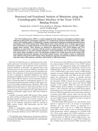
![DNA binding activities through dimerization comes from X-
ray crystallography, pulldown assays, chemical cross-linking,
gel filtration, analytical ultracentrifugation, and mutagenesis
studies and has been inferred from its kinetic profile for DNA
binding (22, 25–27, 42, 44, 45, 47, 69, 72, 81). Consistent with
a dimer autorepression mechanism, TBP mutations that impair
dimerization cause transcriptional derepression in vivo (24,
45). Their effect is dominant to wild-type TBP but is partially
suppressed by wild-type TBP overexpression (45). TBP over-
expression might drive unstable dimer mutants into somewhat
more stable heterodimers with wild-type TBP.
Despite several observations that support a physiological
role for TBP dimerization (45), the topic remains controversial
(20). Ultimately, we wish to understand how a variety of TBP
inhibitory mechanisms are coordinated to regulate TBP. A
necessary step in this effort is the mapping of surfaces on TBP
that are targeted for inhibition in vivo and the identification of
factors responsible for that inhibition. In an effort to more
completely examine the potential physiological significance of
TBP dimerization, we mutated 24 amino acids that comprise
the crystallographic dimer interface. Using in vitro pulldown
and electrophoretic mobility assays, we characterized their
abilities to dimerize, bind TAND, and bind TATA DNA. The
binding data were in good agreement with the crystallographic
and NMR structures of these or related complexes.
Next, we sought to determine whether mutations along the
crystallographic dimer interface affect a variety of phenotypes
in Saccharomyces cerevisiae that we previously linked to defects
in dimerization (24, 45). These include increased TBP turn-
over, transcriptional derepression, partial dominant inhibition
of cell growth (toxicity), and synthetic toxicity in a ⌬TAND
strain. With the collection of mutations throughout the dimer
interface, we observed a correlation between these phenotypes
and relative dimer stability measured in vitro. Mutations that
knock out interfaces between TBP and other inhibitors did not
give similar phenotypes. Taken together, the data provide fur-
ther support for the notion that TBP dimerizes in vivo and is a
physiologically important negative regulator of gene expres-
sion.
MATERIALS AND METHODS
Plasmids. All TBP mutations were created by oligonucleotide-directed mu-
tagenesis. The mutations and the integrity of the entire open reading frame were
confirmed in both the bacterial and yeast expression vectors by DNA sequencing.
Escherichia coli plasmids expressing the His-TBP mutants were designated
pET16b-yTBP(xxx), where xxx indicates the mutation. The plasmid pGEX-
TFIID-C, which encodes human GST-TBP(core), has been described previously
(41). Plasmid pGEX-yTBP(181C) has been described previously (45). The plas-
mid pGEX-scTAF1(10-88) was constructed by PCR amplification of the TAF1
gene and was inserted in frame with the glutathione S-transferase (GST)-coding
sequence of pGEX-3X. Derivatives of this plasmid, pGEX-scTAF1(10-88,
D66K) and pGEX-scTAF1(10-88, F23K, D66K), were constructed by oligonu-
cleotide-directed mutagenesis. The integrity of the entire TAND coding region
was verified by DNA sequencing. Plasmids expressing the HA3-TBP mutants in
S. cerevisiae were designated pCALF-T(xxx)(PGK), where CALF refers to the
CEN/ARS origin, LEU2 marker, Flu3 (or HA3) tagged; T indicates TBP; xxx is
the mutation; and PGK is the promoter controlling HA3-TBP expression. The
plasmid is derived from pDP15-flu3-yTBP (73), in which the SPT15 promoter was
replaced by the PGK1 promoter, as described previously (45). The plasmid
pCALF-T(K145E)(GAL) contains the GAL10 promoter in place of the PGK1
promoter as described previously (24). pADH1-lacZ contains the core (lacking
the upstream activation sequence) ADH1 promoter downstream of four glucose-
repressible Gal4 binding sites (15). The plasmids TAF1/Ura, TAF1/Trp, and
TAF1(⌬TAND)/Trp have been described previously as pYN1/TAF145, pYN2/
TAF145, and pYN2/taf145(⌬10-73), respectively (52).
Strains. YTW22 [MAT␣ ura3-52 trp1-⌬1 his3-⌬200 leu2-⌬1 lys2-801amber
ade2-
101ocher
⌬spt15::TRP1(pCW16-TBP-WT)] is a TBP plasmid shuffle strain (74).
YPH252 (MAT␣ ura3-52 trp1-⌬1 his3-⌬200 leu2-⌬1 lys2-801amber
ade2-101ocher
)
is wild type for TBP (SPT15) and has been described previously (74). In vivo
studies with TAF1 and taf1(⌬TAND) employed the strain Y13.2 (MAT␣ ura3-52
trp1-⌬63 leu2,3-112 his3-609 ⌬taf145 pYN1/TAF145), in which pYN1/TAF145
was replaced by either pYN2/TAF145 or pYN2/taf145(⌬10-73) by using the
plasmid shuffle assay (52).
Protein purification. All His-tagged TBP mutants were purified as follows.
Recombinant E. coli (BL21) cells (500 ml) were grown in YT medium containing
0.2 g of ampicillin per liter at 37°C to an optical density at 595 nm (OD595) of 0.7
and induced with 20 mg of isopropylthio--D-galactoside per liter for 45 min at
30°C. Cells were harvested by centrifugation, washed and resuspended to a
volume of 10 ml in lysis buffer (25 mM HEPES [pH 7.5], 200 mM potassium
chloride, 12.5 mM magnesium chloride, 10% glycerol, 0.05 mM phenylmethyl-
sulfonyl fluoride), and quickly frozen in liquid nitrogen. Cells were thawed and
mixed with 0.8 mg of lysozyme per ml for 10 min at 4°C, with 2 M potassium
chloride for 15 min, and with 0.2% IGEPAL-CA630 for 5 min. Extracts were
sonicated to reduce viscosity and then centrifuged in an SS34 rotor (RC5C
centrifuge) at 15,000 rpm for 30 min at 4°C. Supernatants were mixed with 10
mM imidazole and 0.5 ml of Ni-nitrilotriacetic acid–agarose for 60 min at 4°C.
The slurry was then transferred to a column and then washed with wash buffer
(20 mM HEPES [pH 7.5], 1 M potassium chloride, 12.5 mM magnesium chlo-
ride, 10% glycerol, 60 mM imidazole, 0.05 mM phenylmethylsulfonyl fluoride).
TBP was eluted with TSB buffer (20 mM Tris-acetate [pH 7.5], 0.2 M potassium
glutamate, 2 mM magnesium chloride, 20% glycerol, 0.05 mM phenylmethylsul-
fonyl fluoride) containing 1 M imidazole and dialyzed against TSB buffer con-
taining 60 mM imidazole, 1 mM dithiothreitol, and 0.5 mM phenylmethylsulfonyl
fluoride. TBP aliquots were frozen in liquid nitrogen and stored at Ϫ80°C. TBP
was judged to be ϳ50% pure by sodium dodecyl sulfate-polyacrylamide gel
electrophoresis (SDS-PAGE) followed by silver staining (see Fig. 2). Proteins
concentrations were estimated from these gels by using highly purified TBP
standards whose concentrations were determined by total amino acid analysis.
Human and yeast GST-TBP(core) were expressed from pGEX-TFIID-C and
pGEX-yTBP(181C), respectively, and purified from 3 liters of recombinant E.
coli cells as described above, with the following exceptions. Proteins were ex-
tracted with 1 M potassium chloride and 0.1% IGEPAL-CA630 (rather than 2 M
and 0.2%, respectively). Glutathione agarose (1 ml) (Sigma) was used in place of
nickel agarose. The resin was washed with H buffer (20 mM HEPES [pH 7.5], 2
mM magnesium chloride, 10% glycerol, 0.1 mM phenylmethylsulfonyl fluoride,
and 1 mM dithiothreitol) containing 1 M potassium chloride (H1 buffer) and
then with TSB buffer. The resin-bound GST-TBP(core) was aliquoted, quick-
frozen in liquid nitrogen, and stored at Ϫ80°C.
GST-TAND was expressed from pGEX-scTAF1(10-88) and purified from
recombinant E. coli DH5␣ cells as described above, with the following excep-
tions. Induction with isopropylthio--D-galactoside was for 2 h. Proteins were
extracted with 0.1 M potassium chloride and 0.07% IGEPAL-CA630 (rather
than 2 M and 0.2%, respectively). Glutathione agarose (0.75 ml) (Sigma) was
used in place of nickel agarose. The resin was washed sequentially with H.35, H1,
and then H.35 buffers. Proteins were eluted in H.35 containing 0.1 M reduced
glutathione and dialyzed into H.35 buffer. Aliquots were frozen in liquid nitrogen
and stored at Ϫ80°C. GST-TAND mutants were purified similarly. Proteins were
judged to be approximately 90% pure by SDS-PAGE and silver staining.
GST-TBP(core) pulldown assay. Reaction mixtures contained 20 mM Tris-
acetate (pH 7.5), 75 mM potassium glutamate, 4 mM magnesium chloride, 5%
glycerol, 0.1 g of bovine serum albumin per ml, 4 mM spermidine, 0.025%
IGEPAL-CA630, 0.5 g of heparin per ml, a 5 nM concentration of the indicated
His-tagged TBP mutant, and 20 nM GST-TBP(core) or GST bound to 2 l of
glutathione agarose resin, in 500 l. Reaction mixtures were incubated at 4°C for
45 min with mixing. Resins were washed three times, each with 500 l of reaction
buffer. Bound proteins were eluted and subjected to SDS-PAGE, and TBP was
probed by immunoblotting with TBP antibodies. Reactions were typically per-
formed at least six times, and representative data are shown. TBP was quanti-
tated by densitometric scanning of autoradiograms. Relative pulldown was de-
termined by subtracting local background and normalizing to a wild-type TBP
pulldown present on the same gel.
GST-TAND pulldown assay. Reaction mixtures contained 20 mM Tris-Cl (pH
8.3), 150 mM potassium chloride, 12.5 mM magnesium chloride, 10% glycerol,
50 g of bovine serum albumin per ml, 1 mM dithiothreitol, a 300 nM concen-
tration of the indicated His-tagged TBP mutant, and 300 nM GST-TAND,
GST-TAND(F23K D66K), or GST-TAND(D66K) bound to 10 l of glutathione
VOL. 23, 2003 STRUCTURE AND FUNCTION OF TBP DIMERS 3187
atScientificLibrary,NCI-FCRDConApril20,2009mcb.asm.orgDownloadedfrom](data:image/gif;base64,R0lGODlhAQABAIAAAAAAAP///yH5BAEAAAAALAAAAAABAAEAAAIBRAA7)