SlideShare a Scribd company logo
1 of 12
Download to read offline
ORIGINAL ARTICLE
cIAP1 regulates TNF-mediated cdc42 activation and filopodia
formation
A Marivin1,2,9
, J Berthelet1,2,9
, J Cartier1,2
, C Paul2,3
, S Gemble1,2
, A Morizot4
, W Boireau5
, M Saleh4
, J Bertoglio6,7
, E Solary7,8
and L Dubrez1,2
Tumour necrosis factor-a (TNF) is a cytokine endowed with multiple functions, depending on the cellular and environmental
context. TNF receptor engagement induces the formation of a multimolecular complex including the TNFR-associated factor TRAF2,
the receptor-interaction protein kinase RIP1 and the cellular inhibitor of apoptosis cIAP1, the latter being essential for NF-kB
activation. Here, we show that cIAP1 also regulates TNF-induced actin cytoskeleton reorganization through a cdc42-dependent,
NF-kB-independent pathway. Deletion of cIAP1 prevents TNF-induced filopodia and cdc42 activation. The expression of cIAP1 or its
E3-ubiquitin ligase-defective mutant restores the ability of cIAP1À / À
MEFs to produce filopodia, whereas a cIAP1 mutant unable to
bind TRAF2 does not. Accordingly, the silencing of TRAF2 inhibits TNF-mediated filopodia formation, whereas silencing of RIP1 does
not. cIAP1 directly binds cdc42 and promotes its RhoGDIa-mediated stabilization. TNF decreases cIAP1-cdc42 interaction,
suggesting that TNF-induced recruitment of cIAP1/TRAF2 to the receptor releases cdc42, which in turn triggers actin remodeling.
cIAP1 also regulates cdc42 activation in response to EGF and HRas-V12 expression. A downregulation of cIAP1 altered the cell
polarization, the cell adhesion to endothelial cells and cell intercalation, which are cdc42-dependent processes. Finally, we
demonstrated that the deletion of cIAP1 regulated the HRas-V12-mediated transformation process, including anchorage-
dependent cell growth, tumour growth in a xenograft model and the development of experimental metastasis in the lung.
Oncogene (2014) 33, 5534–5545; doi:10.1038/onc.2013.499; published online 25 November 2013
Keywords: IAPs; TNF; RhoGTPases; cdc42; metastasis; Ras
INTRODUCTION
Tumour necrosis factor-a (TNF) is a mediator of immune and
inflammatory response produced by activated monocytes and
macrophages. This cytokine promotes cell proliferation, cell
differentiation, cytokine secretion and cell death, depending on
the cellular and environmental context.1
TNF also affects cell shape
and cell movement, which may contribute to the recruitment of
fibroblasts or neutrophils to the site of tissue injury.2
Such
morphogenetic modifications involve a dynamic rearrangement of
the actin cytoskeleton controlled by small GTPases of the Rho
family, which includes rhoA, rac1 and cdc42. For example, in
endothelial cells, TNF induces the sequential activation of rac1, rhoA
and cdc42, which leads to the formation of stress fibers and to cell
contraction.2,3
In fibroblasts and macrophages, TNF triggers the
activation of cdc42, which is responsible for the transient formation
of the actin-rich protrusions known as filopodia.2,4,5
Rho GTPases act
as molecular switches that transduce the signal from membrane
receptors to downstream effectors by shuttling between a GTP-
bound active state and a GDP-bound inactive state. Once activated,
they are either rapidly recycled into the inactive form or degraded
by the ubiquitin-proteasome machinery. The Rho GTPase activation
cycle is controlled by guanine-nucleotide exchange factors (GEFs),
which catalyse the transfer of GDP-bound forms into GTP-bound
forms, by GTPase-activation proteins (GAPs), which inactivate Rho
GTPases by hydrolysing the GTP, and by guanine-nucleotide
dissociation inhibitors (GDIs), which are chaperones that stabilize
Rho GTPases in a cytosolic inactive state.2,6,7
TNF binds two related membrane surface receptors. TNFR1,
whose expression is ubiquitous, mediates most of the biological
effects of the cytokine, whereas TNFR2 expression is restricted
mostly to lymphocytes and endothelial cells. Upon ligand
stimulation, TNFR1 recruits, in a membrane-associated complex,
the cytosolic adaptor TNFR1-associated death domain protein
(TRADD), the TNFR-associated factors (TRAFs), the receptor-
interaction protein kinase 1 (RIPK1) and the cellular inhibitors of
apoptosis (cIAPs).8–11
This molecular platform activates ubiquitin-
dependent signaling pathways, resulting in the nuclear factor-
kappaB (NF-kB), mitogen-activated protein kinase (MAPK)
activations and the expression of genes encoding for cytokines,
adhesion molecules, survival and differentiation factors.11–13
In the
absence of cIAPs or when the NF-kB signaling is blocked,
secondary cytoplasmic complexes leading to cell death are
generated from the first one.14–17
cIAPs, including cIAP1 and cIAP2, are E3-ubiquitin ligases
formed as a result of the presence of a C-terminal RING domain.
In addition to the RING, they own three baculovirus IAP repeat
(BIR) domains mediating protein–protein interactions, a ubiquitin-
binding-associated (UBA) domain that allows the recruitment of
1
Institut National dela Sante´ et de la Recherche Me´dicale (Inserm) UMR866, Faculty of Medicine, Dijon, France; 2
Universite´ de Bourgogne; Institut Fe´de´ratif de Recherche (IFR) 100,
Dijon, France; 3
Ecole Pratique des hautes e´tudes (EPHE), Dijon, France; 4
Department of Medicine, McGill University, Montreal, Quebec, Canada; 5
Institut FEMTO-ST, Universite´ de
Franche-Comte´, CLIPP, Besanc¸on, France; 6
Inserm UMR749, Institut Fe´de´ratif de Recherche (IFR) 54, Villejuif, France; 7
Institut Gustave Roussy, Institut Fe´de´ratif de Recherche (IFR)
54, Villejuif, France and 8
UMR1009, Institut Fe´de´ratif de Recherche (IFR) 54, Villejuif, France. Correspondence: Dr L Dubrez, Institut National dela Sante´ et de la Recherche Me´dicale
(Inserm) UMR866, Faculty of Medicine, 7 Boulevard Jeanne d’Arc, 21079 Dijon cedex, France.
E-mail: ldubrez@u-bourgogne.fr
9
These authors contributed equally to this work.
Received 5 April 2013; revised 20 September 2013; accepted 21 October 2013; published online 25 November 2013
Oncogene (2014) 33, 5534–5545
& 2014 Macmillan Publishers Limited All rights reserved 0950-9232/14
www.nature.com/onc
cIAPs into protein complexes18
and a caspase recruitment domain
(CARD) that regulates the E3 activity of the RING domain.19
Numerous ubiquitination targets and/or partners of cIAP1 have
been identified, including signaling molecules, regulators of NF-kB
activating pathways20
and transcriptional regulators such as
Mad121
and E2F1.22
In the TNFR1 signaling pathway, cIAP1
promotes its own ubiquitination and the ubiquitination of the
kinase RIP1 required for the activation and survival of NF-kB ,
whereas it inhibits the formation of the RIP1-containing secondary
intracellular platforms that trigger cell death.9,11,12,14–16
cIAP1 is
also a potent inhibitor of the alternative NF-kB activating pathway
through ubiquitination leading to degradation of NIK.23,24
The present study demonstrates that cIAP1 also regulates
filopodia formation in response to TNF, EGF and oncogenic HRas-
V12 expression. cIAP1 directly interacts with cdc42 and regulates
its activity by promoting its RhoGDI-mediated stabilization. We
show that TNF stimulation disrupts the cIAP1/cdc42 interaction
and facilitates cdc42-mediated actin rearrangement, indepen-
dently of NF-kB activation. A downregulation of cIAP1 affected
cdc42-regulated processes including cell polarization, cell adhe-
sion, cell intercalation and HRas-V12-mediated cell transformation.
RESULTS
Deletion of cIAP1 alters actin cytoskeleton organization.
A previous work from Oberoi et al.25
demonstrated that murine
embryonic fibrobasts (MEFs) from birc2-deficient mice (cIAP1À / À
)
displayed an altered morphology when compared with their wild-
type counterparts. Accordingly, serum-starved cIAP1À / À
MEFs
Figure 1. The organization of the actin cytokeleton is altered in cIAP1À / À
MEFs. (a) Microscopic analysis of serum-starved wt or cIAP1À / À
MEFs. Left panel: phase contrast images. Right panel: F-actin staining using AlexaFluor488-conjugated Phalloidin. (b) Filopodia-like structures
were quantified by counting cells showing more than five filopodia-like structures. More than 100 cells were analysed. The mean
reflects ±s.d. of at least three independent experiments. Statistical analysis was performed using the Student t-test. ***Po0.001; **Po0.01;
*Po0.05. (c) The expression of cIAP1 or cIAP1 mutants, cdc42-N17, IkB-super-repressor (SR), and the efficiency of control (si-Co) or NIK-
targeted siRNAs (si-NIK) were checked by western blot analysis. HSC70 was used as the loading control.
cIAP1 regulates cdc42
A Marivin et al
5535
& 2014 Macmillan Publishers Limited Oncogene (2014) 5534 – 5545
exhibited elongated and contracted cell bodies and the presence
of subtle fine extended protrusions similar to filopodia (Figure 1a),
an effect quantified by counting cells harbouring more than five
filopodia-like structures (Figure 1b). Filopodia is specifically
controlled by RhoGTPase cdc42.6
The expression of cIAP1 or a
dominant-negative form of cdc42 (cdc42-N17) abolished the
filopodia-like structures in cIAP1À / À
MEFs (Figures 1b and c),
demonstrating a cdc42-dependent phenomenon. The adaptor
TRAF2 is a well-known partner of cIAP1 that is required for cIAP1
recruitment into the TNFR complex. The expression of the
cIAP1L47A
mutant, which cannot interact with TRAF222
(Supple-
mentary Figure 1), or the E3-ubiquitin ligase activity-defective
mutant (cIAP1H588A
), also impeded filopodia-like formation
(Figures 1b and c). Depletion of cIAP1 leads to a stabilization of
the NF-kB-inducing kinase NIK and a constitutive activation of
NF-kB.17,26
Neither the silencing of NIK nor the expression of IkB-
super repressor (IkB-SR) abolished spontaneous filopodia in
cIAP1À / À
MEFs significantly (Figures 1b and c). The expression
of IkB-super repressor (IkB-SR) did not alter the capacity of cIAP1
to suppress filopodia either (Figures 1b and c), demonstrating that
the reorganization of actin cytoskeleton in cIAP1À / À
MEFs was
independent of NF-kB activation.
cIAP1 mediates cdc42 stabilization through RhoGDIa
A western blot analysis of Rho GTPases demonstrated a reduced
expression of cdc42 in cIAP1-depleted cells (Figures 2a and b).
However, the GTP-bound/total cdc42 ratio was increased
(Figures 2b and c), demonstrating a constitutive activation of
cdc42, which was associated with an increase in the phosphoryla-
tion of the direct cdc42 effector PAK1 (Figure 2c). Once activated,
RhoGTPAses can be degraded by the proteasome system. The
Figure 2. cIAP1 controls cdc42 stability. (a) Immunoblot analysis of cdc42, Rac1 and RhoA in MEFs from wt or cIAP1-deleted mice, treated or
not with 40 mM MG132 for 4 h. HSC70 was used as the loading control. (b) Immmunoblot analysis of cdc42 in NIH3T3 cells transfected with
cIAP1 targeting siRNA and treated or not with 40 mM MG132 for 4 h. Activated GTP-bound cdc42 was pulled down using the GST-PAK1-CRIB
domain before immunoblot analysis. HSC70 was used as the loading control. (c) Immunoblot analysis of total or GTP-bound cdc42, and total
and phosphorylated form of PAK1 in wt or cIAP1À / À
MEFs. HSC70 was used as the loading control. Upper panel: The ratio GTP-bound/total
cdc42 as quantified by a densitometric analysis of immunoblot. The mean reflects ± s.d. of three independent experiments. (d) Western blot
analysis of total or GTP-bound cdc42 and RhoGDIa in NIH3T3 transfected with cIAP1 encoding vector. HSC70 was used as the loading control.
(e) Immunoblot analysis of cdc42, RhoGDIa and cIAP1 in MEFs from cIAP1-deleted mice transfected with control or cIAP1 encoding vector
plus control or RhoGDIa-targeting siRNA. HSC70 was used as the loading control. (f) Immunoblot analysis of cIAP1 and cdc42 in cytosol- (Cyto)
and membrane (Mb)-enriched fractions from cIAP1-deleted mice transfected with control or cIAP1-encoding vector. GM130 was used as a
control for membrane enrichment, and HSC70 was used as the loading control. Right panel: The ratio cytosolic/membrane cdc42 expression
as quantified by a densitometric analysis of immunoblots. The mean reflects ±s.d. of two independent experiments.
(g) Overexpression of cIAP1 increases the cdc42-RhoGDIa interaction. HEK293T cells were transfected with HA-cdc42 with or without
cIAP1 encoding vectors. RhoGDIa was immunoprecipitated and co-precipitated and revealed by immunoblotting using indicated antibodies.
cIAP1 regulates cdc42
A Marivin et al
5536
Oncogene (2014) 5534 – 5545 & 2014 Macmillan Publishers Limited
proteasome inhibitor MG132 prevented the downregulation of
cdc42 (Figures 2a and b). Conversely, overexpression of cIAP1 in
NIH3T3 or restoration of cIAP1 in cIAP1À / À
MEF increased the total
level of cdc42 (Figures 2d and e, Supplementary Figure 2), mostly in
the cytosol-enriched fraction (Figure 2f), and, to a lesser extent, the
GTP-bound cdc42 (Figure 2d). On the whole, our data suggest that
cIAP1 could stabilize cdc42, mainly in its cytosolic inactive state.
RhoGDIa is an important regulator of Rho GTPases that directly
binds the proteins and stabilizes the cytosolic inactive forms.7,27
Silencing of RhoGDIa prevented the cIAP1-mediated upregulation
of cdc42 (Figure 2e, Supplementary Figure 2), and overexpression of
cIAP1 increased the proportion of RhoGDIa associated with cdc42;
that is, co-precipitation experiments showed that an equivalent
quantity of RhoGDIa pulled down more cdc42 in cIAP1-over-
expressing cells (Figure 2g), demonstrating the importance of
RhoGDIa in cIAP1-mediated cdc42 stabilization.
cIAP1 interacts with cdc42.
We demonstrated an interaction of cIAP1 with cdc42, RhoA and
Rac1 in a glutathione S-transferase (GST) pull-down assay
(Figure 3a). Similar results were obtained in a co-immunoprecipa-
tion experiment using Myc-cIAP1 and EGFP-Rho GTPases
(Supplementary Figure 3A). Cdc42 bound to cIAP1 BIR-containing
constructs (BIR1–3, BIR 1–2 and BIR 2–3) and to the single BIR2
domain with high affinity, whereas TRAF2 bound to BIR1
(Figure 3b). Both GTP- and GDP-loaded cdc42 bound cIAP1,
whereas only the GTP form bound the cdc42 effector PAK1
(Figure 3c, Supplementary Figure 3B). These results were
confirmed using the Surface Plasmon Resonance technology
(Biacore system) showing that the BIR domains of cIAP1 directly
bound GDP- and GTP-loaded recombinant cdc42 with an affinity
quite similar (Figure 3d). An interaction of cdc42 with cIAP1,
RhoGDIa and TRAF2 was observed in vivo in co-immunoprecipita-
tion experiments (Figure 3e). The use of recombinant proteins
demonstrated that cIAP1, but not TRAF2, could directly interact
with cdc42. However, TRAF2 bound GST-cdc42 in the presence of
cIAP1 (Figure 3f).
cIAP1 is required for TNF-induced filopodia formation and Cdc42
activation
In fibroblasts, TNF induces the activation of cdc42 leading to the
formation of filopodia.4,5,28,29
Accordingly, we observed filopodia
in serum-starved NIH3T3 fibroblasts within 15 min of TNF
treatment (Figures 4a and b). Simultaneously, TNF activated
Figure 3. cIAP1 can interact with cdc42. (a–c, f) GST pull-down assay. (a) Cell lysates from HEK293T cells transfected with FLAG or FLAG-cIAP1
encoding vector were deposited onto GST-RhoA, -Rac1 or -cdc42 proteins. Interactions were analysis by immunoblotting analysis using anti-
cIAP1-specific antibody. (b) Lysates from HEK293T cells transfected with GFP-cdc42 encoding vector were incubated with GST-cIAP1 construct
fusion proteins as indicated. Interactions were analysed by a western blot using anti-GFP, anti-TRAF2 or anti-GST antibodies. (c) GST-cdc42 was
immobilized onto glutathione sepharose beads and charged with GDP or GTP before incubating with HEK293T cell lysates. Interactions were
revealed by immunoblotting using specific ant-cIAP1 or anti-PAK1 antibodies. (d) Surface Plasmon Resonance (Biacore) analysis of the
interaction of the BIR domains of cIAP1 with uncharged, GDP- and GTP-charged cdc42 recombinant protein. (e) Co-immunoprecipitation
analysis of the interaction of HA-cdc42 with cIAP1, TRAF2 and RhoGDI in HEK293T cells transfected with HA-cdc42. Interactions were revealed
by immunoblotting using indicated antibodies. (f) Recombinant cIAP1 or TRAF2 was incubated with GST-cdc42 immobilized on glutathione
sepharose beads. Interactions were evaluated by cIAP1 or TRAF2 immunoblotting.
cIAP1 regulates cdc42
A Marivin et al
5537
& 2014 Macmillan Publishers Limited Oncogene (2014) 5534 – 5545
cdc42 and Rac1 Rho GTPases, as indicated by the appearance of
GTP-bound Rho GTPases, and induced the dephosphorylation of
the downstream effectors GSK3 (serine/threonine protein kinase
glycogen synthase kinase-3) and the actin-binding protein
cofilin30
(Figure 4c). siRNA-mediated silencing of cIAP1 prevented
the formation of filopodia (Figures 4a and b), the activation of
cdc42 and Rac1 and the modifications of Rho GTPase downstream
effectors (Figure 4c). cIAP2 siRNA also reduced TNF-induced
reorganization of the actin cytoskeleton (Supplementary Figure 4).
In MEFs, the TNF-induced filopodia were larger and more
abundant compared with the spontaneous filopodia-like struc-
tures observed in cIAP1À / À
MEFs (see Figures 1a and 5a), which
was confirmed by counting the number of filopodia per cell
(Figure 5b). TNF treatment did not induce the formation of new
filopodia, and even significantly decreased spontaneous filopodia
structures, in cIAP1À / À
MEFs (Figures 5a and b), whereas a
prolonged exposure induced cell death in cIAP1À / À
MEFs but not
in wt MEFs17
(Supplementary Figure 5). We did not observe
filopodia formation in response to TNF in cIAP1/cIAP2 double
knockout MEFs either (Figure 5c). The ability of cIAP1À / À
MEFs to
form filopodia in response to TNF was restored by the expression
of wild-type cIAP1 as well as the E3-defective mutant cIAP1H588A
(Figures 5d and e). In contrast, the cIAP1L47A
mutant did not
restore TNF-induced filopodia formation in cIAP1À / À
MEFs
(Figures 5d and e), suggesting a role of TRAF2 in this process.
Accordingly, silencing of TRAF2 in wt MEFs inhibited TNF-
mediated filopodia formation (Figure 5f), whereas silencing of
RIP1, which is the cIAP1-ubiquitin target in the TNF signaling
pathway, did not (Figure 5f). The TNF-mediated filopodia formation
in cIAP1-reconstituted cIAP1À / À
MEFs was totally inhibited by the
expression of the dominant-negative form of cdc42 (cdc42-N17),
but was not affected by the expression of IkB-SR (Figure 5e).
Overall, TNF induced filopodia formation via a cdc42-, cIAP1- and
TRAF2-dependent but RIP1- and NF-kB-independent signaling
pathway. Moreover, the cIAP1 E3-ubiquitin ligase required for
TNF-induced ubiquitination of RIP1 and activation of NF-kB was
dispensable. Of note, the expression of the cIAP1L47A
mutant in wt
MEFs inhibited TNF-induced filopodia formation (Figure 5e),
Figure 4. Silencing of cIAP1 inhibits TNF-mediated Filopodia formation. NIH3T3 fibroblasts were transfected with control (si-Co) or cIAP1
(si-cIAP1)-targeting siRNAs, serum starved for 16 h and stimulated for an indicated time with 100 ng/ml TNF. (a, b) Immunoflorescence analysis
of F-actin staining was conducted using AlexaFluor488-conjugated Phalloidin. Filopodia were quantified by counting cells harbouring more
than five filopodia (b). More than 100 cells were analysed. The mean reflects±s.d. of at least three independent experiments. Statistical
analysis was performed using the Student t-test. ***P ¼ 0.004; *P ¼ 0.028. (c) Western blot analysis of cdc42 and Rac1, total and
phosphorylated forms of cofilin and GSK3a and b. Activated GTP-bound cdc42 and Rac1 were pulled down using the GST-PAK1-CRIB domain
before immunoblot analysis. One representative experiment is shown. HSC70 was used as the loading control.
cIAP1 regulates cdc42
A Marivin et al
5538
Oncogene (2014) 5534 – 5545 & 2014 Macmillan Publishers Limited
suggesting a dominant-negative effect. A co-immunoprecipitation
experiment demonstrated that this mutant interacted with
endogenous cIAP1 (Supplementary Figure 6).
We then analysed the activation of cdc42, Rac1 and RhoA in
response to TNF stimulation in MEFs (Figure 6a). As expected,
TNF did not stimulate cdc42 in cIAP1À / À
MEFs. The activation of
Rac1, which occurs downstream of cdc42 in the TNF-signaling
pathway,4
was also abolished. The expression of the cIAP1L47A
mutant also decreased TNF-induced cdc42 activation and PAK1
phosphorylation, confirming the dominant-negative effect of this
mutant (Figure 6b). Altogether, our results suggest that cIAP1 is
required for cdc42 activation in response to TNF. Co-immunopre-
cipitation experiments showed that TNF exposure decreased the
interaction of cIAP1 and TRAF2 with cdc42 (Figure 6c).
Unlike TNF, which induces cdc42-dependent Rac1 activation,4
EGF activates cdc42 and Rac1 in an independent manner. Deletion
Figure 5. TNF-mediated filopodia formation is impaired in MEF cIAP1À / À
. MEF wt, cIAP1À / À
(a, b, d–f) or cIAP1À / À
/cIAP2À / À
(c) was serum
starved for 16 h, and stimulated for 10 min with 100 ng/ml TNF. (a) Microscopic analysis of F-actin stained using AlexaFluor488-conjugated
Phalloidin. (b) The number of filopodia per cells was counted. More than 100 cells were analysed. One representative experiment is shown.
(c) Filopodia in cIAP1/cIAP2 double knockout MEFs were detected as in A and counted. More than 100 cells were analysed. Results were
expressed as fold filopodia induction/untreated cells (UT). The mean±s.d. reflects at least three independent experiments. The expression of
cIAP1 and cIAP2 is checked by immunoblotting analysis (right panel). (d–f) Wt or cIAP1À / À
MEFs were transfected with encoding plasmid
vector (d, e) or si-RNAs (f), serum starved and treated for 10 min with 100 ng/ml TNF. The expression of the constructs and the silencing
efficiency were checked by immunoblotting analysis (d, f: lower panel). Filopodia were detected as in A and counted (e, f). More than 100 cells
were analysed. Results were expressed as fold filopodia induction/untreated cells (UT). The mean±s.d. reflects at least three independent
experiments (d, e). Statistical analysis was performed using the Student t-test. ***Po0.001; **Po0.01; *Po0.05. N45 (d) or N ¼ 3 (e).
cIAP1 regulates cdc42
A Marivin et al
5539
& 2014 Macmillan Publishers Limited Oncogene (2014) 5534 – 5545
of cIAP1 inhibited EGF-induced cdc42, but not Rac-1 activation
(Figure 6a). Moreover, deletion of cIAP1 did not alter serum-
induced RhoA activation (Figure 6a), suggesting a specific
regulation of cdc42 activation by cIAP1. The EGF-induced filopodia
formation was abolished in cIAP1À / À
MEF and was restored by
the expression of cIAP1 (Supplementary Figure 7). Silencing of
TRAF2 did not inhibit EGF-induced filopodia formation
(Supplementary Figure 7), suggesting a general regulation of
cdc42 by cIAP1, whereas TRAF2 specifically regulated the TNF
signaling pathway.
cIAP1 regulates cdc42 functions
Next, we examined the importance of cIAP1 in the cellular
functions of cdc42. cdc42 is a specific regulator of cell polariza-
tion.31,32
A downregulation of cIAP1 significantly affected cell
polarization, as evaluated by analysing the Golgi apparatus
reorientation to face the leading edge, 5 hours after fibroblast
scratching (Figure 7a). cdc42 is also an important intermediate in
the Ras-mediated cell anchorage-independent cell growth and
transformation pathways.33–36
The expression of oncogenic HRas-
V12 induced the activation of cdc42, which was completely
blocked in cIAP1À / À
MEFs (Figure 7b). Deletion of cIAP1
significantly decreased HRas-V12-mediated cell growth in soft
agar medium (Figure 7c), inhibited the growth of tumour cells
when subcutaneously injected into nude mice (Figure 7d) and
delayed the apparition of lung cancer foci after injection of cells
into the tail vein (Figure 7e, Supplementary Figure 8).
Several steps of the metastatic process are controlled by
RhoGTPases, including the dissemination of tumour cells through
the lymph or the blood, their adhesion to vessel endothelium and
their subsequent migration through the endothelium to colonize
adjacent organs. A recent report from Reymond et al. demon-
strated that the attachment of cancer cells to the endothelial
monolayer and their transendothelial migration is more specifi-
cally controlled by cdc42.37
As demonstrated for cdc42,37
silencing
of cIAP1 significantly reduced the adhesion of PC3 cells to the
HUVEC monolayer and their intercalation between endothelial
cells (Figure 7f). We then analysed the capacity of MEFs,
untransformed or transformed by HRas-V12, to adhere to and
Figure 6. Deletion of cIAP1 prevents cdc42 activation. (a) Immunoblotting analysis of cdc42, Rac1 and RhoA in MEFs from wt or cIAP1-deleted
mice. Cells were serum starved for 16 h, and then stimulated for 10 min with 100 ng/ml TNF or EGF or 10% FBS as indicated. Activated
GTP-bound cdc42 and Rac1 were pulled down with the GST-PAK1-CRIB domain and GTP-RhoA with a GST-Rhotekin-Rho binding domain
before immunoblot analysis. One representative experiment is shown. The ratio GTP-bound/total GTPases is evaluated by a densitometric
analysis of the shown immunoblot. Because of the differential basal level of expression of cdc42 as observed in Figure 3, cell lysates from wt
and cIAP1À / À
MEFs were deposited onto a separate gel and revealed separately. (b) Immunoblotting analysis of cdc42 (GTP-bound and total)
and PAK1 (phosphorylated form and total) in NIH3T3 cells transfected with cIAP1L47A
encoding plasmid vector 24 h before serum starvation.
Cells were stimulated for 10 min or for the indicated time with 100 ng/ml TNF. HSC70 was used as a loading control. (c) HEK293T cells were
transfected with HA-cdc42 and FLAG-cIAP1 and treated or not for 10 min with 100 ng/ml TNF. Immunoprecipitation was performed using an
anti-HA antibody and interactions were revealed by immunoblotting using anti-cIAP1, TRAF2 and HA antibodies.
cIAP1 regulates cdc42
A Marivin et al
5540
Oncogene (2014) 5534 – 5545 & 2014 Macmillan Publishers Limited
Figure 7. cIAP1 regulates cdc42 functions. (a) NIH3T3 cells were transfected with control or cIAP1-targeting siRNA. Wound healings
were induced by scratching the cell monolayer with a pipette tip. Golgi-apparatus staining (red) was performed in NIH3T3 cells and wt and
cIAP1À / À
MEFs with an anti-GM130, and nucleus labelling with Hoechst 5 h after scratching. Right panel. The percentage of cells with Golgi
oriented towards the wound was quantified by counting at least 100 cells (mean ± s.d. of three independent experiments). (b) Cdc42
activation in wt or cIAP1À / À
MEFs transduced with the HRas-V12 construct. Activated GTP-bound cdc42 was pulled down with the GST-PAK1-
CRIB domain before immunoblotting analysis. (c) Anchorage-independent growth of HRas-V12-infected wt or cIAP1À / À
MEFs. Cells were
cultured in soft-agar medium. The number of colonies was evaluated after 10 days of culture. The T-test was used for statistical analysis
(*Po0.05; N ¼ 5). (d) HRas-V12-infected wt or cIAP1À / À
MEFs were injected subcutaneously into nude mice. The tumour was analysed 10 days
later (two independent experiments are shown, n ¼ 5 per group). The Mann–Whitney test was used for statistical analysis (*Po0.05). (e) HRas-
V12-infected wt or cIAP1À / À
MEFs were injected into the tail veins of nude mice. Lung cancer foci were quantified 2 weeks later (two
independent experiments are shown, n ¼ 4 per group). The Mann–Whitney test was used for statistical analysis (***P ¼ 0.0006). (f) CSFE-
labelled PC3 cells, transfected with control or cIAP1-targeting siRNAs, were added on the HUVEC confluent monolayer. PC3 cell adhesion was
quantified by flow cytometry (mean ± s.d. of two independent experiments) (left panel). The efficiency of siRNAs was checked by western blot
analysis. (Upper panel). PC3 cell intercalation was evaluated by counting cells that display a non-round shape and a low phase-bright by time-
lapse microscopy. (4100 cells were analysed. The mean reflects±s.d. of at least three independent experiments) (right panel). Statistical
analysis was performed using an ANOVA test (***Po0.001; **0.001oPo0.01). (g) CFSE-labelled wt or cIAP1À / À
MEFs, transduced or not with
the HRas-V12 construct, were added on the HUVEC confluent monolayer. MEF adhesion (left panel) and intercalation (right panel) were
evaluated as in (f) (4100 cells were analyzed. The mean reflects±s.d. of at least three independent experiments). Statistical analysis was
performed using an ANOVA test (***Po0.001).
cIAP1 regulates cdc42
A Marivin et al
5541
& 2014 Macmillan Publishers Limited Oncogene (2014) 5534 – 5545
intercalate between endothelial cells (supplementary Figure 9).
Accordingly, with the presence of filopodia-like structures on the
cell surface, the cIAP1À / À
MEFs spontaneously adhered to the
HUVEC monolayer more strongly compared with the wt counter-
part (Figure 7g, left panel). HRas-V12 expression stimulated the
attachment of MEFs to the HUVEC monolayer (Figure 7g, left
panel). Both the adhesion and the intercalation were decreased in
HRas-V12-transformed cIAP1À / À
MEFs compared with HRas-V12/
wt MEFs (Figure 7g).
DISCUSSION
cIAP1 is a key determinant of the cellular response to TNF. The
protein is recruited to the TNFR1 through the adaptor TRAF2 and
mediates the activation of the canonical NF-kB survival pathway
while inhibiting the assembly of the secondary cytoplasmic
caspase-activating platform, leading to cell death.20
We
demonstrate here that cIAP1 also regulates actin-cytoskeleton
reorganization upon TNF stimulation through a cdc42-dependent,
NF-kB-independent pathway. We propose a model (Figure 8) in
which cIAP1 could regulate the cycle of cdc42 activation by
stabilizing the interaction of cdc42 with its regulator RhoGDIa. The
recruitment of TRAF2/cIAP1 to the TNF membrane receptor after
TNF stimulation could release cdc42 and allow its activation,
leading to the actin cytoskeleton reorganization.
In fibroblasts, TNF induces the rapid and transient formation of
filopodia,38
which requires the Rho GTPase cdc42.5,39,40
The
pathway connecting the TNFR1 to Rho GTPases remains poorly
understood but appeared to be independent of NF-kB and MAPK
activation.5,29,38
Accordingly, we observed that TNF induced
filopodia formation in the presence of an IkB super-repressor.
TNFR engagement triggers the TRAF2-dependent recruitment of
cIAP1, which induces an autoubiquitination and the ubiquitination
of RIP1 necessary for NF-kB activation.20
We demonstrate that
TNF-induced filopodia formation requires cIAP1 and TRAF2,
whereas RIP1 and the E3-ubiquitin ligase of cIAP1 are
dispensable, suggesting that the TNFR-associated complex can
generate independent signaling pathways leading to NF-kB
activation or filopodia formation. cIAP1 can directly bind to
cdc42, whereas TRAF2 can bind to cdc42 only in the presence of
cIAP1. Both cIAP1 and TRAF2 co-precipitate with cdc42 in resting
cells, and TNF treatment decreased this interaction. Our
hypothesis is that the recruitment of cIAP1/TRAF2 to TNFR
makes cdc42 activation easier, leading to actin cytoskeleton
reorganization (Figure 8). Secondary molecular events are likely
required for the full activation of cdc42, as the activation of Rho
GTPases requires a GEF. Vav is a Rac1-GEF in TNFa signaling in
fibroblasts,29
and the GEF Ect2 has been involved in the TNF-like
weak inducer of apoptosis (TWEAK)-induced cdc42 activation in
glioblastoma;41
however, their role in cdc42 activation has to be
explored in this cell context.
cIAP1 appears to fine-tune cdc42 activation. On one hand, cIAP1
deletion inhibits cdc42 activation in response to TNF and EGF
stimulation and HRas-V12 expression; on the other hand, down-
regulation of cIAP1 decreases the expression of the whole cdc42
in a proteasome-dependent manner but increases the expression
of the activated fraction of cdc42. The downregulation of cIAP1
also promotes the phosphorylation of PAK1, a cdc42 effector, and
cIAP1À / À
MEFs spontaneously form filopodia-like structures.
Interestingly, RhoGDIa depletion infers similar modifications,
including a decreased cdc42 protein level that can be prevented
by inhibiting the proteasome, and an increased ratio of active to
total cdc42.27
RhoGDIa is a Rho chaperone; that is, it maintains a
pool of cytoplamic GTPases in a GDP-bound inactive state and
protects them from proteasomal degradation.27,42
The expression
of cIAP1 increases the cytosolic cdc42 expression in a RhoGDIa-
dependent manner and enhances the fraction of the cdc42 bound
to RhoGDIa. Overall, these data argue for a RhoGDIa-mediated
regulation of cdc42 by cIAP1.
The regulation of RhoGTPases by IAPs is evolutionarily
conserved; that is, it has been observed in Drosophila43
and
Zebrafish.25
Identical to RhoGDIs,42
cIAP1 (present work), XIAP25
and DIAP143
can bind to both GDP- and GTP-bound GTPases.
DIAP1 controls Rac activity independently of its E3-ubiquitine
ligase domain,43
whereas XIAP promotes the ubiquitination and
degradation of the active GTP-bound form of Rac1.25
We observed
an interaction of cIAP1 with Rac1, RhoA and cdc42. In our study,
cIAP1 failed to ubiquitinate cdc42 (Supplementary Figure 10),
Figure 8. Model for the regulation of cdc42 by cIAP1/TRAF2. cIAP1 interacts with TRAF2 via the BIR1 (B1) domain and with cdc42 via the BIR2
(B2) domain. (1) In resting cells, cIAP1 binds cdc42. It stabilizes its interaction with its regulator RhoGDIa and then regulates cdc42 activation.
(2) The recruitment of TRAF2/cIAP1 to the receptor after TNF stimulation releases cdc42 and makes its activation easier, leading to
cytoskeleton reorganization and filopodia formation. (3) Depletion of cIAP1 induces a loss of control of cdc42 and increases the activation/
degradation cycle, leading to cytoskeleton modifications and a filopodia-like structure.
cIAP1 regulates cdc42
A Marivin et al
5542
Oncogene (2014) 5534 – 5545 & 2014 Macmillan Publishers Limited
suggesting that IAPs could differentially modulate various Rho
GTPases. RhoA, Rac1 and cdc42 compete for the binding to and
chaperoning by RhoGDIa, and the stabilization of the interaction
of one Rho GTPase with RhoGDIa favours the degradation of the
others.27
cIAP1 becomes a part of this crosstalk regulatory
mechanism by stabilizing cdc42–RhoGDIa interaction (present
work) and catalysing the proteasomal degradation of Rac1.25
Moreover, an inhibition of the SUMOylation of RhoGDIa by XIAP
has been described44
and a recent study identified RhoGDI2 as a
potential IAP neddylation substrate.45
As central regulators of Rho
GTPase homeostasis, IAPs could regulate their sequential
activation in many biological processes such as intracellular
vesicular traffic, morphogenesis, tissue repair, cell shape,
plasticity, polarization, adhesion and motility.42
In drosophila,
overexpression of DIAP1 compensates the invalidation of rac in
the control of border cell migration during oogenesis.43
In
mammals, the depletion of cIAPs or XIAP alters the invasive
properties of cancer cells, including their migration,19,25,44,46
their
adhesion to the endothelium and their intercalation between
endothelial cells, which can be related to the deregulation of Rho
GTPase homeostasis.25,43,44
Our results indicate a regulatory function for cIAP1 in cdc42-
controlled filopodia formation, cell polarization and adhesion.39,47
The contribution of cdc42 to some oncogenic processes,6
for
example, HRas-V12-driven cell transformation33,35,36
and
metastatic invasion,37
may account for the anti-tumour activity
of small molecule inhibitors of IAPs, such as Smac mimetics.
MATERIALS AND METHODS
Cell culture and treatments
Mouse embryonic immortalized (SV40) fibroblasts (MEF) wt and cIAP1À / À
and cIAP1À / À
/cIAP2À / À
(J. Silke, Melbourne, Australia), mouse NIH3T3
fibroblasts and HEK293T cells were cultured in DMEM medium
(Lonza, Verviers, Belgium) supplemented with 10% fetal bovine serum
(FBS) (Lonza). Cells were serum starved for 16 h before stimulation with
100 ng/ml TNF or EGF (Shenandoah Biotechnology Inc, Warwick, PA, USA).
MG132 (Millipore, Calbiochem, Billerica, MA, USA) was used at 40 mM for 4
hours. PC3 cells were cultivated in RPMI-1640 medium (Lonza) supple-
mented with 10% FBS (Lonza). Primary HUVECs (Lonza) were maintained in
EBM2 medium (Lonza).
Plasmid constructs, siRNAs, cell transfection and viral transduction
NIH3T3 fibroblasts were transfected using Lipofectamine 2000 (Life
technologies, Invitrogen, Carlsbad, CA, USA), and MEFs and HEK293T were
transfected using JET PEI (Polyplus transfection, Illkirch, France). Lipofecta-
mine 2000 (Invitrogen) was used for the transfection of siRNA targeting
mouse NIK (Thermo Fisher Scientific, Waltham, MA, USA), cIAP1,
TRAF2, RIP1 or RhoGDIa (designed and provided by Qiagen, Venlo, The
Netherlands). The DNA constructs used were pCR3-Flag-cIAP1, pCR3-flag-
cIAP1L47A
, pCI-cIAP1, pCI-cIAP1H588A
, pCI-cIAP1L47A
,22
pGEX-cIAP1wt
,
pGEX-cIAP1BIR1–3
(amino-acid 1–483), pGEX-cIAP1CARD-RING
(amino-acid
452–618), pGEX–cIAP1BIR1–2
(amino-acid 1–258), pGEX-cIAP1BIR2–3
(amino-acid 181–363), pGEXcIAP1BIR1
(amino-acid 34–129), pGEX-cIAP1BIR2
(amino-acid 170–260), pGEX-cIAP1BIR3
(amino-acid 256–358),48
pMT90-
Myc-cdc42N17
, pCDNA-HA-cdc42WT
, pEGFP-cdc42, pEGFP-RhoA,
pEGFP-Rac1, pGEX-RhoA, pGEX-Rac1, pGEX-cdc42, pRcCMV-IkB-SR and
pBABE-HRasV12
. HRas-V12 expressing MEFs were generated by retroviral
transduction. Phoenix-Eco cells (Invitrogen), which constitutively produced
gag-pol and ecotropic envelope proteins, were transfected using Jet PEI
(Polyplus transfection) with pMSCV-HRas-V12. MEFs were transduced
overnight with the retroviral-containing supernatant supplemented with
1 mg/ml polybrene (Sigma-Aldrich, St-Louis, MO, USA). Populations of
transduced cells were selected by puromycin exposure. The efficiency of
infection was checked by western blot analysis.
Immunofluorescence analysis of filopodia and cell polarity
Cells were grown and transfected on a chamber slide (Labtek, Thermo
Fisher Scientific, Nunc), serum starved for 16 h and stimulated with TNF or
EGF. Cells were then washed twice with pre-warmed PBS, fixed for 10 min
in 4% paraformaldehyde/PBS, permeabilized using 0.1% triton X-100
(10 min) and saturated for 20 min in 2% bovine serum albumin. Actin
cytoskeleton was labelled with AlexaFluor488-Phalloidin (Invitrogen) in
PBS/BSA 0.5% for 30 min. Cells were mounted on glass slides using
FluorSave (Millipore, Billerica, MA, USA) and examined using a fluorescence
(Nikon Eclipse 80i, Nikon, Champigly, France) or a confocal (Leica TCS SP2;
Leica, Bron, France) microscope. Filopodia were quantified by counting
cells displaying more than five filopodia or by counting the number of
filopodia/cell. More than 100 cells were analysed.
Cell polarity was assessed by an analysis of the Golgi apparatus
orientation in a wound-scratch test. Briefly, after wounding, cell mono-
layers were fixed and subjected to nucleus and Golgi staining using
Hoechst 33258 (Sigma-Aldrich) and AlexaFluor568-conjugated anti-GM130
(Becton, Dickinson and Company, Franklin Lakes, NJ, USA), respectively.
The percentage of cells (4150) with their Golgi orientated towards the
wound was evaluated.
Cellular extracts, cell fractionation, immunoprecipitation and
western blot analysis
Cells were lysed in RIPA (Tris-HCl 50 mM pH 7.5, NaCl 150 mM, NP-40 1%,
DOC 0.5%, SDS 0.1%) or Phospho (Tris-HCl 50 mM pH 7.5, NaCl 100 mM,
NP-40 2%, Glycerol 10%, MgCl2 10 mM, NaF 10 mM, Sodium orthovanadate
1 mM, phosphatase inhibitor phosphatase 2 and 3) buffers complemented
with EDTA-free protease inhibitor cocktail (Sigma-Aldrich, St. Louis, MO
USA). Primary antibodies used for western blotting were goat anti-cIAP1
(R&D Systems, Minneapolis, MN, USA) and GST (Rockland Immunochem-
icals, Philadelphia, Pennsylvania, USA), rabbit anti-GFP (Becton, Dickinson
and Company, Franklin Lakes, NJ, USA), RhoGDIa (Santa Cruz Biotechnol-
ogy Inc, Santa Cruz, CA, USA), PAK1, p-PAK1, Cofilin, p-Cofilin, GSK3a/b,
P-GSK3a/b (Cell Signaling Technology Inc, Danvers, MA, USA) and TRAF2
(Millipore Corporation, Upstate, Billerica, Massachusetts, USA), and mouse
anti-HA (Covance), Rac1 (Upstate), cdc42 (Becton, Dickinson and
Company), RhoA (Cytoskeleton Inc, Denver, CO, USA), cIAP2, pan-cIAPs
(R&D Systems, Cyclex), GM130 (Becton, Dickinson and Company) and
HSC70 (Santa Cruz Biotechnology Inc). The western blot analysis was
performed as previously described.22
Cell fractionation experiments were
performed using the Subcellular Protein Fractionation Kit for Cultured
Cells (Thermo Fisher Scientific) according to the manufacturer’s
instructions.
For immunoprecipitations, cells were lysed in IP buffer (50 mM TrisHCl
pH7.5, NaCl 100 mM, NP-40 2%, Glycerol 10%, MgCl2 10 mM, NaF 10 mM,
Sodium orthovanadate 1 mM protease inhibitor cocktail) and incubated for
4 h at 4 C in the presence of rabbit polyclonal anti-RhoGDIa or mouse anti-
HA and then for 1 h in the presence of mixed A þ G agarose beads
(Millipore). Beads were washed in IP buffer and denaturated in Laemmli
buffer 2X before immunoblot analysis.
Rho GTPase activation assays
Cells were lysed in GTPase buffer (Tris-HCl 50 mM pH 7.5, NaCl 300 mM,
NP-40 2, Glycerol 10%, 10 mM MgCl2, protease inhibitor cocktail). The active
forms of RhoA, rac1 or cdc42 were selectively pulled down by the
GST-Rhotekin-Rho binding domain or GST-PAK1-CRIB domain fused to
glutathione-Sepharose beads (GE Healthcare, Amersham Biosciences,
Fairfield, CT, USA). Beads were washed three times and eluted in Laemmli
2X, and precipitated GTP-RhoA or Rac or Cdc42 was detected by western
blot analysis using an anti-RhoA or Rac1 or Cdc42 antibody.
GST pull-down assay
GST fusion proteins were produced in Escherichia coli, immobilized on
glutathione-Sepharose (Amersham Biosciences, Fairfield, CT, USA) and
incubated with either tagged protein-expressing HEK293T cell lysates or
recombinant cIAP1 or TRAF2 (SignalChem, Richmond, Canada). The pull-
down proteins were revealed by western blot analysis. Recombinant cIAP1
protein was produced using a TNT-quick coupled transcription/translation
system (Promega, Madison, WI, USA) according to the manufacturer’s
instructions. For the analysis of the interaction with the GDP- or GTP-
bound form of cdc42, GST-cdc42 or cdc42 from cell lysates was charged
with GDP (1 mM) or GTPgS (0.1 mM) (Millipore) in 0.5 M EDTA for 15 min at
30 1C under agitation before the pull-down assay. The reaction was
stopped by adding 60 mM MgCl2.
cIAP1 regulates cdc42
A Marivin et al
5543
& 2014 Macmillan Publishers Limited Oncogene (2014) 5534 – 5545
Surface Plasmon Resonance (Biacore) analysis
Design and fabrication of homemade chips compatible with Surface
Plasmon Resonance was routinely performed with the help of the
MIMENTO technological platform, Besanc¸on, France. The cIAP1-BIRs chips
fabricated in this study consisted in the covalent grafting of cIAP1 entities
on a chemically activated self-assembled monolayer following the
procedure of protein chip building recently published.49
This procedure
was performed in a 10 mM acetate buffer (pH4.5) and led to a surface
coverage of approximately 8 fmol/mm2
of cIAP1-BIRs per spot. Biacore
experiments were performed with the Biacore 2000 apparatus at 25 1C
with a flow rate between 2 and 30 ml/min. Purified cdc42 was charged with
GDP (1 mM) or GTPgS (0.1 mM) (Millipore) in 0.5 M EDTA for 15 min at 30 1C
under agitation and injected into the Biacore. Protein–protein interaction
was monitored using Biacore 200 control software (GE-Healthcare, Little
Chalfont, UK) and analysed using Biaevaluation 3.2 RCI software (GE-
Healthcare, Little Chalfont, UK).
Soft agar colony formation
Cells (50 000 cells/well) were cultured in 0.45% agarose in growth media
and layered on top of 0.75% agarose growth media in a six-well dish.
Colonies were counted under a light microscope 2–3 weeks post plating.
For each experiment, cells were seeded in triplicate and three fields per
well were quantified.
Mouse tumoural models
Exponentially growing HRas-V12 and control-transduced MEF cells (1 Â 106
/100 ml PBS) were s.c. injected into the flank of nude mice. Tumour growth
was monitored by measuring with calipers in two perpendicular diameters,
and tumour volumes were calculated using the formula v ¼ a2´b/2 (aob).
For the analysis of lung colonization, HRas-V12-transduced MEFs (1 Â 106
/
100 ml PBS) were injected into the tail veins of nude mice. The mice were
killed 2 weeks later, and the number of tumour foci at the lung surface was
counted. Lungs were fixed and sections were stained with haematoxylin
and eosin and observed using AxioZOOM V16 (Carl Zeiss, Oberkoren,
Germany). The experiments were performed twice n ¼ 4–5 per group.
Cell adhesion and intercalation
Cell adhesion and intercalation assay was performed as previously
described.37
Briefly, 130 000 CSFE-labelled PC3 cells or MEFs were added
onto confluent HUVECs in 24-well plates and washed twice with PBS. Cells
were trypsinized and adherent cells were quantified using a LSRII flow
cytometer (Becton, Dickinson and Company, Franklin Lakes, NJ, USA). For
the analysis of intercalation, 150 000 PC3 cells or MEFs were added onto
confluent HUVECs in 6-well plates. Cells were monitored by time-lap
microscopy in a humidified chamber at 37 C and 5% CO2 with an inverted
microscope AxioVert 200 M (Carl Zeiss) equipped with a motorized stage
with a 10x objective lens and using AxioVison software (Carl Zeiss). Cells
were tracked manually using AxioVision software and cells were
considered as intercalated when they were no more round, when they
were no longer phase-bright and were clearly part of the HUVEC
monolayer, as shown in Supplementary Figure 9.
Stastitical analysis
Student’s t test, ANOVA or the Mann–Whitney test was used for statistical
analysis
CONFLICT OF INTEREST
The authors declare no conflict of interest.
ACKNOWLEDGEMENTS
We thank Dr J Silke, Dr E Lemichez, Dr S Gasman, Dr CL Day, Dr S Ansieu, Dr R Weil
and S Monier for kindly providing plasmids and cell lines. We are grateful to Lydie
Desoche, Aziza Aznague, Cedric Seignez and Benoit Simon (FEMTO-ST, CLIPP
platform) for their technical assistance. We thank A Bouchot and B Gasquet
(CellImaP Imagery Facility), A Hammann (Cytometry platform), V Saint-Giorgio
(Animal Facility), A Oudot and B Collin (Precilinal imagery platform, Georges-Franc¸ois
Leclerc Center) for the use of the imagery, cytometry and animal facilities. We thank
P Meier, K Rajalingam, J Bre´ard, M David and S Ansieu for helpful discussions.
This work was supported by grants from the ‘Comite´ de Coˆte d’Or of the Ligue Contre
le Cancer’ (LD), the ’Association pour la Recherche sur le Cancer’ (ARC to LD), the
Association ‘Cent pour sang la Vie’ (LD), the European Union and the ‘Conseil
Re´gional de Bourgogne’, a French Government grant managed by the French
National Research Agency under the program ‘Investissements d’Avenir’ with
reference ANR-11-LABX-0021’, and fellowships from the ‘Ministe`re de l’Enseignement
Supe´rieur et de la Recherche’ of France (to AM, JB, JC), ARC (JC) and the ‘Socie´te´
Franc¸aise d’He´matologie’ (AM).
AUTHOR CONTRIBUTIONS
AM and JB performed most of the experiments and analysed the data. JB
performed the in vivo experiment and analysis. JC performed additional
experiments and data analysis. CP and AM contributed to the in vivo analysis.
SG and WB performed the biacore experiments and analysis. MS and JB
provided valuable materials and expert evaluation. ES provided expert
evaluation and corrected the paper and LD conceived and supervised the
project, analysed the data and wrote the paper with input from all authors.
REFERENCES
1 Parameswaran N, Patial S. Tumor necrosis factor-alpha signaling in macrophages.
Crit Rev Eukaryot Gene Expr 2010; 20: 87–103.
2 Mathew SJ, Haubert D, Kronke M, Leptin M. Looking beyond death: a morpho-
genetic role for the TNF signalling pathway. J Cell Sci 2009; 122: 1939–1946.
3 McKenzie JA, Ridley AJ. Roles of Rho/ROCK and MLCK in TNF-alpha-induced
changes in endothelial morphology and permeability. J Cell Physiol 2007; 213:
221–228.
4 Peppelenbosch M, Boone E, GE Jones, van Deventer SJ, Haegeman G, Fiers W
et al. Multiple signal transduction pathways regulate TNF-induced actin reorga-
nization in macrophages: inhibition of cdc42-mediated filopodium formation by
TNF. J Immunol 1999; 162: 837–845.
5 Puls A, Eliopoulos AG, Nobes CD, Bridges T, Young LS, Hall A. Activation of the
small GTPase Cdc42 by the inflammatory cytokines TNF(alpha) and IL-1, and by
the Epstein-Barr virus transforming protein LMP1. J Cell Sci 1999; 112: 2983–2992.
6 Stengel K, Zheng Y. Cdc42 in oncogenic transformation, invasion, and tumor-
igenesis. Cell Signal 2011; 23: 1415–1423.
7 Garcia-Mata R, Boulter E, Burridge K. The ’invisible hand’: regulation of RHO
GTPases by RHOGDIs. Nat Rev Mol Cell Biol 2011; 12: 493–504.
8 Micheau O, Tschopp J. Induction of TNF receptor I-mediated apoptosis via two
sequential signaling complexes. Cell 2003; 114: 181–190.
9 Bertrand MJ, Milutinovic S, Dickson KM, Ho WC, Boudreault A, Durkin J et al. cIAP1
and cIAP2 facilitate cancer cell survival by functioning as E3 ligases that promote
RIP1 ubiquitination. Mol Cell 2008; 30: 689–700.
10 Dynek JN, Goncharov T, Dueber EC, Fedorova AV, Izrael-Tomasevic A, Phu L et al.
c-IAP1 and UbcH5 promote K11-linked polyubiquitination of RIP1 in TNF
signalling. Embo J 2010; 29: 4198–4209.
11 Haas TL, Emmerich CH, Gerlach B, Schmukle AC, Cordier SM, Rieser E et al.
Recruitment of the linear ubiquitin chain assembly complex stabilizes the TNF-R1
signaling complex and is required for TNF-mediated gene induction. Mol Cell
2009; 36: 831–844.
12 Varfolomeev E, Goncharov T, Fedorova AV, Dynek JN, Zobel K, Deshayes K et al.
c-IAP1 and c-IAP2 are critical mediators of tumor necrosis factor alpha (TNFalpha)-
induced NF-kappaB activation. J Biol Chem 2008; 283: 24295–24299.
13 Silke J. The regulation of TNF signalling: what a tangled web we weave. Curr Opin
Immunol 2011; 23: 620–626.
14 Feoktistova M, Geserick P, Kellert B, Dimitrova DP, Langlais C, Hupe M et al. cIAPs
block ripoptosome formation, a RIP1/caspase-8 containing intracellular cell death
complex differentially regulated by cFLIP isoforms. Mol Cell 2011; 43: 449–463.
15 Wang L, Du F, Wang X. TNF-alpha induces two distinct caspase-8 activation
pathways. Cell 2008; 133: 693–703.
16 Vanlangenakker N, Vanden Berghe T, Bogaert P, Laukens B, Zobel K, Deshayes K
et al. cIAP1 and TAK1 protect cells from TNF-induced necrosis by preventing
RIP1/RIP3-dependent reactive oxygen species production. Cell Death Differ 2011;
18: 656–665.
17 Vince JE, Wong WW, Khan N, Feltham R, Chau D, Ahmed AU et al. IAP antagonists
target cIAP1 to induce TNFalpha-dependent apoptosis. Cell 2007; 131: 682–693.
18 Gyrd-Hansen M, Darding M, Miasari M, Santoro MM, Zender L, Xue W et al. IAPs
contain an evolutionarily conserved ubiquitin-binding domain that regulates NF-
kappaB as well as cell survival and oncogenesis. Nat Cell Biol 2008; 10: 1309–1317.
19 Lopez J, John SW, Tenev T, Rautureau GJ, Hinds MG, Francalanci F et al. CARD-
Mediated Autoinhibition of cIAP1’s E3 Ligase Activity Suppresses Cell Proliferation
and Migration. Mol Cell 2011; 42: 569–583.
20 Gyrd-Hansen M, Meier P. IAPs: from caspase inhibitors to modulators of
NF-kappaB, inflammation and cancer. Nat Rev Cancer 2010; 10: 561–574.
cIAP1 regulates cdc42
A Marivin et al
5544
Oncogene (2014) 5534 – 5545 & 2014 Macmillan Publishers Limited
21 Xu L, Zhu J, Hu X, Zhu H, Kim HT, LaBaer J et al. c-IAP1 cooperates with Myc by
acting as a ubiquitin ligase for Mad1. Mol Cell 2007; 28: 914–922.
22 Cartier J, Berthelet J, Marivin A, Gemble S, Edmond V, Plenchette S et al.
Cellular inhibitor of apoptosis protein-1 (cIAP1) can regulate E2F1 transcription
factor-mediated control of cyclin transcription. J Biol Chem 2011; 286:
26406–26417.
23 Zarnegar BJ, Wang Y, Mahoney DJ, Dempsey PW, Cheung HH, He J et al. Non-
canonical NF-kappaB activation requires coordinated assembly of a regulatory
complex of the adaptors cIAP1, cIAP2, TRAF2 and TRAF3 and the kinase NIK. Nat
Immunol 2008; 9: 1371–1378.
24 Vallabhapurapu S, Matsuzawa A, Zhang W, Tseng PH, Keats JJ, Wang H et al.
Nonredundant and complementary functions of TRAF2 and TRAF3 in a ubiquiti-
nation cascade that activates NIK-dependent alternative NF-kappaB signaling. Nat
Immunol 2008; 9: 1364–1370.
25 Oberoi TK, Dogan T, Hocking JC, Scholz RP, Mooz J, Anderson CL et al. IAPs
regulate the plasticity of cell migration by directly targeting Rac1 for degradation.
Embo J 2011; 31: 14–28.
26 Varfolomeev E, Blankenship JW, Wayson SM, Fedorova AV, Kayagaki N, Garg P
et al. IAP antagonists induce autoubiquitination of c-IAPs, NF-kappaB activation,
and TNFalpha-dependent apoptosis. Cell 2007; 131: 669–681.
27 Boulter E, Garcia-Mata R, Guilluy C, Dubash A, Rossi G, Brennwald PJ et al. Reg-
ulation of Rho GTPase crosstalk, degradation and activity by RhoGDI1. Nat Cell Biol
2010; 12: 477–483.
28 Haubert D, Gharib N, Rivero F, Wiegmann K, Hosel M, Kronke M et al. PtdIns(4,5)P-
restricted plasma membrane localization of FAN is involved in TNF-induced actin
reorganization. Embo J 2007; 26: 3308–3321.
29 Kant S, Swat W, Zhang S, Zhang ZY, Neel BG, Flavell RA et al. TNF-stimulated MAP
kinase activation mediated by a Rho family GTPase signaling pathway. Genes Dev
2011; 25: 2069–2078.
30 Van Troys M, Huyck L, Leyman S, Dhaese S, Vandekerkhove J, Ampe C. Ins and
outs of ADF/cofilin activity and regulation. Eur J Cell Biol 2008; 87: 649–667.
31 Iden S, Collard JG. Crosstalk between small GTPases and polarity proteins in cell
polarization. Nat Rev Mol Cell Biol 2008; 9: 846–859.
32 Etienne-Manneville S. Cdc42--the centre of polarity. J Cell Sci 2004; 117:
1291–1300.
33 Stengel KR, Zheng Y. Essential role of cdc42 in ras-induced transformation
revealed by gene targeting. PLoS One 2012; 7: e37317.
34 Makrodouli E, Oikonomou E, Koc M, Andera L, Sasazuki T, Shirasawa S et al. BRAF
and RAS oncogenes regulate Rho GTPase pathways to mediate migration and
invasion properties in human colon cancer cells: a comparative study. Mol Cancer
2011; 10: 118–138.
35 Cheng CM, Li H, Gasman S, Huang J, Schiff R, Chang EC. Compartmentalized Ras
proteins transform NIH 3T3 cells with different efficiencies. Mol Cell Biol 2011; 31:
983–997.
36 Qiu RG, Abo A, McCormick F, Symons M. Cdc42 regulates anchorage-independent
growth and is necessary for Ras transformation. Mol Cell Biol 1997; 17: 3449–3458.
37 Reymond N, Im JH, Garg R, Vega FM, Borda d’Agua B, Riou P et al. Cdc42 promotes
transendothelial migration of cancer cells through beta1 integrin. The Journal of
cell biology. [Research Support, Non-U.S. Gov’t] 2012; 199: 653–668.
38 Gadea G, Roger L, Anguille C, de Toledo M, Gire V, Roux P. TNFalpha induces
sequential activation of Cdc42- and p38/p53-dependent pathways that antag-
onistically regulate filopodia formation. J Cell Sci 2004; 117: 6355–6364.
39 Yang L, Wang L, Zheng Y. Gene targeting of Cdc42 and Cdc42GAP affirms the
critical involvement of Cdc42 in filopodia induction, directed migration, and
proliferation in primary mouse embryonic fibroblasts. Mol Biol Cell 2006; 17:
4675–4685.
40 Allen WE, Jones GE, Pollard JW, Ridley AJ. Rho, Rac and Cdc42 regulate actin
organization and cell adhesion in macrophages. J Cell Sci 1997; 110: 707–720.
41 Fortin SP, Ennis MJ, Schumacher CA, Zylstra-Diegel CR, Williams BO, Ross JT et al.
Cdc42 and the guanine nucleotide exchange factors Ect2 and trio mediate Fn14-
induced migration and invasion of glioblastoma cells. Mol Cancer Res 2012; 10:
958–968.
42 Boulter E, Estrach S, Garcia-Mata R, Feral CC. Off the beaten paths: alternative and
crosstalk regulation of Rho GTPases. FASEB J. 2012; 26: 469–479.
43 Geisbrecht ER, Montell DJ. A role for drosophila IAP1-mediated caspase inhibition
in Rac-dependent cell migration. Cell 2004; 118: 111–125.
44 Liu J, Zhang D, Luo W, Yu Y, Yu J, Li J et al. X-linked inhibitor of apoptosis protein
(XIAP) mediates cancer cell motility via Rho GDP dissociation inhibitor (RhoGDI)-
dependent regulation of the cytoskeleton. J Biol Chem 2011; 286: 15630–15640.
45 Zhuang M, Guan S, Wang H, Burlingame AL, Wells JA. Substrates of IAP ubiquitin
ligases identified with a designed orthogonal E3 ligase, the neddylator. Mol Cell
2013; 49: 273–282.
46 Dogan T, Harms GS, Hekman M, Karreman C, Oberoi TK, Alnemri ES et al. X-linked
and cellular IAPs modulate the stability of C-RAF kinase and cell motility. Nat Cell
Biol 2008; 10: 1447–1455.
47 Hehnly H, Xu W, Chen JL, Stamnes M. Cdc42 regulates microtubule-dependent
golgi positioning. Traffic 2010; 11: 1067–1078.
48 Mace PD, Smits C, Vaux DL, Silke J, Day CL. Asymmetric recruitment of cIAPs by
TRAF2. J Mol Biol 2010; 400: 8–15.
49 Grandclement C, Pallandre JR, Valmary Degano S, Viel E, Bouard A, Balland J et al.
Neuropilin-2 expression promotes TGF-beta1-mediated epithelial to mesenchy-
mal transition in colorectal cancer cells. PLoS One 2011; 6: e20444.
Supplementary Information accompanies this paper on the Oncogene website (http://www.nature.com/onc)
cIAP1 regulates cdc42
A Marivin et al
5545
& 2014 Macmillan Publishers Limited Oncogene (2014) 5534 – 5545

More Related Content

What's hot

Power Point Cell Cycle & Apoptosis
Power Point Cell Cycle & ApoptosisPower Point Cell Cycle & Apoptosis
Power Point Cell Cycle & ApoptosisFirdika Arini
 
Cancer signal-transduction
Cancer signal-transductionCancer signal-transduction
Cancer signal-transductionDeepika Tripathi
 
J. Biol. Chem.-2008-Lin-15003-14
J. Biol. Chem.-2008-Lin-15003-14J. Biol. Chem.-2008-Lin-15003-14
J. Biol. Chem.-2008-Lin-15003-14Da-Wei Lin
 
Cancer Res-2014-Chakraborty-3489-500
Cancer Res-2014-Chakraborty-3489-500Cancer Res-2014-Chakraborty-3489-500
Cancer Res-2014-Chakraborty-3489-500Rachel Stupay
 
Fibroblast growth factors
Fibroblast growth factorsFibroblast growth factors
Fibroblast growth factorsMisbah Akram
 
A high copy number screen of the trs23Δ99C mutant
A high copy number screen of the trs23Δ99C mutantA high copy number screen of the trs23Δ99C mutant
A high copy number screen of the trs23Δ99C mutantGabriel Bendavit
 
Activation of rat alveolar macrophage derived latent transforming growth fact...
Activation of rat alveolar macrophage derived latent transforming growth fact...Activation of rat alveolar macrophage derived latent transforming growth fact...
Activation of rat alveolar macrophage derived latent transforming growth fact...Tiensae Teshome
 
Immunohistochemistry seminar
Immunohistochemistry seminarImmunohistochemistry seminar
Immunohistochemistry seminarTesfalem Korga
 
project- Liron Schwartz
project- Liron Schwartzproject- Liron Schwartz
project- Liron SchwartzLiron Schwartz
 
The Thiazide-sensitive NaCl
The Thiazide-sensitive NaClThe Thiazide-sensitive NaCl
The Thiazide-sensitive NaClAvin Snyder
 
Sindrome di rett
Sindrome di rettSindrome di rett
Sindrome di rettiva martini
 
Cupid Peptides presentation wjr
Cupid Peptides presentation wjrCupid Peptides presentation wjr
Cupid Peptides presentation wjrCupid Peptides
 

What's hot (20)

Power Point Cell Cycle & Apoptosis
Power Point Cell Cycle & ApoptosisPower Point Cell Cycle & Apoptosis
Power Point Cell Cycle & Apoptosis
 
Cancer signal-transduction
Cancer signal-transductionCancer signal-transduction
Cancer signal-transduction
 
J. Biol. Chem.-2008-Lin-15003-14
J. Biol. Chem.-2008-Lin-15003-14J. Biol. Chem.-2008-Lin-15003-14
J. Biol. Chem.-2008-Lin-15003-14
 
Cancer Res-2014-Chakraborty-3489-500
Cancer Res-2014-Chakraborty-3489-500Cancer Res-2014-Chakraborty-3489-500
Cancer Res-2014-Chakraborty-3489-500
 
Fibroblast growth factors
Fibroblast growth factorsFibroblast growth factors
Fibroblast growth factors
 
A high copy number screen of the trs23Δ99C mutant
A high copy number screen of the trs23Δ99C mutantA high copy number screen of the trs23Δ99C mutant
A high copy number screen of the trs23Δ99C mutant
 
Activation of rat alveolar macrophage derived latent transforming growth fact...
Activation of rat alveolar macrophage derived latent transforming growth fact...Activation of rat alveolar macrophage derived latent transforming growth fact...
Activation of rat alveolar macrophage derived latent transforming growth fact...
 
Debanjan summer
Debanjan summerDebanjan summer
Debanjan summer
 
Immunohistochemistry seminar
Immunohistochemistry seminarImmunohistochemistry seminar
Immunohistochemistry seminar
 
project- Liron Schwartz
project- Liron Schwartzproject- Liron Schwartz
project- Liron Schwartz
 
The Thiazide-sensitive NaCl
The Thiazide-sensitive NaClThe Thiazide-sensitive NaCl
The Thiazide-sensitive NaCl
 
cancerres
cancerrescancerres
cancerres
 
ICC Final Paper
ICC Final PaperICC Final Paper
ICC Final Paper
 
Fulltext
FulltextFulltext
Fulltext
 
Hematopoiesis nature
Hematopoiesis natureHematopoiesis nature
Hematopoiesis nature
 
Lepow Day Poster
Lepow Day PosterLepow Day Poster
Lepow Day Poster
 
Sindrome di rett
Sindrome di rettSindrome di rett
Sindrome di rett
 
Final UROP poster 2013
Final UROP poster 2013Final UROP poster 2013
Final UROP poster 2013
 
Cupid Peptides presentation wjr
Cupid Peptides presentation wjrCupid Peptides presentation wjr
Cupid Peptides presentation wjr
 
Momen A et al 2014 J Receptors Signal Transduction
Momen A et al 2014  J Receptors Signal TransductionMomen A et al 2014  J Receptors Signal Transduction
Momen A et al 2014 J Receptors Signal Transduction
 

Similar to cIAP1 regulates TNF-induced cdc42 activation and filopodia formation

Multifunctional nucleolus in Plant cell growth and development.
Multifunctional nucleolus in Plant cell growth and development.Multifunctional nucleolus in Plant cell growth and development.
Multifunctional nucleolus in Plant cell growth and development.Bhawna Mishra
 
CELLULAR REPROGRAMMING: Current Technology, Perspectives and Generation of iP...
CELLULAR REPROGRAMMING: Current Technology, Perspectives and Generation of iP...CELLULAR REPROGRAMMING: Current Technology, Perspectives and Generation of iP...
CELLULAR REPROGRAMMING: Current Technology, Perspectives and Generation of iP...Munna Yadav
 
N fk b signaling in cancer
N fk b signaling in cancerN fk b signaling in cancer
N fk b signaling in cancerSrilaxmiMenon
 
Silencing c-Myc translation as a therapeutic strategy through targeting PI3Kd...
Silencing c-Myc translation as a therapeutic strategy through targeting PI3Kd...Silencing c-Myc translation as a therapeutic strategy through targeting PI3Kd...
Silencing c-Myc translation as a therapeutic strategy through targeting PI3Kd...Mark Lipstein
 
2011 - Cellular inhibitor of apoptosis protein-1 (cIAP1) can regulate E2F1 tr...
2011 - Cellular inhibitor of apoptosis protein-1 (cIAP1) can regulate E2F1 tr...2011 - Cellular inhibitor of apoptosis protein-1 (cIAP1) can regulate E2F1 tr...
2011 - Cellular inhibitor of apoptosis protein-1 (cIAP1) can regulate E2F1 tr...Simon Gemble
 
ROR1 ADCs in Clinical Trials MK-2140, NBE-002 & CS5001.pdf
ROR1 ADCs in Clinical Trials MK-2140, NBE-002 & CS5001.pdfROR1 ADCs in Clinical Trials MK-2140, NBE-002 & CS5001.pdf
ROR1 ADCs in Clinical Trials MK-2140, NBE-002 & CS5001.pdfDoriaFang
 
48x36_horizontal7_23_14
48x36_horizontal7_23_1448x36_horizontal7_23_14
48x36_horizontal7_23_14Angela DiNardo
 
The CDKN2A
The CDKN2AThe CDKN2A
The CDKN2ARpat8312
 
Mitochondrial dynamics and cancer (2)
Mitochondrial dynamics and cancer (2)Mitochondrial dynamics and cancer (2)
Mitochondrial dynamics and cancer (2)soumik1997
 

Similar to cIAP1 regulates TNF-induced cdc42 activation and filopodia formation (20)

Multifunctional nucleolus in Plant cell growth and development.
Multifunctional nucleolus in Plant cell growth and development.Multifunctional nucleolus in Plant cell growth and development.
Multifunctional nucleolus in Plant cell growth and development.
 
CELLULAR REPROGRAMMING: Current Technology, Perspectives and Generation of iP...
CELLULAR REPROGRAMMING: Current Technology, Perspectives and Generation of iP...CELLULAR REPROGRAMMING: Current Technology, Perspectives and Generation of iP...
CELLULAR REPROGRAMMING: Current Technology, Perspectives and Generation of iP...
 
N fk b signaling in cancer
N fk b signaling in cancerN fk b signaling in cancer
N fk b signaling in cancer
 
Tlr signaling 2013-lim-
Tlr signaling 2013-lim-Tlr signaling 2013-lim-
Tlr signaling 2013-lim-
 
Silencing c-Myc translation as a therapeutic strategy through targeting PI3Kd...
Silencing c-Myc translation as a therapeutic strategy through targeting PI3Kd...Silencing c-Myc translation as a therapeutic strategy through targeting PI3Kd...
Silencing c-Myc translation as a therapeutic strategy through targeting PI3Kd...
 
Inflammation
InflammationInflammation
Inflammation
 
2011 - Cellular inhibitor of apoptosis protein-1 (cIAP1) can regulate E2F1 tr...
2011 - Cellular inhibitor of apoptosis protein-1 (cIAP1) can regulate E2F1 tr...2011 - Cellular inhibitor of apoptosis protein-1 (cIAP1) can regulate E2F1 tr...
2011 - Cellular inhibitor of apoptosis protein-1 (cIAP1) can regulate E2F1 tr...
 
JBC2
JBC2JBC2
JBC2
 
ROR1 ADCs in Clinical Trials MK-2140, NBE-002 & CS5001.pdf
ROR1 ADCs in Clinical Trials MK-2140, NBE-002 & CS5001.pdfROR1 ADCs in Clinical Trials MK-2140, NBE-002 & CS5001.pdf
ROR1 ADCs in Clinical Trials MK-2140, NBE-002 & CS5001.pdf
 
publication 3
publication 3publication 3
publication 3
 
3 nod
3 nod3 nod
3 nod
 
PI3 kinase pathway
PI3 kinase pathwayPI3 kinase pathway
PI3 kinase pathway
 
Diabetic nephropathy
Diabetic nephropathyDiabetic nephropathy
Diabetic nephropathy
 
Cell cycle regulation
Cell cycle regulationCell cycle regulation
Cell cycle regulation
 
Cell cycle regulation
Cell cycle regulationCell cycle regulation
Cell cycle regulation
 
Cell cycle regulation
Cell cycle regulationCell cycle regulation
Cell cycle regulation
 
48x36_horizontal7_23_14
48x36_horizontal7_23_1448x36_horizontal7_23_14
48x36_horizontal7_23_14
 
Aacr2009 rn ai
Aacr2009 rn aiAacr2009 rn ai
Aacr2009 rn ai
 
The CDKN2A
The CDKN2AThe CDKN2A
The CDKN2A
 
Mitochondrial dynamics and cancer (2)
Mitochondrial dynamics and cancer (2)Mitochondrial dynamics and cancer (2)
Mitochondrial dynamics and cancer (2)
 

cIAP1 regulates TNF-induced cdc42 activation and filopodia formation

  • 1. ORIGINAL ARTICLE cIAP1 regulates TNF-mediated cdc42 activation and filopodia formation A Marivin1,2,9 , J Berthelet1,2,9 , J Cartier1,2 , C Paul2,3 , S Gemble1,2 , A Morizot4 , W Boireau5 , M Saleh4 , J Bertoglio6,7 , E Solary7,8 and L Dubrez1,2 Tumour necrosis factor-a (TNF) is a cytokine endowed with multiple functions, depending on the cellular and environmental context. TNF receptor engagement induces the formation of a multimolecular complex including the TNFR-associated factor TRAF2, the receptor-interaction protein kinase RIP1 and the cellular inhibitor of apoptosis cIAP1, the latter being essential for NF-kB activation. Here, we show that cIAP1 also regulates TNF-induced actin cytoskeleton reorganization through a cdc42-dependent, NF-kB-independent pathway. Deletion of cIAP1 prevents TNF-induced filopodia and cdc42 activation. The expression of cIAP1 or its E3-ubiquitin ligase-defective mutant restores the ability of cIAP1À / À MEFs to produce filopodia, whereas a cIAP1 mutant unable to bind TRAF2 does not. Accordingly, the silencing of TRAF2 inhibits TNF-mediated filopodia formation, whereas silencing of RIP1 does not. cIAP1 directly binds cdc42 and promotes its RhoGDIa-mediated stabilization. TNF decreases cIAP1-cdc42 interaction, suggesting that TNF-induced recruitment of cIAP1/TRAF2 to the receptor releases cdc42, which in turn triggers actin remodeling. cIAP1 also regulates cdc42 activation in response to EGF and HRas-V12 expression. A downregulation of cIAP1 altered the cell polarization, the cell adhesion to endothelial cells and cell intercalation, which are cdc42-dependent processes. Finally, we demonstrated that the deletion of cIAP1 regulated the HRas-V12-mediated transformation process, including anchorage- dependent cell growth, tumour growth in a xenograft model and the development of experimental metastasis in the lung. Oncogene (2014) 33, 5534–5545; doi:10.1038/onc.2013.499; published online 25 November 2013 Keywords: IAPs; TNF; RhoGTPases; cdc42; metastasis; Ras INTRODUCTION Tumour necrosis factor-a (TNF) is a mediator of immune and inflammatory response produced by activated monocytes and macrophages. This cytokine promotes cell proliferation, cell differentiation, cytokine secretion and cell death, depending on the cellular and environmental context.1 TNF also affects cell shape and cell movement, which may contribute to the recruitment of fibroblasts or neutrophils to the site of tissue injury.2 Such morphogenetic modifications involve a dynamic rearrangement of the actin cytoskeleton controlled by small GTPases of the Rho family, which includes rhoA, rac1 and cdc42. For example, in endothelial cells, TNF induces the sequential activation of rac1, rhoA and cdc42, which leads to the formation of stress fibers and to cell contraction.2,3 In fibroblasts and macrophages, TNF triggers the activation of cdc42, which is responsible for the transient formation of the actin-rich protrusions known as filopodia.2,4,5 Rho GTPases act as molecular switches that transduce the signal from membrane receptors to downstream effectors by shuttling between a GTP- bound active state and a GDP-bound inactive state. Once activated, they are either rapidly recycled into the inactive form or degraded by the ubiquitin-proteasome machinery. The Rho GTPase activation cycle is controlled by guanine-nucleotide exchange factors (GEFs), which catalyse the transfer of GDP-bound forms into GTP-bound forms, by GTPase-activation proteins (GAPs), which inactivate Rho GTPases by hydrolysing the GTP, and by guanine-nucleotide dissociation inhibitors (GDIs), which are chaperones that stabilize Rho GTPases in a cytosolic inactive state.2,6,7 TNF binds two related membrane surface receptors. TNFR1, whose expression is ubiquitous, mediates most of the biological effects of the cytokine, whereas TNFR2 expression is restricted mostly to lymphocytes and endothelial cells. Upon ligand stimulation, TNFR1 recruits, in a membrane-associated complex, the cytosolic adaptor TNFR1-associated death domain protein (TRADD), the TNFR-associated factors (TRAFs), the receptor- interaction protein kinase 1 (RIPK1) and the cellular inhibitors of apoptosis (cIAPs).8–11 This molecular platform activates ubiquitin- dependent signaling pathways, resulting in the nuclear factor- kappaB (NF-kB), mitogen-activated protein kinase (MAPK) activations and the expression of genes encoding for cytokines, adhesion molecules, survival and differentiation factors.11–13 In the absence of cIAPs or when the NF-kB signaling is blocked, secondary cytoplasmic complexes leading to cell death are generated from the first one.14–17 cIAPs, including cIAP1 and cIAP2, are E3-ubiquitin ligases formed as a result of the presence of a C-terminal RING domain. In addition to the RING, they own three baculovirus IAP repeat (BIR) domains mediating protein–protein interactions, a ubiquitin- binding-associated (UBA) domain that allows the recruitment of 1 Institut National dela Sante´ et de la Recherche Me´dicale (Inserm) UMR866, Faculty of Medicine, Dijon, France; 2 Universite´ de Bourgogne; Institut Fe´de´ratif de Recherche (IFR) 100, Dijon, France; 3 Ecole Pratique des hautes e´tudes (EPHE), Dijon, France; 4 Department of Medicine, McGill University, Montreal, Quebec, Canada; 5 Institut FEMTO-ST, Universite´ de Franche-Comte´, CLIPP, Besanc¸on, France; 6 Inserm UMR749, Institut Fe´de´ratif de Recherche (IFR) 54, Villejuif, France; 7 Institut Gustave Roussy, Institut Fe´de´ratif de Recherche (IFR) 54, Villejuif, France and 8 UMR1009, Institut Fe´de´ratif de Recherche (IFR) 54, Villejuif, France. Correspondence: Dr L Dubrez, Institut National dela Sante´ et de la Recherche Me´dicale (Inserm) UMR866, Faculty of Medicine, 7 Boulevard Jeanne d’Arc, 21079 Dijon cedex, France. E-mail: ldubrez@u-bourgogne.fr 9 These authors contributed equally to this work. Received 5 April 2013; revised 20 September 2013; accepted 21 October 2013; published online 25 November 2013 Oncogene (2014) 33, 5534–5545 & 2014 Macmillan Publishers Limited All rights reserved 0950-9232/14 www.nature.com/onc
  • 2. cIAPs into protein complexes18 and a caspase recruitment domain (CARD) that regulates the E3 activity of the RING domain.19 Numerous ubiquitination targets and/or partners of cIAP1 have been identified, including signaling molecules, regulators of NF-kB activating pathways20 and transcriptional regulators such as Mad121 and E2F1.22 In the TNFR1 signaling pathway, cIAP1 promotes its own ubiquitination and the ubiquitination of the kinase RIP1 required for the activation and survival of NF-kB , whereas it inhibits the formation of the RIP1-containing secondary intracellular platforms that trigger cell death.9,11,12,14–16 cIAP1 is also a potent inhibitor of the alternative NF-kB activating pathway through ubiquitination leading to degradation of NIK.23,24 The present study demonstrates that cIAP1 also regulates filopodia formation in response to TNF, EGF and oncogenic HRas- V12 expression. cIAP1 directly interacts with cdc42 and regulates its activity by promoting its RhoGDI-mediated stabilization. We show that TNF stimulation disrupts the cIAP1/cdc42 interaction and facilitates cdc42-mediated actin rearrangement, indepen- dently of NF-kB activation. A downregulation of cIAP1 affected cdc42-regulated processes including cell polarization, cell adhe- sion, cell intercalation and HRas-V12-mediated cell transformation. RESULTS Deletion of cIAP1 alters actin cytoskeleton organization. A previous work from Oberoi et al.25 demonstrated that murine embryonic fibrobasts (MEFs) from birc2-deficient mice (cIAP1À / À ) displayed an altered morphology when compared with their wild- type counterparts. Accordingly, serum-starved cIAP1À / À MEFs Figure 1. The organization of the actin cytokeleton is altered in cIAP1À / À MEFs. (a) Microscopic analysis of serum-starved wt or cIAP1À / À MEFs. Left panel: phase contrast images. Right panel: F-actin staining using AlexaFluor488-conjugated Phalloidin. (b) Filopodia-like structures were quantified by counting cells showing more than five filopodia-like structures. More than 100 cells were analysed. The mean reflects ±s.d. of at least three independent experiments. Statistical analysis was performed using the Student t-test. ***Po0.001; **Po0.01; *Po0.05. (c) The expression of cIAP1 or cIAP1 mutants, cdc42-N17, IkB-super-repressor (SR), and the efficiency of control (si-Co) or NIK- targeted siRNAs (si-NIK) were checked by western blot analysis. HSC70 was used as the loading control. cIAP1 regulates cdc42 A Marivin et al 5535 & 2014 Macmillan Publishers Limited Oncogene (2014) 5534 – 5545
  • 3. exhibited elongated and contracted cell bodies and the presence of subtle fine extended protrusions similar to filopodia (Figure 1a), an effect quantified by counting cells harbouring more than five filopodia-like structures (Figure 1b). Filopodia is specifically controlled by RhoGTPase cdc42.6 The expression of cIAP1 or a dominant-negative form of cdc42 (cdc42-N17) abolished the filopodia-like structures in cIAP1À / À MEFs (Figures 1b and c), demonstrating a cdc42-dependent phenomenon. The adaptor TRAF2 is a well-known partner of cIAP1 that is required for cIAP1 recruitment into the TNFR complex. The expression of the cIAP1L47A mutant, which cannot interact with TRAF222 (Supple- mentary Figure 1), or the E3-ubiquitin ligase activity-defective mutant (cIAP1H588A ), also impeded filopodia-like formation (Figures 1b and c). Depletion of cIAP1 leads to a stabilization of the NF-kB-inducing kinase NIK and a constitutive activation of NF-kB.17,26 Neither the silencing of NIK nor the expression of IkB- super repressor (IkB-SR) abolished spontaneous filopodia in cIAP1À / À MEFs significantly (Figures 1b and c). The expression of IkB-super repressor (IkB-SR) did not alter the capacity of cIAP1 to suppress filopodia either (Figures 1b and c), demonstrating that the reorganization of actin cytoskeleton in cIAP1À / À MEFs was independent of NF-kB activation. cIAP1 mediates cdc42 stabilization through RhoGDIa A western blot analysis of Rho GTPases demonstrated a reduced expression of cdc42 in cIAP1-depleted cells (Figures 2a and b). However, the GTP-bound/total cdc42 ratio was increased (Figures 2b and c), demonstrating a constitutive activation of cdc42, which was associated with an increase in the phosphoryla- tion of the direct cdc42 effector PAK1 (Figure 2c). Once activated, RhoGTPAses can be degraded by the proteasome system. The Figure 2. cIAP1 controls cdc42 stability. (a) Immunoblot analysis of cdc42, Rac1 and RhoA in MEFs from wt or cIAP1-deleted mice, treated or not with 40 mM MG132 for 4 h. HSC70 was used as the loading control. (b) Immmunoblot analysis of cdc42 in NIH3T3 cells transfected with cIAP1 targeting siRNA and treated or not with 40 mM MG132 for 4 h. Activated GTP-bound cdc42 was pulled down using the GST-PAK1-CRIB domain before immunoblot analysis. HSC70 was used as the loading control. (c) Immunoblot analysis of total or GTP-bound cdc42, and total and phosphorylated form of PAK1 in wt or cIAP1À / À MEFs. HSC70 was used as the loading control. Upper panel: The ratio GTP-bound/total cdc42 as quantified by a densitometric analysis of immunoblot. The mean reflects ± s.d. of three independent experiments. (d) Western blot analysis of total or GTP-bound cdc42 and RhoGDIa in NIH3T3 transfected with cIAP1 encoding vector. HSC70 was used as the loading control. (e) Immunoblot analysis of cdc42, RhoGDIa and cIAP1 in MEFs from cIAP1-deleted mice transfected with control or cIAP1 encoding vector plus control or RhoGDIa-targeting siRNA. HSC70 was used as the loading control. (f) Immunoblot analysis of cIAP1 and cdc42 in cytosol- (Cyto) and membrane (Mb)-enriched fractions from cIAP1-deleted mice transfected with control or cIAP1-encoding vector. GM130 was used as a control for membrane enrichment, and HSC70 was used as the loading control. Right panel: The ratio cytosolic/membrane cdc42 expression as quantified by a densitometric analysis of immunoblots. The mean reflects ±s.d. of two independent experiments. (g) Overexpression of cIAP1 increases the cdc42-RhoGDIa interaction. HEK293T cells were transfected with HA-cdc42 with or without cIAP1 encoding vectors. RhoGDIa was immunoprecipitated and co-precipitated and revealed by immunoblotting using indicated antibodies. cIAP1 regulates cdc42 A Marivin et al 5536 Oncogene (2014) 5534 – 5545 & 2014 Macmillan Publishers Limited
  • 4. proteasome inhibitor MG132 prevented the downregulation of cdc42 (Figures 2a and b). Conversely, overexpression of cIAP1 in NIH3T3 or restoration of cIAP1 in cIAP1À / À MEF increased the total level of cdc42 (Figures 2d and e, Supplementary Figure 2), mostly in the cytosol-enriched fraction (Figure 2f), and, to a lesser extent, the GTP-bound cdc42 (Figure 2d). On the whole, our data suggest that cIAP1 could stabilize cdc42, mainly in its cytosolic inactive state. RhoGDIa is an important regulator of Rho GTPases that directly binds the proteins and stabilizes the cytosolic inactive forms.7,27 Silencing of RhoGDIa prevented the cIAP1-mediated upregulation of cdc42 (Figure 2e, Supplementary Figure 2), and overexpression of cIAP1 increased the proportion of RhoGDIa associated with cdc42; that is, co-precipitation experiments showed that an equivalent quantity of RhoGDIa pulled down more cdc42 in cIAP1-over- expressing cells (Figure 2g), demonstrating the importance of RhoGDIa in cIAP1-mediated cdc42 stabilization. cIAP1 interacts with cdc42. We demonstrated an interaction of cIAP1 with cdc42, RhoA and Rac1 in a glutathione S-transferase (GST) pull-down assay (Figure 3a). Similar results were obtained in a co-immunoprecipa- tion experiment using Myc-cIAP1 and EGFP-Rho GTPases (Supplementary Figure 3A). Cdc42 bound to cIAP1 BIR-containing constructs (BIR1–3, BIR 1–2 and BIR 2–3) and to the single BIR2 domain with high affinity, whereas TRAF2 bound to BIR1 (Figure 3b). Both GTP- and GDP-loaded cdc42 bound cIAP1, whereas only the GTP form bound the cdc42 effector PAK1 (Figure 3c, Supplementary Figure 3B). These results were confirmed using the Surface Plasmon Resonance technology (Biacore system) showing that the BIR domains of cIAP1 directly bound GDP- and GTP-loaded recombinant cdc42 with an affinity quite similar (Figure 3d). An interaction of cdc42 with cIAP1, RhoGDIa and TRAF2 was observed in vivo in co-immunoprecipita- tion experiments (Figure 3e). The use of recombinant proteins demonstrated that cIAP1, but not TRAF2, could directly interact with cdc42. However, TRAF2 bound GST-cdc42 in the presence of cIAP1 (Figure 3f). cIAP1 is required for TNF-induced filopodia formation and Cdc42 activation In fibroblasts, TNF induces the activation of cdc42 leading to the formation of filopodia.4,5,28,29 Accordingly, we observed filopodia in serum-starved NIH3T3 fibroblasts within 15 min of TNF treatment (Figures 4a and b). Simultaneously, TNF activated Figure 3. cIAP1 can interact with cdc42. (a–c, f) GST pull-down assay. (a) Cell lysates from HEK293T cells transfected with FLAG or FLAG-cIAP1 encoding vector were deposited onto GST-RhoA, -Rac1 or -cdc42 proteins. Interactions were analysis by immunoblotting analysis using anti- cIAP1-specific antibody. (b) Lysates from HEK293T cells transfected with GFP-cdc42 encoding vector were incubated with GST-cIAP1 construct fusion proteins as indicated. Interactions were analysed by a western blot using anti-GFP, anti-TRAF2 or anti-GST antibodies. (c) GST-cdc42 was immobilized onto glutathione sepharose beads and charged with GDP or GTP before incubating with HEK293T cell lysates. Interactions were revealed by immunoblotting using specific ant-cIAP1 or anti-PAK1 antibodies. (d) Surface Plasmon Resonance (Biacore) analysis of the interaction of the BIR domains of cIAP1 with uncharged, GDP- and GTP-charged cdc42 recombinant protein. (e) Co-immunoprecipitation analysis of the interaction of HA-cdc42 with cIAP1, TRAF2 and RhoGDI in HEK293T cells transfected with HA-cdc42. Interactions were revealed by immunoblotting using indicated antibodies. (f) Recombinant cIAP1 or TRAF2 was incubated with GST-cdc42 immobilized on glutathione sepharose beads. Interactions were evaluated by cIAP1 or TRAF2 immunoblotting. cIAP1 regulates cdc42 A Marivin et al 5537 & 2014 Macmillan Publishers Limited Oncogene (2014) 5534 – 5545
  • 5. cdc42 and Rac1 Rho GTPases, as indicated by the appearance of GTP-bound Rho GTPases, and induced the dephosphorylation of the downstream effectors GSK3 (serine/threonine protein kinase glycogen synthase kinase-3) and the actin-binding protein cofilin30 (Figure 4c). siRNA-mediated silencing of cIAP1 prevented the formation of filopodia (Figures 4a and b), the activation of cdc42 and Rac1 and the modifications of Rho GTPase downstream effectors (Figure 4c). cIAP2 siRNA also reduced TNF-induced reorganization of the actin cytoskeleton (Supplementary Figure 4). In MEFs, the TNF-induced filopodia were larger and more abundant compared with the spontaneous filopodia-like struc- tures observed in cIAP1À / À MEFs (see Figures 1a and 5a), which was confirmed by counting the number of filopodia per cell (Figure 5b). TNF treatment did not induce the formation of new filopodia, and even significantly decreased spontaneous filopodia structures, in cIAP1À / À MEFs (Figures 5a and b), whereas a prolonged exposure induced cell death in cIAP1À / À MEFs but not in wt MEFs17 (Supplementary Figure 5). We did not observe filopodia formation in response to TNF in cIAP1/cIAP2 double knockout MEFs either (Figure 5c). The ability of cIAP1À / À MEFs to form filopodia in response to TNF was restored by the expression of wild-type cIAP1 as well as the E3-defective mutant cIAP1H588A (Figures 5d and e). In contrast, the cIAP1L47A mutant did not restore TNF-induced filopodia formation in cIAP1À / À MEFs (Figures 5d and e), suggesting a role of TRAF2 in this process. Accordingly, silencing of TRAF2 in wt MEFs inhibited TNF- mediated filopodia formation (Figure 5f), whereas silencing of RIP1, which is the cIAP1-ubiquitin target in the TNF signaling pathway, did not (Figure 5f). The TNF-mediated filopodia formation in cIAP1-reconstituted cIAP1À / À MEFs was totally inhibited by the expression of the dominant-negative form of cdc42 (cdc42-N17), but was not affected by the expression of IkB-SR (Figure 5e). Overall, TNF induced filopodia formation via a cdc42-, cIAP1- and TRAF2-dependent but RIP1- and NF-kB-independent signaling pathway. Moreover, the cIAP1 E3-ubiquitin ligase required for TNF-induced ubiquitination of RIP1 and activation of NF-kB was dispensable. Of note, the expression of the cIAP1L47A mutant in wt MEFs inhibited TNF-induced filopodia formation (Figure 5e), Figure 4. Silencing of cIAP1 inhibits TNF-mediated Filopodia formation. NIH3T3 fibroblasts were transfected with control (si-Co) or cIAP1 (si-cIAP1)-targeting siRNAs, serum starved for 16 h and stimulated for an indicated time with 100 ng/ml TNF. (a, b) Immunoflorescence analysis of F-actin staining was conducted using AlexaFluor488-conjugated Phalloidin. Filopodia were quantified by counting cells harbouring more than five filopodia (b). More than 100 cells were analysed. The mean reflects±s.d. of at least three independent experiments. Statistical analysis was performed using the Student t-test. ***P ¼ 0.004; *P ¼ 0.028. (c) Western blot analysis of cdc42 and Rac1, total and phosphorylated forms of cofilin and GSK3a and b. Activated GTP-bound cdc42 and Rac1 were pulled down using the GST-PAK1-CRIB domain before immunoblot analysis. One representative experiment is shown. HSC70 was used as the loading control. cIAP1 regulates cdc42 A Marivin et al 5538 Oncogene (2014) 5534 – 5545 & 2014 Macmillan Publishers Limited
  • 6. suggesting a dominant-negative effect. A co-immunoprecipitation experiment demonstrated that this mutant interacted with endogenous cIAP1 (Supplementary Figure 6). We then analysed the activation of cdc42, Rac1 and RhoA in response to TNF stimulation in MEFs (Figure 6a). As expected, TNF did not stimulate cdc42 in cIAP1À / À MEFs. The activation of Rac1, which occurs downstream of cdc42 in the TNF-signaling pathway,4 was also abolished. The expression of the cIAP1L47A mutant also decreased TNF-induced cdc42 activation and PAK1 phosphorylation, confirming the dominant-negative effect of this mutant (Figure 6b). Altogether, our results suggest that cIAP1 is required for cdc42 activation in response to TNF. Co-immunopre- cipitation experiments showed that TNF exposure decreased the interaction of cIAP1 and TRAF2 with cdc42 (Figure 6c). Unlike TNF, which induces cdc42-dependent Rac1 activation,4 EGF activates cdc42 and Rac1 in an independent manner. Deletion Figure 5. TNF-mediated filopodia formation is impaired in MEF cIAP1À / À . MEF wt, cIAP1À / À (a, b, d–f) or cIAP1À / À /cIAP2À / À (c) was serum starved for 16 h, and stimulated for 10 min with 100 ng/ml TNF. (a) Microscopic analysis of F-actin stained using AlexaFluor488-conjugated Phalloidin. (b) The number of filopodia per cells was counted. More than 100 cells were analysed. One representative experiment is shown. (c) Filopodia in cIAP1/cIAP2 double knockout MEFs were detected as in A and counted. More than 100 cells were analysed. Results were expressed as fold filopodia induction/untreated cells (UT). The mean±s.d. reflects at least three independent experiments. The expression of cIAP1 and cIAP2 is checked by immunoblotting analysis (right panel). (d–f) Wt or cIAP1À / À MEFs were transfected with encoding plasmid vector (d, e) or si-RNAs (f), serum starved and treated for 10 min with 100 ng/ml TNF. The expression of the constructs and the silencing efficiency were checked by immunoblotting analysis (d, f: lower panel). Filopodia were detected as in A and counted (e, f). More than 100 cells were analysed. Results were expressed as fold filopodia induction/untreated cells (UT). The mean±s.d. reflects at least three independent experiments (d, e). Statistical analysis was performed using the Student t-test. ***Po0.001; **Po0.01; *Po0.05. N45 (d) or N ¼ 3 (e). cIAP1 regulates cdc42 A Marivin et al 5539 & 2014 Macmillan Publishers Limited Oncogene (2014) 5534 – 5545
  • 7. of cIAP1 inhibited EGF-induced cdc42, but not Rac-1 activation (Figure 6a). Moreover, deletion of cIAP1 did not alter serum- induced RhoA activation (Figure 6a), suggesting a specific regulation of cdc42 activation by cIAP1. The EGF-induced filopodia formation was abolished in cIAP1À / À MEF and was restored by the expression of cIAP1 (Supplementary Figure 7). Silencing of TRAF2 did not inhibit EGF-induced filopodia formation (Supplementary Figure 7), suggesting a general regulation of cdc42 by cIAP1, whereas TRAF2 specifically regulated the TNF signaling pathway. cIAP1 regulates cdc42 functions Next, we examined the importance of cIAP1 in the cellular functions of cdc42. cdc42 is a specific regulator of cell polariza- tion.31,32 A downregulation of cIAP1 significantly affected cell polarization, as evaluated by analysing the Golgi apparatus reorientation to face the leading edge, 5 hours after fibroblast scratching (Figure 7a). cdc42 is also an important intermediate in the Ras-mediated cell anchorage-independent cell growth and transformation pathways.33–36 The expression of oncogenic HRas- V12 induced the activation of cdc42, which was completely blocked in cIAP1À / À MEFs (Figure 7b). Deletion of cIAP1 significantly decreased HRas-V12-mediated cell growth in soft agar medium (Figure 7c), inhibited the growth of tumour cells when subcutaneously injected into nude mice (Figure 7d) and delayed the apparition of lung cancer foci after injection of cells into the tail vein (Figure 7e, Supplementary Figure 8). Several steps of the metastatic process are controlled by RhoGTPases, including the dissemination of tumour cells through the lymph or the blood, their adhesion to vessel endothelium and their subsequent migration through the endothelium to colonize adjacent organs. A recent report from Reymond et al. demon- strated that the attachment of cancer cells to the endothelial monolayer and their transendothelial migration is more specifi- cally controlled by cdc42.37 As demonstrated for cdc42,37 silencing of cIAP1 significantly reduced the adhesion of PC3 cells to the HUVEC monolayer and their intercalation between endothelial cells (Figure 7f). We then analysed the capacity of MEFs, untransformed or transformed by HRas-V12, to adhere to and Figure 6. Deletion of cIAP1 prevents cdc42 activation. (a) Immunoblotting analysis of cdc42, Rac1 and RhoA in MEFs from wt or cIAP1-deleted mice. Cells were serum starved for 16 h, and then stimulated for 10 min with 100 ng/ml TNF or EGF or 10% FBS as indicated. Activated GTP-bound cdc42 and Rac1 were pulled down with the GST-PAK1-CRIB domain and GTP-RhoA with a GST-Rhotekin-Rho binding domain before immunoblot analysis. One representative experiment is shown. The ratio GTP-bound/total GTPases is evaluated by a densitometric analysis of the shown immunoblot. Because of the differential basal level of expression of cdc42 as observed in Figure 3, cell lysates from wt and cIAP1À / À MEFs were deposited onto a separate gel and revealed separately. (b) Immunoblotting analysis of cdc42 (GTP-bound and total) and PAK1 (phosphorylated form and total) in NIH3T3 cells transfected with cIAP1L47A encoding plasmid vector 24 h before serum starvation. Cells were stimulated for 10 min or for the indicated time with 100 ng/ml TNF. HSC70 was used as a loading control. (c) HEK293T cells were transfected with HA-cdc42 and FLAG-cIAP1 and treated or not for 10 min with 100 ng/ml TNF. Immunoprecipitation was performed using an anti-HA antibody and interactions were revealed by immunoblotting using anti-cIAP1, TRAF2 and HA antibodies. cIAP1 regulates cdc42 A Marivin et al 5540 Oncogene (2014) 5534 – 5545 & 2014 Macmillan Publishers Limited
  • 8. Figure 7. cIAP1 regulates cdc42 functions. (a) NIH3T3 cells were transfected with control or cIAP1-targeting siRNA. Wound healings were induced by scratching the cell monolayer with a pipette tip. Golgi-apparatus staining (red) was performed in NIH3T3 cells and wt and cIAP1À / À MEFs with an anti-GM130, and nucleus labelling with Hoechst 5 h after scratching. Right panel. The percentage of cells with Golgi oriented towards the wound was quantified by counting at least 100 cells (mean ± s.d. of three independent experiments). (b) Cdc42 activation in wt or cIAP1À / À MEFs transduced with the HRas-V12 construct. Activated GTP-bound cdc42 was pulled down with the GST-PAK1- CRIB domain before immunoblotting analysis. (c) Anchorage-independent growth of HRas-V12-infected wt or cIAP1À / À MEFs. Cells were cultured in soft-agar medium. The number of colonies was evaluated after 10 days of culture. The T-test was used for statistical analysis (*Po0.05; N ¼ 5). (d) HRas-V12-infected wt or cIAP1À / À MEFs were injected subcutaneously into nude mice. The tumour was analysed 10 days later (two independent experiments are shown, n ¼ 5 per group). The Mann–Whitney test was used for statistical analysis (*Po0.05). (e) HRas- V12-infected wt or cIAP1À / À MEFs were injected into the tail veins of nude mice. Lung cancer foci were quantified 2 weeks later (two independent experiments are shown, n ¼ 4 per group). The Mann–Whitney test was used for statistical analysis (***P ¼ 0.0006). (f) CSFE- labelled PC3 cells, transfected with control or cIAP1-targeting siRNAs, were added on the HUVEC confluent monolayer. PC3 cell adhesion was quantified by flow cytometry (mean ± s.d. of two independent experiments) (left panel). The efficiency of siRNAs was checked by western blot analysis. (Upper panel). PC3 cell intercalation was evaluated by counting cells that display a non-round shape and a low phase-bright by time- lapse microscopy. (4100 cells were analysed. The mean reflects±s.d. of at least three independent experiments) (right panel). Statistical analysis was performed using an ANOVA test (***Po0.001; **0.001oPo0.01). (g) CFSE-labelled wt or cIAP1À / À MEFs, transduced or not with the HRas-V12 construct, were added on the HUVEC confluent monolayer. MEF adhesion (left panel) and intercalation (right panel) were evaluated as in (f) (4100 cells were analyzed. The mean reflects±s.d. of at least three independent experiments). Statistical analysis was performed using an ANOVA test (***Po0.001). cIAP1 regulates cdc42 A Marivin et al 5541 & 2014 Macmillan Publishers Limited Oncogene (2014) 5534 – 5545
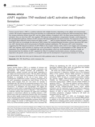
  • 9. intercalate between endothelial cells (supplementary Figure 9). Accordingly, with the presence of filopodia-like structures on the cell surface, the cIAP1À / À MEFs spontaneously adhered to the HUVEC monolayer more strongly compared with the wt counter- part (Figure 7g, left panel). HRas-V12 expression stimulated the attachment of MEFs to the HUVEC monolayer (Figure 7g, left panel). Both the adhesion and the intercalation were decreased in HRas-V12-transformed cIAP1À / À MEFs compared with HRas-V12/ wt MEFs (Figure 7g). DISCUSSION cIAP1 is a key determinant of the cellular response to TNF. The protein is recruited to the TNFR1 through the adaptor TRAF2 and mediates the activation of the canonical NF-kB survival pathway while inhibiting the assembly of the secondary cytoplasmic caspase-activating platform, leading to cell death.20 We demonstrate here that cIAP1 also regulates actin-cytoskeleton reorganization upon TNF stimulation through a cdc42-dependent, NF-kB-independent pathway. We propose a model (Figure 8) in which cIAP1 could regulate the cycle of cdc42 activation by stabilizing the interaction of cdc42 with its regulator RhoGDIa. The recruitment of TRAF2/cIAP1 to the TNF membrane receptor after TNF stimulation could release cdc42 and allow its activation, leading to the actin cytoskeleton reorganization. In fibroblasts, TNF induces the rapid and transient formation of filopodia,38 which requires the Rho GTPase cdc42.5,39,40 The pathway connecting the TNFR1 to Rho GTPases remains poorly understood but appeared to be independent of NF-kB and MAPK activation.5,29,38 Accordingly, we observed that TNF induced filopodia formation in the presence of an IkB super-repressor. TNFR engagement triggers the TRAF2-dependent recruitment of cIAP1, which induces an autoubiquitination and the ubiquitination of RIP1 necessary for NF-kB activation.20 We demonstrate that TNF-induced filopodia formation requires cIAP1 and TRAF2, whereas RIP1 and the E3-ubiquitin ligase of cIAP1 are dispensable, suggesting that the TNFR-associated complex can generate independent signaling pathways leading to NF-kB activation or filopodia formation. cIAP1 can directly bind to cdc42, whereas TRAF2 can bind to cdc42 only in the presence of cIAP1. Both cIAP1 and TRAF2 co-precipitate with cdc42 in resting cells, and TNF treatment decreased this interaction. Our hypothesis is that the recruitment of cIAP1/TRAF2 to TNFR makes cdc42 activation easier, leading to actin cytoskeleton reorganization (Figure 8). Secondary molecular events are likely required for the full activation of cdc42, as the activation of Rho GTPases requires a GEF. Vav is a Rac1-GEF in TNFa signaling in fibroblasts,29 and the GEF Ect2 has been involved in the TNF-like weak inducer of apoptosis (TWEAK)-induced cdc42 activation in glioblastoma;41 however, their role in cdc42 activation has to be explored in this cell context. cIAP1 appears to fine-tune cdc42 activation. On one hand, cIAP1 deletion inhibits cdc42 activation in response to TNF and EGF stimulation and HRas-V12 expression; on the other hand, down- regulation of cIAP1 decreases the expression of the whole cdc42 in a proteasome-dependent manner but increases the expression of the activated fraction of cdc42. The downregulation of cIAP1 also promotes the phosphorylation of PAK1, a cdc42 effector, and cIAP1À / À MEFs spontaneously form filopodia-like structures. Interestingly, RhoGDIa depletion infers similar modifications, including a decreased cdc42 protein level that can be prevented by inhibiting the proteasome, and an increased ratio of active to total cdc42.27 RhoGDIa is a Rho chaperone; that is, it maintains a pool of cytoplamic GTPases in a GDP-bound inactive state and protects them from proteasomal degradation.27,42 The expression of cIAP1 increases the cytosolic cdc42 expression in a RhoGDIa- dependent manner and enhances the fraction of the cdc42 bound to RhoGDIa. Overall, these data argue for a RhoGDIa-mediated regulation of cdc42 by cIAP1. The regulation of RhoGTPases by IAPs is evolutionarily conserved; that is, it has been observed in Drosophila43 and Zebrafish.25 Identical to RhoGDIs,42 cIAP1 (present work), XIAP25 and DIAP143 can bind to both GDP- and GTP-bound GTPases. DIAP1 controls Rac activity independently of its E3-ubiquitine ligase domain,43 whereas XIAP promotes the ubiquitination and degradation of the active GTP-bound form of Rac1.25 We observed an interaction of cIAP1 with Rac1, RhoA and cdc42. In our study, cIAP1 failed to ubiquitinate cdc42 (Supplementary Figure 10), Figure 8. Model for the regulation of cdc42 by cIAP1/TRAF2. cIAP1 interacts with TRAF2 via the BIR1 (B1) domain and with cdc42 via the BIR2 (B2) domain. (1) In resting cells, cIAP1 binds cdc42. It stabilizes its interaction with its regulator RhoGDIa and then regulates cdc42 activation. (2) The recruitment of TRAF2/cIAP1 to the receptor after TNF stimulation releases cdc42 and makes its activation easier, leading to cytoskeleton reorganization and filopodia formation. (3) Depletion of cIAP1 induces a loss of control of cdc42 and increases the activation/ degradation cycle, leading to cytoskeleton modifications and a filopodia-like structure. cIAP1 regulates cdc42 A Marivin et al 5542 Oncogene (2014) 5534 – 5545 & 2014 Macmillan Publishers Limited
  • 10. suggesting that IAPs could differentially modulate various Rho GTPases. RhoA, Rac1 and cdc42 compete for the binding to and chaperoning by RhoGDIa, and the stabilization of the interaction of one Rho GTPase with RhoGDIa favours the degradation of the others.27 cIAP1 becomes a part of this crosstalk regulatory mechanism by stabilizing cdc42–RhoGDIa interaction (present work) and catalysing the proteasomal degradation of Rac1.25 Moreover, an inhibition of the SUMOylation of RhoGDIa by XIAP has been described44 and a recent study identified RhoGDI2 as a potential IAP neddylation substrate.45 As central regulators of Rho GTPase homeostasis, IAPs could regulate their sequential activation in many biological processes such as intracellular vesicular traffic, morphogenesis, tissue repair, cell shape, plasticity, polarization, adhesion and motility.42 In drosophila, overexpression of DIAP1 compensates the invalidation of rac in the control of border cell migration during oogenesis.43 In mammals, the depletion of cIAPs or XIAP alters the invasive properties of cancer cells, including their migration,19,25,44,46 their adhesion to the endothelium and their intercalation between endothelial cells, which can be related to the deregulation of Rho GTPase homeostasis.25,43,44 Our results indicate a regulatory function for cIAP1 in cdc42- controlled filopodia formation, cell polarization and adhesion.39,47 The contribution of cdc42 to some oncogenic processes,6 for example, HRas-V12-driven cell transformation33,35,36 and metastatic invasion,37 may account for the anti-tumour activity of small molecule inhibitors of IAPs, such as Smac mimetics. MATERIALS AND METHODS Cell culture and treatments Mouse embryonic immortalized (SV40) fibroblasts (MEF) wt and cIAP1À / À and cIAP1À / À /cIAP2À / À (J. Silke, Melbourne, Australia), mouse NIH3T3 fibroblasts and HEK293T cells were cultured in DMEM medium (Lonza, Verviers, Belgium) supplemented with 10% fetal bovine serum (FBS) (Lonza). Cells were serum starved for 16 h before stimulation with 100 ng/ml TNF or EGF (Shenandoah Biotechnology Inc, Warwick, PA, USA). MG132 (Millipore, Calbiochem, Billerica, MA, USA) was used at 40 mM for 4 hours. PC3 cells were cultivated in RPMI-1640 medium (Lonza) supple- mented with 10% FBS (Lonza). Primary HUVECs (Lonza) were maintained in EBM2 medium (Lonza). Plasmid constructs, siRNAs, cell transfection and viral transduction NIH3T3 fibroblasts were transfected using Lipofectamine 2000 (Life technologies, Invitrogen, Carlsbad, CA, USA), and MEFs and HEK293T were transfected using JET PEI (Polyplus transfection, Illkirch, France). Lipofecta- mine 2000 (Invitrogen) was used for the transfection of siRNA targeting mouse NIK (Thermo Fisher Scientific, Waltham, MA, USA), cIAP1, TRAF2, RIP1 or RhoGDIa (designed and provided by Qiagen, Venlo, The Netherlands). The DNA constructs used were pCR3-Flag-cIAP1, pCR3-flag- cIAP1L47A , pCI-cIAP1, pCI-cIAP1H588A , pCI-cIAP1L47A ,22 pGEX-cIAP1wt , pGEX-cIAP1BIR1–3 (amino-acid 1–483), pGEX-cIAP1CARD-RING (amino-acid 452–618), pGEX–cIAP1BIR1–2 (amino-acid 1–258), pGEX-cIAP1BIR2–3 (amino-acid 181–363), pGEXcIAP1BIR1 (amino-acid 34–129), pGEX-cIAP1BIR2 (amino-acid 170–260), pGEX-cIAP1BIR3 (amino-acid 256–358),48 pMT90- Myc-cdc42N17 , pCDNA-HA-cdc42WT , pEGFP-cdc42, pEGFP-RhoA, pEGFP-Rac1, pGEX-RhoA, pGEX-Rac1, pGEX-cdc42, pRcCMV-IkB-SR and pBABE-HRasV12 . HRas-V12 expressing MEFs were generated by retroviral transduction. Phoenix-Eco cells (Invitrogen), which constitutively produced gag-pol and ecotropic envelope proteins, were transfected using Jet PEI (Polyplus transfection) with pMSCV-HRas-V12. MEFs were transduced overnight with the retroviral-containing supernatant supplemented with 1 mg/ml polybrene (Sigma-Aldrich, St-Louis, MO, USA). Populations of transduced cells were selected by puromycin exposure. The efficiency of infection was checked by western blot analysis. Immunofluorescence analysis of filopodia and cell polarity Cells were grown and transfected on a chamber slide (Labtek, Thermo Fisher Scientific, Nunc), serum starved for 16 h and stimulated with TNF or EGF. Cells were then washed twice with pre-warmed PBS, fixed for 10 min in 4% paraformaldehyde/PBS, permeabilized using 0.1% triton X-100 (10 min) and saturated for 20 min in 2% bovine serum albumin. Actin cytoskeleton was labelled with AlexaFluor488-Phalloidin (Invitrogen) in PBS/BSA 0.5% for 30 min. Cells were mounted on glass slides using FluorSave (Millipore, Billerica, MA, USA) and examined using a fluorescence (Nikon Eclipse 80i, Nikon, Champigly, France) or a confocal (Leica TCS SP2; Leica, Bron, France) microscope. Filopodia were quantified by counting cells displaying more than five filopodia or by counting the number of filopodia/cell. More than 100 cells were analysed. Cell polarity was assessed by an analysis of the Golgi apparatus orientation in a wound-scratch test. Briefly, after wounding, cell mono- layers were fixed and subjected to nucleus and Golgi staining using Hoechst 33258 (Sigma-Aldrich) and AlexaFluor568-conjugated anti-GM130 (Becton, Dickinson and Company, Franklin Lakes, NJ, USA), respectively. The percentage of cells (4150) with their Golgi orientated towards the wound was evaluated. Cellular extracts, cell fractionation, immunoprecipitation and western blot analysis Cells were lysed in RIPA (Tris-HCl 50 mM pH 7.5, NaCl 150 mM, NP-40 1%, DOC 0.5%, SDS 0.1%) or Phospho (Tris-HCl 50 mM pH 7.5, NaCl 100 mM, NP-40 2%, Glycerol 10%, MgCl2 10 mM, NaF 10 mM, Sodium orthovanadate 1 mM, phosphatase inhibitor phosphatase 2 and 3) buffers complemented with EDTA-free protease inhibitor cocktail (Sigma-Aldrich, St. Louis, MO USA). Primary antibodies used for western blotting were goat anti-cIAP1 (R&D Systems, Minneapolis, MN, USA) and GST (Rockland Immunochem- icals, Philadelphia, Pennsylvania, USA), rabbit anti-GFP (Becton, Dickinson and Company, Franklin Lakes, NJ, USA), RhoGDIa (Santa Cruz Biotechnol- ogy Inc, Santa Cruz, CA, USA), PAK1, p-PAK1, Cofilin, p-Cofilin, GSK3a/b, P-GSK3a/b (Cell Signaling Technology Inc, Danvers, MA, USA) and TRAF2 (Millipore Corporation, Upstate, Billerica, Massachusetts, USA), and mouse anti-HA (Covance), Rac1 (Upstate), cdc42 (Becton, Dickinson and Company), RhoA (Cytoskeleton Inc, Denver, CO, USA), cIAP2, pan-cIAPs (R&D Systems, Cyclex), GM130 (Becton, Dickinson and Company) and HSC70 (Santa Cruz Biotechnology Inc). The western blot analysis was performed as previously described.22 Cell fractionation experiments were performed using the Subcellular Protein Fractionation Kit for Cultured Cells (Thermo Fisher Scientific) according to the manufacturer’s instructions. For immunoprecipitations, cells were lysed in IP buffer (50 mM TrisHCl pH7.5, NaCl 100 mM, NP-40 2%, Glycerol 10%, MgCl2 10 mM, NaF 10 mM, Sodium orthovanadate 1 mM protease inhibitor cocktail) and incubated for 4 h at 4 C in the presence of rabbit polyclonal anti-RhoGDIa or mouse anti- HA and then for 1 h in the presence of mixed A þ G agarose beads (Millipore). Beads were washed in IP buffer and denaturated in Laemmli buffer 2X before immunoblot analysis. Rho GTPase activation assays Cells were lysed in GTPase buffer (Tris-HCl 50 mM pH 7.5, NaCl 300 mM, NP-40 2, Glycerol 10%, 10 mM MgCl2, protease inhibitor cocktail). The active forms of RhoA, rac1 or cdc42 were selectively pulled down by the GST-Rhotekin-Rho binding domain or GST-PAK1-CRIB domain fused to glutathione-Sepharose beads (GE Healthcare, Amersham Biosciences, Fairfield, CT, USA). Beads were washed three times and eluted in Laemmli 2X, and precipitated GTP-RhoA or Rac or Cdc42 was detected by western blot analysis using an anti-RhoA or Rac1 or Cdc42 antibody. GST pull-down assay GST fusion proteins were produced in Escherichia coli, immobilized on glutathione-Sepharose (Amersham Biosciences, Fairfield, CT, USA) and incubated with either tagged protein-expressing HEK293T cell lysates or recombinant cIAP1 or TRAF2 (SignalChem, Richmond, Canada). The pull- down proteins were revealed by western blot analysis. Recombinant cIAP1 protein was produced using a TNT-quick coupled transcription/translation system (Promega, Madison, WI, USA) according to the manufacturer’s instructions. For the analysis of the interaction with the GDP- or GTP- bound form of cdc42, GST-cdc42 or cdc42 from cell lysates was charged with GDP (1 mM) or GTPgS (0.1 mM) (Millipore) in 0.5 M EDTA for 15 min at 30 1C under agitation before the pull-down assay. The reaction was stopped by adding 60 mM MgCl2. cIAP1 regulates cdc42 A Marivin et al 5543 & 2014 Macmillan Publishers Limited Oncogene (2014) 5534 – 5545
  • 11. Surface Plasmon Resonance (Biacore) analysis Design and fabrication of homemade chips compatible with Surface Plasmon Resonance was routinely performed with the help of the MIMENTO technological platform, Besanc¸on, France. The cIAP1-BIRs chips fabricated in this study consisted in the covalent grafting of cIAP1 entities on a chemically activated self-assembled monolayer following the procedure of protein chip building recently published.49 This procedure was performed in a 10 mM acetate buffer (pH4.5) and led to a surface coverage of approximately 8 fmol/mm2 of cIAP1-BIRs per spot. Biacore experiments were performed with the Biacore 2000 apparatus at 25 1C with a flow rate between 2 and 30 ml/min. Purified cdc42 was charged with GDP (1 mM) or GTPgS (0.1 mM) (Millipore) in 0.5 M EDTA for 15 min at 30 1C under agitation and injected into the Biacore. Protein–protein interaction was monitored using Biacore 200 control software (GE-Healthcare, Little Chalfont, UK) and analysed using Biaevaluation 3.2 RCI software (GE- Healthcare, Little Chalfont, UK). Soft agar colony formation Cells (50 000 cells/well) were cultured in 0.45% agarose in growth media and layered on top of 0.75% agarose growth media in a six-well dish. Colonies were counted under a light microscope 2–3 weeks post plating. For each experiment, cells were seeded in triplicate and three fields per well were quantified. Mouse tumoural models Exponentially growing HRas-V12 and control-transduced MEF cells (1 Â 106 /100 ml PBS) were s.c. injected into the flank of nude mice. Tumour growth was monitored by measuring with calipers in two perpendicular diameters, and tumour volumes were calculated using the formula v ¼ a2´b/2 (aob). For the analysis of lung colonization, HRas-V12-transduced MEFs (1 Â 106 / 100 ml PBS) were injected into the tail veins of nude mice. The mice were killed 2 weeks later, and the number of tumour foci at the lung surface was counted. Lungs were fixed and sections were stained with haematoxylin and eosin and observed using AxioZOOM V16 (Carl Zeiss, Oberkoren, Germany). The experiments were performed twice n ¼ 4–5 per group. Cell adhesion and intercalation Cell adhesion and intercalation assay was performed as previously described.37 Briefly, 130 000 CSFE-labelled PC3 cells or MEFs were added onto confluent HUVECs in 24-well plates and washed twice with PBS. Cells were trypsinized and adherent cells were quantified using a LSRII flow cytometer (Becton, Dickinson and Company, Franklin Lakes, NJ, USA). For the analysis of intercalation, 150 000 PC3 cells or MEFs were added onto confluent HUVECs in 6-well plates. Cells were monitored by time-lap microscopy in a humidified chamber at 37 C and 5% CO2 with an inverted microscope AxioVert 200 M (Carl Zeiss) equipped with a motorized stage with a 10x objective lens and using AxioVison software (Carl Zeiss). Cells were tracked manually using AxioVision software and cells were considered as intercalated when they were no more round, when they were no longer phase-bright and were clearly part of the HUVEC monolayer, as shown in Supplementary Figure 9. Stastitical analysis Student’s t test, ANOVA or the Mann–Whitney test was used for statistical analysis CONFLICT OF INTEREST The authors declare no conflict of interest. ACKNOWLEDGEMENTS We thank Dr J Silke, Dr E Lemichez, Dr S Gasman, Dr CL Day, Dr S Ansieu, Dr R Weil and S Monier for kindly providing plasmids and cell lines. We are grateful to Lydie Desoche, Aziza Aznague, Cedric Seignez and Benoit Simon (FEMTO-ST, CLIPP platform) for their technical assistance. We thank A Bouchot and B Gasquet (CellImaP Imagery Facility), A Hammann (Cytometry platform), V Saint-Giorgio (Animal Facility), A Oudot and B Collin (Precilinal imagery platform, Georges-Franc¸ois Leclerc Center) for the use of the imagery, cytometry and animal facilities. We thank P Meier, K Rajalingam, J Bre´ard, M David and S Ansieu for helpful discussions. This work was supported by grants from the ‘Comite´ de Coˆte d’Or of the Ligue Contre le Cancer’ (LD), the ’Association pour la Recherche sur le Cancer’ (ARC to LD), the Association ‘Cent pour sang la Vie’ (LD), the European Union and the ‘Conseil Re´gional de Bourgogne’, a French Government grant managed by the French National Research Agency under the program ‘Investissements d’Avenir’ with reference ANR-11-LABX-0021’, and fellowships from the ‘Ministe`re de l’Enseignement Supe´rieur et de la Recherche’ of France (to AM, JB, JC), ARC (JC) and the ‘Socie´te´ Franc¸aise d’He´matologie’ (AM). AUTHOR CONTRIBUTIONS AM and JB performed most of the experiments and analysed the data. JB performed the in vivo experiment and analysis. JC performed additional experiments and data analysis. CP and AM contributed to the in vivo analysis. SG and WB performed the biacore experiments and analysis. MS and JB provided valuable materials and expert evaluation. ES provided expert evaluation and corrected the paper and LD conceived and supervised the project, analysed the data and wrote the paper with input from all authors. REFERENCES 1 Parameswaran N, Patial S. Tumor necrosis factor-alpha signaling in macrophages. Crit Rev Eukaryot Gene Expr 2010; 20: 87–103. 2 Mathew SJ, Haubert D, Kronke M, Leptin M. Looking beyond death: a morpho- genetic role for the TNF signalling pathway. J Cell Sci 2009; 122: 1939–1946. 3 McKenzie JA, Ridley AJ. Roles of Rho/ROCK and MLCK in TNF-alpha-induced changes in endothelial morphology and permeability. J Cell Physiol 2007; 213: 221–228. 4 Peppelenbosch M, Boone E, GE Jones, van Deventer SJ, Haegeman G, Fiers W et al. Multiple signal transduction pathways regulate TNF-induced actin reorga- nization in macrophages: inhibition of cdc42-mediated filopodium formation by TNF. J Immunol 1999; 162: 837–845. 5 Puls A, Eliopoulos AG, Nobes CD, Bridges T, Young LS, Hall A. Activation of the small GTPase Cdc42 by the inflammatory cytokines TNF(alpha) and IL-1, and by the Epstein-Barr virus transforming protein LMP1. J Cell Sci 1999; 112: 2983–2992. 6 Stengel K, Zheng Y. Cdc42 in oncogenic transformation, invasion, and tumor- igenesis. Cell Signal 2011; 23: 1415–1423. 7 Garcia-Mata R, Boulter E, Burridge K. The ’invisible hand’: regulation of RHO GTPases by RHOGDIs. Nat Rev Mol Cell Biol 2011; 12: 493–504. 8 Micheau O, Tschopp J. Induction of TNF receptor I-mediated apoptosis via two sequential signaling complexes. Cell 2003; 114: 181–190. 9 Bertrand MJ, Milutinovic S, Dickson KM, Ho WC, Boudreault A, Durkin J et al. cIAP1 and cIAP2 facilitate cancer cell survival by functioning as E3 ligases that promote RIP1 ubiquitination. Mol Cell 2008; 30: 689–700. 10 Dynek JN, Goncharov T, Dueber EC, Fedorova AV, Izrael-Tomasevic A, Phu L et al. c-IAP1 and UbcH5 promote K11-linked polyubiquitination of RIP1 in TNF signalling. Embo J 2010; 29: 4198–4209. 11 Haas TL, Emmerich CH, Gerlach B, Schmukle AC, Cordier SM, Rieser E et al. Recruitment of the linear ubiquitin chain assembly complex stabilizes the TNF-R1 signaling complex and is required for TNF-mediated gene induction. Mol Cell 2009; 36: 831–844. 12 Varfolomeev E, Goncharov T, Fedorova AV, Dynek JN, Zobel K, Deshayes K et al. c-IAP1 and c-IAP2 are critical mediators of tumor necrosis factor alpha (TNFalpha)- induced NF-kappaB activation. J Biol Chem 2008; 283: 24295–24299. 13 Silke J. The regulation of TNF signalling: what a tangled web we weave. Curr Opin Immunol 2011; 23: 620–626. 14 Feoktistova M, Geserick P, Kellert B, Dimitrova DP, Langlais C, Hupe M et al. cIAPs block ripoptosome formation, a RIP1/caspase-8 containing intracellular cell death complex differentially regulated by cFLIP isoforms. Mol Cell 2011; 43: 449–463. 15 Wang L, Du F, Wang X. TNF-alpha induces two distinct caspase-8 activation pathways. Cell 2008; 133: 693–703. 16 Vanlangenakker N, Vanden Berghe T, Bogaert P, Laukens B, Zobel K, Deshayes K et al. cIAP1 and TAK1 protect cells from TNF-induced necrosis by preventing RIP1/RIP3-dependent reactive oxygen species production. Cell Death Differ 2011; 18: 656–665. 17 Vince JE, Wong WW, Khan N, Feltham R, Chau D, Ahmed AU et al. IAP antagonists target cIAP1 to induce TNFalpha-dependent apoptosis. Cell 2007; 131: 682–693. 18 Gyrd-Hansen M, Darding M, Miasari M, Santoro MM, Zender L, Xue W et al. IAPs contain an evolutionarily conserved ubiquitin-binding domain that regulates NF- kappaB as well as cell survival and oncogenesis. Nat Cell Biol 2008; 10: 1309–1317. 19 Lopez J, John SW, Tenev T, Rautureau GJ, Hinds MG, Francalanci F et al. CARD- Mediated Autoinhibition of cIAP1’s E3 Ligase Activity Suppresses Cell Proliferation and Migration. Mol Cell 2011; 42: 569–583. 20 Gyrd-Hansen M, Meier P. IAPs: from caspase inhibitors to modulators of NF-kappaB, inflammation and cancer. Nat Rev Cancer 2010; 10: 561–574. cIAP1 regulates cdc42 A Marivin et al 5544 Oncogene (2014) 5534 – 5545 & 2014 Macmillan Publishers Limited
  • 12. 21 Xu L, Zhu J, Hu X, Zhu H, Kim HT, LaBaer J et al. c-IAP1 cooperates with Myc by acting as a ubiquitin ligase for Mad1. Mol Cell 2007; 28: 914–922. 22 Cartier J, Berthelet J, Marivin A, Gemble S, Edmond V, Plenchette S et al. Cellular inhibitor of apoptosis protein-1 (cIAP1) can regulate E2F1 transcription factor-mediated control of cyclin transcription. J Biol Chem 2011; 286: 26406–26417. 23 Zarnegar BJ, Wang Y, Mahoney DJ, Dempsey PW, Cheung HH, He J et al. Non- canonical NF-kappaB activation requires coordinated assembly of a regulatory complex of the adaptors cIAP1, cIAP2, TRAF2 and TRAF3 and the kinase NIK. Nat Immunol 2008; 9: 1371–1378. 24 Vallabhapurapu S, Matsuzawa A, Zhang W, Tseng PH, Keats JJ, Wang H et al. Nonredundant and complementary functions of TRAF2 and TRAF3 in a ubiquiti- nation cascade that activates NIK-dependent alternative NF-kappaB signaling. Nat Immunol 2008; 9: 1364–1370. 25 Oberoi TK, Dogan T, Hocking JC, Scholz RP, Mooz J, Anderson CL et al. IAPs regulate the plasticity of cell migration by directly targeting Rac1 for degradation. Embo J 2011; 31: 14–28. 26 Varfolomeev E, Blankenship JW, Wayson SM, Fedorova AV, Kayagaki N, Garg P et al. IAP antagonists induce autoubiquitination of c-IAPs, NF-kappaB activation, and TNFalpha-dependent apoptosis. Cell 2007; 131: 669–681. 27 Boulter E, Garcia-Mata R, Guilluy C, Dubash A, Rossi G, Brennwald PJ et al. Reg- ulation of Rho GTPase crosstalk, degradation and activity by RhoGDI1. Nat Cell Biol 2010; 12: 477–483. 28 Haubert D, Gharib N, Rivero F, Wiegmann K, Hosel M, Kronke M et al. PtdIns(4,5)P- restricted plasma membrane localization of FAN is involved in TNF-induced actin reorganization. Embo J 2007; 26: 3308–3321. 29 Kant S, Swat W, Zhang S, Zhang ZY, Neel BG, Flavell RA et al. TNF-stimulated MAP kinase activation mediated by a Rho family GTPase signaling pathway. Genes Dev 2011; 25: 2069–2078. 30 Van Troys M, Huyck L, Leyman S, Dhaese S, Vandekerkhove J, Ampe C. Ins and outs of ADF/cofilin activity and regulation. Eur J Cell Biol 2008; 87: 649–667. 31 Iden S, Collard JG. Crosstalk between small GTPases and polarity proteins in cell polarization. Nat Rev Mol Cell Biol 2008; 9: 846–859. 32 Etienne-Manneville S. Cdc42--the centre of polarity. J Cell Sci 2004; 117: 1291–1300. 33 Stengel KR, Zheng Y. Essential role of cdc42 in ras-induced transformation revealed by gene targeting. PLoS One 2012; 7: e37317. 34 Makrodouli E, Oikonomou E, Koc M, Andera L, Sasazuki T, Shirasawa S et al. BRAF and RAS oncogenes regulate Rho GTPase pathways to mediate migration and invasion properties in human colon cancer cells: a comparative study. Mol Cancer 2011; 10: 118–138. 35 Cheng CM, Li H, Gasman S, Huang J, Schiff R, Chang EC. Compartmentalized Ras proteins transform NIH 3T3 cells with different efficiencies. Mol Cell Biol 2011; 31: 983–997. 36 Qiu RG, Abo A, McCormick F, Symons M. Cdc42 regulates anchorage-independent growth and is necessary for Ras transformation. Mol Cell Biol 1997; 17: 3449–3458. 37 Reymond N, Im JH, Garg R, Vega FM, Borda d’Agua B, Riou P et al. Cdc42 promotes transendothelial migration of cancer cells through beta1 integrin. The Journal of cell biology. [Research Support, Non-U.S. Gov’t] 2012; 199: 653–668. 38 Gadea G, Roger L, Anguille C, de Toledo M, Gire V, Roux P. TNFalpha induces sequential activation of Cdc42- and p38/p53-dependent pathways that antag- onistically regulate filopodia formation. J Cell Sci 2004; 117: 6355–6364. 39 Yang L, Wang L, Zheng Y. Gene targeting of Cdc42 and Cdc42GAP affirms the critical involvement of Cdc42 in filopodia induction, directed migration, and proliferation in primary mouse embryonic fibroblasts. Mol Biol Cell 2006; 17: 4675–4685. 40 Allen WE, Jones GE, Pollard JW, Ridley AJ. Rho, Rac and Cdc42 regulate actin organization and cell adhesion in macrophages. J Cell Sci 1997; 110: 707–720. 41 Fortin SP, Ennis MJ, Schumacher CA, Zylstra-Diegel CR, Williams BO, Ross JT et al. Cdc42 and the guanine nucleotide exchange factors Ect2 and trio mediate Fn14- induced migration and invasion of glioblastoma cells. Mol Cancer Res 2012; 10: 958–968. 42 Boulter E, Estrach S, Garcia-Mata R, Feral CC. Off the beaten paths: alternative and crosstalk regulation of Rho GTPases. FASEB J. 2012; 26: 469–479. 43 Geisbrecht ER, Montell DJ. A role for drosophila IAP1-mediated caspase inhibition in Rac-dependent cell migration. Cell 2004; 118: 111–125. 44 Liu J, Zhang D, Luo W, Yu Y, Yu J, Li J et al. X-linked inhibitor of apoptosis protein (XIAP) mediates cancer cell motility via Rho GDP dissociation inhibitor (RhoGDI)- dependent regulation of the cytoskeleton. J Biol Chem 2011; 286: 15630–15640. 45 Zhuang M, Guan S, Wang H, Burlingame AL, Wells JA. Substrates of IAP ubiquitin ligases identified with a designed orthogonal E3 ligase, the neddylator. Mol Cell 2013; 49: 273–282. 46 Dogan T, Harms GS, Hekman M, Karreman C, Oberoi TK, Alnemri ES et al. X-linked and cellular IAPs modulate the stability of C-RAF kinase and cell motility. Nat Cell Biol 2008; 10: 1447–1455. 47 Hehnly H, Xu W, Chen JL, Stamnes M. Cdc42 regulates microtubule-dependent golgi positioning. Traffic 2010; 11: 1067–1078. 48 Mace PD, Smits C, Vaux DL, Silke J, Day CL. Asymmetric recruitment of cIAPs by TRAF2. J Mol Biol 2010; 400: 8–15. 49 Grandclement C, Pallandre JR, Valmary Degano S, Viel E, Bouard A, Balland J et al. Neuropilin-2 expression promotes TGF-beta1-mediated epithelial to mesenchy- mal transition in colorectal cancer cells. PLoS One 2011; 6: e20444. Supplementary Information accompanies this paper on the Oncogene website (http://www.nature.com/onc) cIAP1 regulates cdc42 A Marivin et al 5545 & 2014 Macmillan Publishers Limited Oncogene (2014) 5534 – 5545