1. Oncogene (2005) 24, 5676–5692
& 2005 Nature Publishing Group All rights reserved 0950-9232/05 $30.00
www.nature.com/onc
The role of Smad signaling in hematopoiesis
Jonas Larsson1,2 and Stefan Karlsson*,1
1
Molecular Medicine and Gene Therapy, Institute of Laboratory Medicine and The Lund Strategic Research Center for Stem Cell
Biology and Cell Therapy, Lund University, BMC A12, Lund 221 84, Sweden; 2Center for Regenerative Medicine and Technology,
Massachusetts General Hospital, Harvard Medical School, Boston, MA, USA
The TGF-b family of ligands, including TGF-b, bone or lack-of-function animal models can be used to
morphogenetic protein (BMP) and activin, signal through investigate the role of Smad signaling in vivo and how
Smad pathways to regulate the fate of hematopoietic the findings obtained compare with earlier in vitro
progenitor and stem cells during development and post- findings using active ligands. The outcome of Smad
natally. BMP regulates hematopoietic stem cell (HSC) signaling is very context dependent and will often have a
specification during development, while TGF-b1, 2 and 3 different effect in ex vivo cultures than in the bone
are not essential for the generation of HSCs. BMP4 can marrow (BM) microenvironment where many internal
increase proliferation of human hematopoietic progeni- and external signals cooperate to regulate the fate of
tors, while TGF-b acts as a negative regulator of HSCs and their progeny. The importance of signaling
hematopoietic progenitor and stem cells in vitro. In crosstalk will be discussed as well as how the knowledge
contrast, TGF-b signaling deficiency in vivo does not generated can be used in clinical medicine, for example,
affect proliferation of HSCs and does not affect lineage by expanding stem cells for advanced cell therapy in the
choice either. Therefore, the outcome of Smad signaling is future.
very context dependent in hematopoiesis and regulation of
hematopoietic stem and progenitor cells is more compli-
cated in the bone marrow microenvironment in vivo than is
seen in liquid cultures ex vivo. Smad signaling regulates Ligands and receptors that activate Smad signaling
hematopoiesis by crosstalk with other regulatory signals pathways
and future research will define in more detail how the
various pathways interact and how the knowledge TGF-b is the founding member and prototype of a large
obtained can be used to develop advanced cell therapies. superfamily of structurally related peptide growth
Oncogene (2005) 24, 5676–5692. doi:10.1038/sj.onc.1208920 factors. Other key members of this TGF-b superfamily
are the activins, BMPs and growth and differentiation
Keywords: Smad signaling; hematopoiesis; stem cells factors (Massague, 1998). There are three isoforms of
TGF-b in mammals, TGF-b1 (Derynck et al., 1985),
TGF-b2 (de Martin et al., 1987; Madisen et al., 1988)
and TGF-b3 (Derynck et al., 1988; ten Dijke et al.,
1988), which are encoded by separate genes located at
different chromosomes. These isoforms share 70–80%
Introduction amino-acid sequence identity, bind to the same receptors
and show very similar actions in in vitro culture systems.
TGF-b has been characterized as a well-known reg- However, they have distinct expression patterns in vivo
ulator of hematopoiesis through detailed studies of and mice lacking either TGF-b1, 2 or 3 overlap
hematopoietic progenitors in vitro. A relatively recent phenotypically very little, indicating that there are many
detailed review describes how TGF-b regulates prolif- specific functions for the different isoforms (see below).
eration and differentiation of hematopoietic precursors Members of the TGF-b superfamily transduce their
in vitro, often as a negative regulator of proliferation signals across the plasma membrane through transmem-
(Fortunel et al., 2000). In this review, we will discuss brane serine/threonine kinase receptors. Ligand binding
how various Smad signaling pathways (TGF-b, bone to these signaling receptors is facilitated by certain
morphogenetic protein (BMP), activin) regulate hema- accessory receptors, for example, betaglycan and en-
topoiesis in vitro and in vivo. Smad signaling participates doglin, which have affinity for the ligand but no
in the specification or generation of hematopoietic stem signaling activity (Cheifetz et al., 1992; Lopez-Casillas
cells (HSCs) during development and continues to et al., 1993). There are two classes of signaling receptors,
regulate the fate of HSCs and their progeny after their type I and type II, both of which are required for signal
generation during development and postnatally. We will transduction (reviewed in Massague, 1998). Type I
discuss how genetic approaches using gain-of-function receptors, also known as activin receptor-like kinases
(ALKs), have an inactive kinase domain, while type II
*Correspondence: S Karlsson; E-mail: Stefan.karlsson@molmed.lu.se receptor kinases are constitutively active. The ligand
2. Role of Smad signaling in hematopoiesis
J Larsson and S Karlsson
5677
binds constitutively active type II receptors, whereby the expressed on HSCs (Chen et al., 2002). Antibodies
inactive type I receptors are recruited to form a against endoglin can be used to highly enrich repopulat-
heterotetrameric complex in which the type II receptor ing HSCs by flow cytometry, sorting cells that are
transphosphorylates and activates the kinase domain of endoglin positive, Sca-1 þ and Rhodamine-low (Chen
the type I receptor (Wrana et al., 1994). Activated type I et al., 2003). The role of endoglin, if any, in the
receptor kinase subsequently phosphorylates intracellu- regulation of HSCs is unknown.
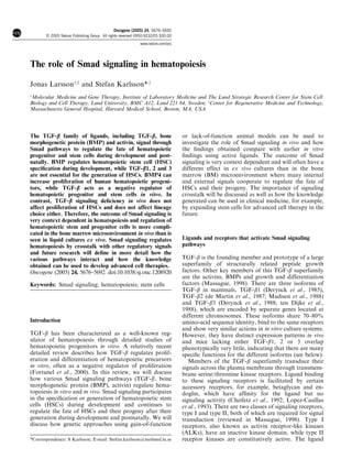
lar mediators known as the Smad proteins (Figure 1).
The activation of the receptors is tightly regulated Smad signaling pathways that regulate hematopoiesis
through ligand traps, soluble proteins that sequester the
ligand and block its access to membrane receptors. The Smad proteins can be divided into three different
These include the TGF-b latency-associated polypep- groups depending on their role in the signal transduc-
tide, decorin and a2-macroglobulin, which bind to free tion: the receptor-activated Smads (R-Smads), the
TGF-b, and follistatin, which binds to activins and common-partner Smad (Smad4) and the inhibitory
BMPs (Shi and Massague, 2003). The regulation of Smads (I-Smads) (Heldin et al., 1997). R-Smads become
receptor activation is also controlled by membrane- phosphorylated by activated type I receptors, whereby
anchored proteins that act as coreceptors and promote they oligomerize with Smad4 to form a heterodimeric
ligand binding to the signaling receptors. Betaglycan, complex that is translocated into the nucleus (Figure 1).
often referred to as the TGF-b type III receptor, R-Smad/Smad4 complexes have been shown to interact
promotes high-affinity presentation of TGF-b to the directly with specific DNA sequences as well as with
signaling receptor and this is particularly critical for transcription factors, coactivators and corepressors to
TGF-b2 (Derynck and Zhang, 2003; Shi and Massague, regulate transcription of target genes in a cell-type-
2003). Betaglycan also facilitates the binding of inhibin specific and ligand dose-dependent manner (Heldin
to activin receptors to block activin signaling. Endoglin et al., 1997; Derynck and Zhang, 2003; ten Dijke and
is another membrane-anchored protein, structurally Hill, 2004). R-Smads and Smad4 bind to specific DNA
related to betaglycan, which facilitates TGF-b signaling sequences with a 100-fold lower affinity than the
in endothelial cells (ECs). Although endoglin does not interacting high-affinity, DNA-binding transcription
bind TGF-b by itself, mutations in endoglin and the factors, yet their DNA binding (except Smad2) is
ALK1 receptor can give rise to hereditary hemorrhagic required for transcriptional activation (Derynck and
telangiectasia (HHT), indicating a common role for Zhang, 2003). The I-Smads act as inhibitors of Smad-
endoglin and ALK1 in maintaining normal vascular mediated signal transduction by interacting with the
development. More recently, a role for endoglin in type I receptor and inhibiting the phosphorylation of
hematopoiesis has been suggested because endoglin is R-Smads (Nakao et al., 1997), by recruiting E3-ubiquitin
TGFβ-1 Activin BMP2
Ligand TGFβ-2 Nodal BMP4
TGFβ-3 BMP7 BMP7
Type II TβR-II ActR-II
BMPR-II
ActR-IIB ActR-II
ActR-IB Endoglin ActR-IIB
Type I TβR-I ALK1 BMPR-IA
BMPR-IB ALK2
Smad6
R-Smads Smad2 Smad3 Smad1 Smad5 Smad8
Smad7
Smad4
Co-Smads
Nucleus
Figure 1 Overview of the Smad signaling pathways. Ligands of the TGF-b superfamily transduce their signals through
heterotetrameric complexes of type I and type II receptors, which phosphorylate the intracellular R-Smad proteins. The inhibitory
Smads are shown in red. The majority of the components shown are expressed on purified murine HSCs (Utsugisawa, Moody and
Karlsson, unpublished studies). ActR-I is also referred to as ALK2, BMPRIA as ALK3, ActRIB as ALK4, TGF-bRI as ALK5 and
BMPRIB as ALK6. ALK1 combines with TGF-bRII to mediate signals in ECs, but has not been shown to play a role in hematopoietic
cells
Oncogene
3. Role of Smad signaling in hematopoiesis
J Larsson and S Karlsson
5678
ligases to degrade activated type I receptors, or by direct Smad3 cooperates with Runx proteins to activate
dephosphorylation and subsequent inactivation of the transcription in epithelial cells, whereas the same
type I receptor (Shi and Massague, 2003; ten Dijke promoters in mesenchymal cells are repressed (Alliston
and Hill, 2004). Alternatively, I-Smads may compete et al., 2001; Derynck and Zhang, 2003).
with Smad4 in binding R-Smads and thereby prevent
the formation of the R-Smad/Smad4 complex (Hata
Duration and intensity of the signal
et al., 1998).
Figure 1 describes the close relationship between the Upon signal stimulation, Smad complexes accumu-
TGF-b, activin and BMP signaling pathways. TGF-b late in the nucleus where they remain for hours (ten
signals through specific type I and type II receptors Dijke and Hill, 2004). The levels of the Smad complexes
known as TbRI (or ALK5) and TbRII, respectively in the nucleus determine the nature and the duration
(Wang et al., 1991; Attisano et al., 1993; Ebner et al., of the signal. In the nucleus, the R-Smads are
1993; Franzen et al., 1993). The activated TGF-b dephosphorylated and are disassociated from Smad4
receptor complex phosphorylates the R-Smads Smad2 and exported from the nucleus. If the receptors are
and Smad3, which also act as mediators of the activin active, Smad signaling continues, but if the receptors
signaling pathway (Heldin et al., 1997). In addition to are inactive, the dephosphorylated Smads accumulate
signaling via ALK5, it has been shown that TGF-b over time in the cytoplasm and signaling stops
can signal through another type I receptor (ALK1) in (Inman et al., 2002; Xu et al., 2002). Apart from
ECs, which phosphorylates a different set of R-Smads: nucleocytoplasmic shuttling of Smads, the duration
Smad1, Smad5 and Smad8 (Roelen et al., 1997; Lux and activity of Smad pathways can be regulated by
et al., 1999; Oh et al., 2000). These Smads are the different receptor internalization routes. Clathrin-
intracellular mediators of the BMP signaling pathway. dependent internalization of receptors into early
As shown in Figure 1, the type I receptors ALK1, ALK2 endosomes promotes Smad signaling, whereas inter-
(ActRI), ALK3 (BMPRIA) and ALK6 (BMPRIB) nalization via lipid raft-caveolar compartments contain-
phosphorylate Smad1, Smad5 and Smad8, whereas ing receptor bound to Smad7-ubiquitin ligase
ALK4 (ActRIB) and ALK5 (TGF-bRI) phosphorylate complexes leads to degradation of receptors (Di
Smad2 and Smad3 (Derynck and Zhang, 2003; Shi and Guglielmo et al., 2003; Shi and Massague, 2003; ten
Massague, 2003; ten Dijke and Hill, 2004). All R-Smads Dijke and Hill, 2004). Therefore, the internalization
from both the activin and BMP groups use Smad4 as a pathway has implications for receptor availability and
partner to form a transcriptionally active complex. The duration of the signal.
I-Smad, Smad7 inhibits the activity of all R-Smads,
while Smad6 shows a more preferential inhibition of the
Smad-dependent and Smad-independent signaling
BMP Smads (Itoh et al., 1998).
crosstalk with other pathways
Specificity of signaling Smads are the most well-characterized transducers of
TGF-b signaling so far, playing a critical role in most
This convergence in the mediation of signals from TGF- TGF-b actions. Increasing evidence suggests, however,
b superfamily members by using the Smad pathway that TGF-b may also send signals though another
raises the question of how specificity in signaling for intracellular signaling pathway, the mitogen-activated
different family members and different isoforms can be protein kinase (MAPK) pathway (reviewed in Massa-
achieved. As mentioned above, expression of accessory gue, 2000). The complexity of intracellular TGF-b signal
proteins like betaglycan and endoglin can affect signal- transduction is further demonstrated by the fact that the
ing specificity. Different combinations of receptor Smad pathway is believed to be part of a larger network
molecules in the tetrameric receptor complexes allow involving many other signaling pathways, which
differential ligand binding and a differential signal are interacting in a so-called ‘signaling crosstalk’
response. For example, activin receptor type II com- (Massague, 2000). Several kinase pathways inhibit or
bines with ALK4 to mediate activin signals, but when it enhance TGF-b-induced nuclear translocation of
combines with ALK3 or ALK6, it mediates signals from Smads. For example, the Erk MAPK pathway, stimu-
BMP4 (Derynck and Zhang, 2003; Shi and Massague, lated by the activation of tyrosine kinase receptors
2003). Heteromeric interactions of activated Smads may and/or Ras, inhibits ligand-induced nuclear transloca-
also determine signaling specificity. Most TGF-b tion of activated Smads (de Caestecker et al., 1998;
responses are mediated by Smad3 and Smad4, whereas Kretzschmar et al., 1999; Funaba et al., 2002; Derynck
activin responses are mediated by Smad2 and Smad4 and Zhang, 2003). The Erk MAPK and calcium-
(Derynck and Zhang, 2003). Some TGF-b responses calmodulin-dependent protein kinase II inhibit TGF-b
require a concomitant activation of Smad2 and Smad3 signaling through phosphorylation of Smads at phos-
together with Smad4 (Feng et al., 2000). The cellular phorylation sites that are different from those that
context is also very important in determining the are phosphorylated by ALKs (Derynck and Zhang,
consequences of Smad signaling responses since differ- 2003; Shi and Massague, 2003). TGF-b signaling can
ent cells express a variety of transcription factors and also be independent of the Smad pathways (reviewed in
cofactors to determine whether a specific set of genes Derynck and Zhang, 2003). Rapid activation (5–15 min)
can be affected, activated or repressed. For example, by TGF-b does not involve Smad-mediated trans-
Oncogene
4. Role of Smad signaling in hematopoiesis
J Larsson and S Karlsson
5679
criptional responses and independence from Smad complex mix of dividing and maturing cells of different
activation has been supported by findings demon- lineages can be found. The process of hematopoiesis can
strating MAPK activation in Smad4-deficient cells and be described as hierarchical with the rare HSCs at the
cells that express dominant-negative Smads (Engel et al., top of the hierarchy giving rise first to progenitors and
1999). Therefore, MAPK pathways can regulate Smad then to precursors with single lineage commitment and
transcriptional responses, but TGF-b can also activate ending in terminally differentiated mature cells of
Erk, JNK (c-Jun N-terminal kinase) and p38 MAPK various lineages (Ogawa, 1993; Orkin, 2000). In between
kinase pathways independent of Smad-mediated tran- the HSC and terminally differentiated cells, there is a
scription. This complex crosstalk by TGF-b family of continuum of progenitors at different stages, which,
ligands with other signaling cascades has also been depending on certain stimuli, can divide and progress
extended to other signaling pathways (reviewed in towards certain lineages (Figure 2). It is generally
Derynck and Zhang, 2003). believed that hematopoietic development divides at an
early stage into a myeloid and a lymphoid branch. The
lymphoid branch gives rise to B, T and natural killer
Hematopoiesis and HSCs cells, while the myeloid branch differentiates to all other
cell types including erythrocytes. Indeed, two distinct
All blood cells can be generated from a common HSC progenitor populations with either lymphoid or myeloid
through an extremely dynamic process called hemato- restricted potential, the so-called common lymphoid
poiesis or blood cell formation. The generation of progenitors and common myeloid progenitors, have
definitive or adult-type HSC during development occurs been isolated from mouse BM (Kondo et al., 1997;
in the aorta-mesonephros-gonad region (AGM) of the Akashi et al., 2000).
embryo (Medvinsky et al., 1993; Cumano et al., 1996; HSCs are pluripotent and should therefore be able to
Medvinsky and Dzierzak, 1996; Cumano et al., 2000), give rise to all hematopoietic lineages. They are
and more recently there have been reports that may operationally defined as cells that can completely
indicate that definitive HSC are also found in the yolk reconstitute a recipient following BM ablation. Simi-
sac and later in placenta (Yoder et al., 1997; Gekas larly, they must have the capacity to self-renew to give
et al., 2005). The fetal liver is seeded with HSC from the rise to other stem cells (Domen and Weissman, 1999).
AGM and they are expanded there and generate a large A distinction is often made between long-term repopu-
amount of progeny cells. Postnatally, hematopoiesis lating HSCs (LT-HSCs) and short-term repopulating
takes place in the BM where the HSCs as well as a HSCs (ST-HSCs) (Morrison and Weissman, 1994). The
LT-HSC “
“LSK (Lineage- Sca1+ c-kit+)in mouse Stem Cell
ST-HSC
Multipotent
progenitor
MPP
Myeloid Lymphoid
CMP CLP
Committed
Pro-B Pro-T progenitor
MEP GMP
Pre-B
Megakaryocyte
Mature
cells
Eosinophil
Neutrophil Basophil
B Cell T Cell NK Cell
Erythrocyte Platelets Macrophage
Dendritic Cell
Figure 2 The hierarchy of hematopoietic cells. LT-HSC, long-term repopulating HSC; ST-HSC, short-term repopulating HSC; MPP,
multipotent progenitor; CMP, common myeloid progenitor; CLP, common lymphoid progenitor; MEP, megakaryocyte/erythroid
progenitor; GMP, granulocyte–macrophage progenitor. The encircled pluripotent population, LT-HSC, ST-HSC and MPP are LinÀ,
Sca-1 þ , c-kit þ as shown
Oncogene
5. Role of Smad signaling in hematopoiesis
J Larsson and S Karlsson
5680
only way to test these criteria is through BM transplan- development
tation experiments into lethally irradiated recipients.
Although the HSC can only be absolutely defined malignant
through BM transplantation assays, advances in cell self-renewal transformation
sorting have allowed near purification of murine HSC.
Early committed progenitor populations, depicted in
Figure 2, can also be purified (Spangrude et al., 1988;
Morrison and Weissman, 1994; Kondo et al., 1997;
Akashi et al., 2000). HSC are highly enriched in a
population that is negative for lineage markers (LinÀ)
quiescence
and positive for Sca-1 and c-kit (LSK cells) (Spangrude apoptosis
et al., 1988). C-kit, which is the receptor for stem cell
factor (SCF), has a wider expression pattern than Sca-1,
marking most multipotent progenitors (MPPs) (Lyman
and Jacobsen, 1998). Sca-1 and c-kit are often used differentiation
migration
together for positive selection of HSCs from LinÀ cells.
The LSK cells contain LT-HSC, ST-HSC and MPPs,
which cannot repopulate recipients. Further enrichment
for LT-HSC has been demonstrated by including Figure 3 HSC fate options. Signals from internal and external
negative selection for the CD34 marker. Indeed, LSK regulatory factors decide whether the HSC are maintained in
CD34À cells contain a very high proportion of HSCs and quiescence, proliferate, undergo apoptosis, migrate out of the BM
space or develop a malignant clone that grows in an uncontrolled
isolated single LSK CD34À cells have been transplanted manner
and have given full long-term hematopoietic recon-
stitution (Osawa et al., 1996). A further separation
between LT-HSC, ST-HSC and MPP has recently been
made by showing that repopulating HSC express the megakaryocytic development and Pax-5 for B-cell
thrombopoietin (Tpo) receptor (c-mpl), whereas MPP development (Pevny et al., 1991; Fujiwara et al., 1996;
do not, and MPPs express Flt3, whereas pluripotent Shivdasani et al., 1997; Nutt et al., 1999; Rolink et al.,
repopulating HSC do not (Adolfsson et al., 2001; Yang 1999). The PU.1 and GATA-1 transcription factors
et al., 2005a). have both been shown to have concentration-dependent
effects on differentiation and lineage choice (Kulessa
et al., 1995; DeKoter and Singh, 2000). Enforced
Regulation of hematopoiesis
expression of HoxB4 has been shown to increase the
Regulation of HSCs is governed by two intimately self-renewal potential of primitive murine and human
involved entities. One is the gene expression pattern in hematopoietic progenitors (Sauvageau et al., 1995;
the cell and the other is the composition of external Antonchuk et al., 2002; Buske et al., 2002) and
signals from the BM microenvironment. Transcription deficiency of HoxB4 causes a mild proliferative defect
factors are internal signals that regulate gene expression, in HSC (Bjornsson et al., 2003; Brun et al., 2004). Apart
while external signals from the BM microenvironment from transcription factors, there are also other intrinsic
can be mediated by cell–cell interactions, cell–extra- factors within the HSCs that are important regulators of
cellular matrix (ECM) interactions and by soluble stem cell fate. These include effector molecules in the cell
growth factors. Figure 3 demonstrates the various fate cycle machinery. For example, high levels of cyclin D2
options upon alteration of internal or external signals have been detected in cycling HSCs, suggesting an
that may promote or antagonize proliferation (self- important role in HSC cycling, while studies of knock-
renewal), quiescence, differentiation, migration, apop- out mice have shown that the cyclin-dependent kinase
tosis or malignant transformation. inhibitor (CDKI) p21 acts to maintain HSCs in a
A number of transcription factors have been shown to quiescent state (Cheshier et al., 1999; Cheng et al., 2000).
be critical intrinsic factors for specification of HSCs Many hematopoietic growth factors have been
during ontogeny. Nascent HSC in the AGM region considered to be key external regulators of HSCs. SCF
express the hematopoietic transcription factors Runx-1, (also known as c-kit ligand) and its receptor c-kit as well
c-Myb, PU.1, SCL and LMO2 (Tavian et al., 1996; as Tpo and its receptor c-mpl are expressed on HSCs
North et al., 1999; Orkin and Zon, 2002). Most of these and efficiently promote survival of primitive murine and
genes have also been found to be essential for human primitive progenitors (Li and Johnson, 1994;
hematopoiesis using mouse knockout models. They are Keller et al., 1995; Borge et al., 1996). They can also
also observed in chromosomal translocations in leuke- synergize with other growth factors, such as IL-3 and
mias or cause leukemia when misexpressed or disrupted IL-6, to induce proliferation of these primitive cells and
in the mouse (reviewed in Orkin, 2000). Studies in are therefore widely used in various in vitro culture
knockout mouse models have elegantly demonstrated systems (Ramsfjell et al., 1996). Important roles in HSC
crucial roles of several transcription factors in early regulation in vivo have been shown through analysis of
hematopoietic commitment steps and lineage outcome. mice lacking either c-kit or c-mpl. These mice have
For example, GATA-1 is required for erythroid and severe HSC deficiencies as demonstrated by reduced LT
Oncogene
6. Role of Smad signaling in hematopoiesis
J Larsson and S Karlsson
5681
repopulating ability following transplantation (Miller occurrence of erythrocytes and ECs in the blood islands
et al., 1996; Kimura et al., 1998). Flt3-ligand (FL) and has led to the hypothesis that they have a common
its receptor Flt3 has been shown to promote in vitro mesodermal precursor, the ‘hemangioblast’ (Mikkola
growth of HSCs (Lyman and Jacobsen, 1998). However, and Orkin, 2002) (Figure 4). By E8–E8.5 precursors
a role of FL in regulating the most primitive HSCs is with definitive hematopoietic potential arise in the yolk
uncertain since Flt3 is not expressed on LTR-HSCs sac as well as intraembryonically along the dorsal aorta
(Adolfsson et al., 2001). Stem cell homeostasis in the in a region called para-aortic splanchnopleura, which
adult may also be under control of morphogens that later forms the AGM (Cumano et al., 1996; Medvinsky
regulate early embryogenesis (Orkin and Zon, 2002). and Dzierzak, 1996; Yoder et al., 1997). Shortly
BMP4 maintains HSC in culture and sonic hedgehog thereafter, hematopoiesis shifts to the fetal liver and
(Shh) may amplify HSC through modulation of the around the time of birth the BM becomes the major
BMP pathway (Bhatia et al., 1999; Bhardwaj et al., site of hematopoiesis. The mechanisms that control the
2001). The Notch signaling pathway and the Wnt sequential induction of mesoderm, formation of the
pathway also regulate HSC homeostasis and both hemangioblast and commitment of hematopoietic
activation of Notch and stimulation by Wnt have been precursors are still not well understood. An important
shown to increase proliferation of HSC (Van Den Berg tool to investigate these early specification events of
et al., 1998; Varnum-Finney et al., 2000; Stier et al., HSCs, in addition to the study of embryos, has been the
2002; Reya et al., 2003). embryonic stem (ES) cell differentiation system. In the
It is generally believed that maintenance of HSCs is absence of leukemia inhibiting factor, ES cells will
controlled by a balance of positively and negatively aggregate and form the so-called embryoid bodies (EBs)
acting signals. Although a number of growth factors that are composed of all three germ layers. These can be
have been identified that exert critical growth promoting differentiated further in a manner that mimics several
effects on HSCs, as described above, much less is known aspects of normal embryonic development in utero,
about negative regulation of HSCs. Apart from TGF-b including formation of hemangioblasts from mesoder-
(see below), both tumor necrosis factor-a (TNF-a) and mal precursors and the generation of primitive and
interferon-g (IFN-g) have been associated with negative definitive hematopoietic cells (Lacaud et al., 2001)
regulation of hematopoietic progenitors (Jacobsen et al., (Figure 4).
1992; Maciejewski et al., 1995; Zhang et al., 1995; Yang A large number of studies in several different species
et al., 2005b). Furthermore, it was recently shown that have implicated a key role of BMP4 in mesoderm
TNF-a through activation of Fas inhibits self-renewal of induction and hematopoietic commitment (reviewed in
murine LTR-HSCs, bringing out TNF-a and Fas as Snyder et al., 2004). For example, studies using Xenopus
major negative regulators of HSCs (Bryder et al., 2001). embryos and explant assays have demonstrated that
BMP4 together with either basic fibroblast growth
factor or activin A can induce the formation of red
blood cells from non-mesodermal structures (Huber
Smad signaling and specification of HSCs during et al., 1998). Furthermore, hematopoietic commitment
development induced by the GATA binding transcription factors
GATA-1 and GATA-2 required intact BMP signaling
The first signs of hematopoiesis in the developing mouse (Maeno et al., 1996; Huber et al., 1998). Mice lacking
embryos are the formation of blood islands from BMP4 die between E7.5 and E9.5 exhibit severe defects
extraembryonic mesoderm in the yolk sac at embryonic in mesoderm formation and the embryos that survive up
day 7.5 (E7.5) (Yoder, 2001). Blood islands are to E9.5 show defective blood islands (Winnier et al.,
composed of primitive, nucleated erythrocytes sur- 1995). Studies using differentiation of mouse ES cells
rounded by ECs. The close spatial and temporal support these findings as BMP4 induces the formation
Connective tissue
Heart
Ectoderm Urogenital system Angioblast
Endoderm
‘‘Hemangioblast’’
Mesoderm Hematopoietic
precursor
BMP-4 BMP-4 Smad5
BMP-4
Smad5
Blastula / Gastrulation /
ES-cell Activin A TGF-β
Embryoid body formation
Smad5
Figure 4 Specification of lineages during development. HSCs are formed from the mesodermal germ layer through an intermediate
precursor with both endothelial and hematopoietic potential, the hemangioblast. The Smad signaling pathways critically regulate
several aspects of the specification and subsequent expansion of HSCs
Oncogene
7. Role of Smad signaling in hematopoiesis
J Larsson and S Karlsson
5682
of mesoderm and hematopoietic precursors and inhibi- marked increased frequency of HPP-CFCs. These data
tion of BMP signaling impairs hematopoietic develop- suggest that Smad5 transduces signals that inhibit the
ment in this setting (Johansson and Wiles, 1995; Park early specification events of hematopoietic precursors as
et al., 2004). In addition, it was recently demonstrated well as their subsequent expansion (Liu et al., 2003).
that BMP4, in combination with cytokines, pro- These findings are somewhat confusing since Smad5 is
motes hematopoietic differentiation of human ES cells primarily thought to be involved in the BMP signaling
(Chadwick et al., 2003). While BMP4 appears to have a cascade and BMP4 has quite an opposite role in this
crucial instructive role for the induction and formation regard. However, Smad5-deficient HPP-CFCs displayed
of blood cell precursors, other factors may regulate their decreased sensitivity to TGF-b1 inhibition, suggesting a
subsequent expansion. Vascular endothelial growth role of Smad5 in mediating TGF-b signals in this setting
factor (VEGF) has been shown to mediate efficient (Liu et al., 2003). Furthermore, in another study,
expansion and differentiation of BMP4 induced hema- inhibition of Smad5 was shown to neutralize the
topoietic progenitors (Park et al., 2004). Interestingly, inhibitory effects of TGF-b on primitive hematopoietic
the VEGF effect is inhibited by TGF-b and augmented progenitors (Bruno et al., 1998). Thus, Smad5 may be an
by activin A implicating these family members in the additional mediator of TGF-b signaling in hematopoie-
regulation of committed blood precursors during devel- tic cells (apart form Smad2 and Smad3), but the
opment (Park et al., 2004) (Figure 4). potential mechanisms for this are not understood. It
The original studies of mouse embryos lacking TGF- has been shown that Smad5 differs from the other
b1 demonstrated severe defects in the yolk sacs, BMP-regulated Smads (Smad1 and Smad8) in its DNA-
including malformed vascular structures and an almost binding abilities and more resembles the TGF-b-specific
complete absence of red blood cells leading to death Smad3 in those aspects (Li et al., 2001). The alternative
around E10 (Dickson et al., 1995). The apparent lack of TGF-b type I receptor, ALK1, can transduce TGF-b
hematopoiesis in these embryos suggested a critical role signals through Smad1, Smad5 and Smad8 in ECs
for TGF-b1 in directing hematopoietic development (Roelen et al., 1997; Lux et al., 1999; Oh et al., 2000). An
during embryogenesis, a notion that was further intriguing possibility would be that the accessory
supported from studies of TbRII-deficient embryos receptor endoglin could play a role in this regard.
(Dickson et al., 1995; Oshima et al., 1996). However, Endoglin and ALK1 seem to mediate highly similar
the mechanism behind the lack of hematopoietic cells responses as mutations in the genes encoding either
was investigated in more detail using TGF-b signaling- endoglin or ALK1 have been directly linked to a human
deficient embryos lacking TbRI. In striking contrast to disease called HHT, which is an autosomal dominant
the severe yolk sac anemia, which was present also in disorder, characterized by recurrent hemorrhages and
these embryos, a large increase in numbers of erythroid vascular dysplasia in multiple organs (McAllister et al.,
colony-forming cells (CFCs) could be detected when 1994; Johnson et al., 1996). While ALK1 is not
yolk sac cells were assayed in vitro (Larsson et al., 2001). expressed in HSCs (our unpublished observations),
These findings demonstrated that induction and com- endoglin was recently shown to be selectively expressed
mitment of hematopoietic precursors during develop- in the most primitive fraction of hematopoietic cells
ment are intact in the absence of TGF-b signaling. Thus, (Chen et al., 2002).
in contrast to BMP4, TGF-b1 does not seem to be
an important regulator of the earliest specification
events of hematopoietic commitment. The increased Smad signaling in adult HSCs
numbers of erythroid progenitors suggested, however,
that TGF-b has an inhibitory function in the early TGF-b signaling regulates multiple aspects of hemato-
expansion of committed hematopoietic precursors poiesis and exerts a wide range of responses in cells at all
which is consistent with the studies mentioned above differentiation stages of the hematopoietic hierarchy.
using ES cell differentiation (Park et al., 2004). Here, we have focused our discussion mainly on the role
The role of Smad5 in embryonic hematopoiesis has of TGF-b and Smad signaling in growth regulation of
been studied using embryos and ES cells with targeted HSCs. For a detailed review on TGF-b signaling in
disruptions of the Smad5 gene. Disruption of the Smad5 hematopoiesis in general see Fortunel et al. (2000).
gene in mice leads to a phenotype very similar to that
observed in knockouts for the TGF-b receptors with
TGF-b1 ligand stimulation of HSCs and progenitors
severe defects in yolk sac circulation and subsequent
death around midgestation (Chang et al., 1999; Yang A critical role for TGF-b in the regulation of HSCs and
et al., 1999). Yolk sacs from these embryos had progenitor cells was demonstrated more than 15 years
increased numbers of high-proliferative potential col- ago. The original findings showed potent inhibition by
ony-forming cells (HPP-CFCs) with enhanced replating TGF-b1 on the growth of early MPPs, while more
potential (Liu et al., 2003). Importantly, similar findings mature progenitors were unaffected (Ohta et al., 1987;
were also demonstrated in EBs derived from in vitro Keller et al., 1988). A large number of studies on both
differentiated Smad5À/À ES cells (Liu et al., 2003). human and murine cells have supported these original
These EBs had increased numbers of blast colony- findings of potent growth inhibitory actions on early
forming cells (BL-CFCs), which are believed to be the in hematopoietic progenitors (Ottmann and Pelus, 1988;
vitro equivalent of hemangioblasts in addition to a Sing et al., 1988; Keller et al., 1990; Jacobsen et al.,
Oncogene
8. Role of Smad signaling in hematopoiesis
J Larsson and S Karlsson
5683
1991a). However, the effects on more mature progenitor those of hematopoietic origin, express TGF-b receptors,
cells are complex and depend on the presence of other they are also likely to respond to exogenous TGF-b
growth factors. For example TGF-b inhibits IL-3- stimulation.
induced granulocyte–macrophage (GM) colony forma-
tion, while GM-CSF-induced GM colony formation is
TGF-b ligand neutralization
stimulated (Ruscetti and Bartelmez, 2001). Thus, the
effects of TGF-b in these in vitro systems are dependent The role of endogenous TGF-b has been investigated in
on the differentiation stage of the target cells and the several studies using TGF-b neutralizing antibodies or
actions of other cytokines. Studies on purified murine antisense oligonucleotides to block TGF-b signaling in
HSCs showed that TGF-b inhibits the initial cell vitro. Such treatment of early human hematopoietic
divisions of these cells demonstrating direct regulatory progenitors releases these cells from quiescence and
effects on HSCs (Sitnicka et al., 1996). Similar direct enhances the frequency of colony formation (Fortunel
inhibitory actions of TGF-b have also been demon- et al., 1998; Hatzfeld et al., 1991). Similar effects have
strated in purified human primitive progenitor cells been demonstrated in the murine system. Culture of
(Garbe et al., 1997; Batard et al., 2000). The effects of mouse BM cells in the presence of an anti-TGF-b
TGF-b on hematopoiesis have also been studied in vivo. antibody resulted in increased long-term in vivo repo-
TGF-b1 was injected into the femoral artery and pulation ability of these cells, suggesting that interfering
potently inhibited multipotent hematopoietic progeni- with TGF-b signaling may cause expansion of HSCs in
tors in the BM (Goey et al., 1989). vitro (Soma et al., 1996). These studies have implied an
The mechanism of TGF-b action on hematopoietic important role of endogenous TGF-b signaling in
progenitors is not fully understood, but the effects are in maintaining quiescence of HSCs. The effects of TGF-b
part due to modulation of cytokine receptors. TGF-b neutralization have further been evaluated in several
downmodulates expression of the receptors for IL-1, studies using retroviral gene transfer to hematopoietic
GM-CSF, IL-3, G-CSF and SCF (Dubois et al., 1990; cells. Release from quiescence is a prerequisite for
Jacobsen et al., 1991b; Dubois et al., 1994). The growth successful gene transfer with oncoretroviral vectors and
inhibitory effects by TGF-b are mainly thought to be a number of studies have demonstrated increased
exerted by cell cycle arrest. This is supported by findings retroviral transduction efficiency of both murine and
demonstrating reversibility in the growth inhibitory human primitive hematopoietic progenitors following
actions of TGF-b, suggesting that TGF-b delays neutralization of TGF-b (Hatzfeld et al., 1996; Imbert
proliferation rather than exerting irreversible negative et al., 1998; Yu et al., 1998). However, when evaluating
effects such as induction of apoptosis (Sitnicka et al., results from such studies, a particular concern is the
1996; Batard et al., 2000). However, a number of reports common use of high concentrations of serum in the
have shown involvement of TGF-b also in apoptosis of culture medium. Indeed, when levels of latent and active
BM progenitors. In fact, both apoptotic and antiapop- TGF-b1 were quantified in serum used for hematopoie-
totic effects of TGF-b have been described (Jacobsen tic cultures, significant levels of both forms could be
et al., 1995; Veiby et al., 1996; Dybedal et al., 1997). detected (Dybedal and Jacobsen, 1995). Thus, observed
Thus, TGF-b may regulate growth of hematopoietic effects could at least partly be due to neutralization of
progenitors through effects on both cell cycling and exogenous TGF-b in the medium rather than blocked
apoptosis. endogenous signaling. Therefore, experimental ap-
A relative drawback when interpreting effects of proaches involving loss of function may be preferred
exogenous ligand stimulation involves the difficulty of to determine the physiological role of a particular
ruling out toxic effects from very biopotent molecules signaling pathway.
like active TGF-b. The actions of TGF-b are known to
be strongly contextual and it is possible that factors like Genetic approaches for inactivation of TGF-b signaling in
the presence or absence of serum or the degree of
HSCs
cytokine stimulation used for in vitro culture may
strongly affect the biological responses observed. While numerous in vitro studies have suggested a critical
Although soluble growth factors can be intravenously role of TGF-b in negatively regulating proliferation of
injected into mice to test also their in vivo functions, it is hematopoietic stem- and early progenitor cells, the role
very difficult to control the concentrations and distribu- of endogenous TGF-b signaling in hematopoiesis in vivo
tion of the growth factor or the efficiency of antibody has until recently remained largely unknown. Although
neutralization. Interpretations of experiments involving genetic manipulation of hematopoietic cells can be used
injection of active TGF-b in vivo are also complicated by effectively to study fate decisions in hematopoietic cells
the fact that active TGF-b is a very potent growth factor in vitro, genetic perturbation of Smad signaling offers a
with a short half-life and is generated from the more particularly promising approach to study the role of
stable latent TGF-b form (Taipale et al., 1994) (Munger Smad pathways in the function and fate options of
et al., 1997; Crawford et al., 1998; Fortunel et al., 2000). hematopoietic cells in vivo. Gain-of-function approaches
Further, the levels and spatial distribution achieved can be used through retroviral transgenesis of BM cells
from exogenous sources of active TGF-b could very well followed by transplantation to restrict the phenotype to
exert effects that are not relevant in the cells physiolo- the hematopoietic system (Sauvageau et al., 1995) or
gical context. Since practically all cell types, including through the use of transgenic mice overexpressing
Oncogene
9. Role of Smad signaling in hematopoiesis
J Larsson and S Karlsson
5684
ligands, mutant receptors or Smads. Overexpression of HSCs under stress, for example, during recovery after
the TGF-b ligands has been used to study liver fibrosis BM transplantation or serial transplantation.
and kidney pathology (Sanderson et al., 1995), but
studies using overexpression of ligands can generate
HSCs in conditional knockout mice for TGF-b receptors
complex phenotypes because many effects are not cell
autonomous. The regulation of T-cell development and The physiological role of TGF-b signaling in adult
function was effectively studied by targeting expression hematopoiesis has been assessed using conditional Cre/
of a dominant-negative TGF-b receptor II to T cells in loxP knockout mice for the TGF-b receptors I and II.
mice (Gorelik and Flavell, 2000; Lucas et al., 2000). By mating the animals to the inducible MxCre
However, for studies of HSCs, overexpression in transgenic mice, the consequences of disrupted TGF-b
transgenic mice has been used to a quite limited extent signaling could be studied in adult hematopoiesis. As
mostly because good tissue-specific promoters for the expected, since both TbRI and TbRII are believed to be
HSC compartment are not available, and even if they necessary for TGF-b signal transduction, knockout
were, they could cause a lethal phenotype during induction of either TbRI or TbRII caused a complete
development and their use would then be limited to block in TGF-b signaling and the phenotypes in these
developmental hematopoiesis. Inducible systems have mice were indistinguishable (Leveen et al., 2002; Larsson
been developed to study genetic regulation of hemato- et al., 2003). The induction of receptor-knockout in
poiesis (Bjornsson et al., 2001), but this approach, while adult mice lead to a lethal disorder by 8–10 weeks after
promising, has not been applied so far to study the induction, in many ways similar to the phenotype in
Smad signaling pathway in hematopoietic development. TGF-b1-deficient mice (Leveen et al., 2002). Histo-
Lack-of-function studies using gene knockout mice pathological examination showed multiple inflamma-
may be the most accurate way to study the physiological tory lesions in several organs. Surprisingly, however,
role of a certain gene or regulatory pathway in vivo. and unlike the TGF-b1 mice, BM from these receptor-
However, conventional knockout models have some deficient animals, taken immediately after induction,
limitations. For example, knocking out genes that are caused inflammation and death by 6–9 weeks post-
important during development often results in embryo- transplantation when transferred in limited numbers to
nic or early lethality. This has been a common normal C57BL/6 recipients. The findings demonstrated
phenotype for many genes associated with stem cell that TGF-b receptor-deficient cells of hematopoietic
regulation, like TGF-b and its receptors (Shull et al., origin, most likely T cells, could induce multifocal
1992; Kulkarni et al., 1993; Kaartinen et al., 1995; inflammatory disease in a dominant manner (Leveen
Oshima et al., 1996; Sanford et al., 1997; Larsson et al., et al., 2002). However, before the onset of disease all
2001). Important insights into the role of TGF-b in hematopoietic parameters were normal in the induced
regulating the immune system came from the TGF-b1 knockout mice (Larsson et al., 2003). Transplantation of
knockout mice (Shull et al., 1992; Kulkarni et al., 1993). BM cells to the immune-deficient nude mice bypassed
These animals exhibited two phenotypes: around 50% the inflammatory disease and provided a model to study
of the mice died during embryogenesis from failed yolk the LT repopulation kinetics of knockout HSCs.
sac circulation as discussed above (Dickson et al., 1995), Surprisingly, when the proliferation kinetics of TGF-b
while the others survived beyond birth, due to maternal signaling-deficient HSCs were measured by competitive
transfer of TGF-b1 via the placenta (Shull et al., 1992; repopulation in this model, no difference compared to
Kulkarni et al., 1993; Letterio et al., 1994). Surviving controls was observed, neither in primary nor in
mice developed a rapid wasting syndrome shortly after secondary recipients (Larsson et al., 2003). The same
birth and died at an age of 3–5 weeks exhibiting massive applied when HSCs were further challenged under
inflammatory infiltrates (mainly lymphocytes and conditions of severe hematopoietic stress triggered by
macrophages) and tissue destruction in several organs, myeloablative treatment or serial transplantations
particularly the heart and lungs. The early lethality and (Larsson et al., 2005). Thus, these experiments clearly
massive inflammatory response made it very difficult to suggested that endogenous TGF-b signaling does not
carry out detailed studies of hematopoiesis and HSC play a critical role in regulating growth and maintenance
function in the TGF-b1 knockout mice. The Cre/loxP of HSCs in vivo. This is in sharp contrast to the
conditional knockout system has become an important numerous reports demonstrating very potent effects of
tool to bypass embryonic lethality and to direct a gene blocking TGF-b in these cells in vitro and could possibly
knock out to a certain tissue, cell type or time point be explained by differences in the experimental ap-
during development and in the adult mouse (Sauer, proach to interfere with TGF-b signaling: transient
1998). The success of this system is highly dependent on block using antibodies or antisense oligonucleotides
specific and efficient Cre delivery from transgenic mice. versus permanent genetic deletion of the receptors.
The IFN-inducible MxCre transgenic mouse has be- However, when TGF-b signaling-deficient HSCs from
come a very useful tool for Cre delivery in the induced receptor-knockout mice were assayed in vitro
hematopoietic system because it expresses Cre in HSC with low cytokine stimulation (SCF alone), these cells
upon induction with IFN (Kuhn et al., 1995; Horcher had a significantly increased recruitment to prolifera-
et al., 2001; Brakebusch et al., 2002; Gerber et al., 2002). tion, consistent with other in vitro studies. This suggests
Thus, this system allows for accurate studies of steady- that differences in the in vitro and in vivo environment
state hematopoiesis in adult mice or the behavior of are the main reason for the different outcomes between
Oncogene
10. Role of Smad signaling in hematopoiesis
J Larsson and S Karlsson
5685
these experimental settings. Cells in the hematopoietic activation of several signaling pathways can result in
microenvironment, including BM stromal fibroblasts, interference. It is not clear, however, whether this occurs
osteoblasts and ECs, present growth regulatory signals at the level of intracellular signaling through specific
to HSC and play an important role for HSC regulation crosstalk between signaling pathways or whether the
in the BM environment (Calvi et al., 2003; Zhang et al., cellular responses from positively acting signals simply
2003) (Figure 5). In addition, interactions between ECM dominate over the effects from inhibitory signals, in this
components and membrane bound molecules on HSCs case endogenous TGF-b signaling. Similar mechanisms
such as the integrins allow for adhesion and homing of could apply in vivo. The degree of cytokine stimulation
circulating HSCs to the BM microenvironment (Potoc- within the BM compartment is highly dependent on the
nik et al., 2000). These interactions are also thought to level of hematopoietic stress. Thus, under conditions
transduce signals to the intracellular space, and act to requiring high demands on the hematopoietic system,
maintain the HSCs in a quiescent state (Krause, 2002) such as the recovery from irradiation or cytotoxic
(Figure 5). Thus, there are numerous signaling entities in treatment, the signals within the BM microenvironment
the in vivo microenvironment that are not present in are quite different than under normal steady-state
standard in vitro culture systems. The extreme expansion hematopoiesis. Thus, the behavior of HSCs following
potential of HSCs requires a tight regulatory network transplantation as studied in the receptor-knockout
with backup mechanisms in order to minimize the risk mice may not be representative for normal steady-state
of uncontrolled growth and differentiation. Crosstalk conditions. It will therefore be important to create
and compensatory mechanisms from other signaling models where development and maintenance of TGF-b
pathways are therefore likely to be more pronounced in signaling-deficient HSCs can be followed over a long
vivo and may very well explain the differences between period of time under normal steady-state conditions
our findings in vivo and those from in vitro studies. An with less stimulation.
important observation when the growth kinetics of
knockout HSCs were studied in vitro under serum-free
The case of TGF-b2
conditions was that increased proliferation recruitment
of HSCs only was evident under low stimulatory The vast majority of studies of TGF-b signaling in
conditions (SCF alone) and could not be observed when hematopoiesis have specifically investigated the role of
additional cytokines were added to the medium (Lars- the TGF-b1 isoform. Although some in vitro studies
son et al., 2003). This suggests that endogenous TGF-b have suggested inhibitory effects of TGF-b2 and TGF-
signaling, in this experimental setting, only executes b3 on the growth of primitive progenitors, the roles of
significant antiproliferative effects when positive acting these TGF-b isoforms in the regulation of hematopoiesis
signals are kept at low levels. The same phenomenon have remained mostly unclear (Ohta et al., 1987;
was seen when primitive human progenitors overexpres- Ottmann and Pelus, 1988; Jacobsen et al., 1991a).
sing a dominant-negative mutant of the TGF-b type II However, very recent work has demonstrated an
receptor were grown under similar conditions with unexpected positive regulatory role of TGF-b2 in HSCs
varying cytokine stimulation (Fan et al., 2002). To- and progenitors. Langer et al. (2004) studied prolifera-
gether, these observations demonstrate the context tion of primitive hematopoietic progenitor cells and
dependency of TGF-b signaling in HSCs and how found a biphasic dose response to TGF-b2 where low
integrins
cytoskeleton
M
soluble growth factors
EC
HSC
membrane bound
Osteoblasts
growth factors
Fibroblasts
Endothelial cells
Bone
Figure 5 The principal regulatory entities within the HSC niche in the BM microenvironment. Stromal fibroblasts, osteoblasts and
ECs as well as ECM components present growth regulatory signals to HSCs
Oncogene
11. Role of Smad signaling in hematopoiesis
J Larsson and S Karlsson
5686
concentrations exerted stimulatory effects, while higher mice for the ligands and receptors. Conditional knock-
concentrations were inhibitory. Interestingly, the re- out mice for the BMP type I receptor, BMPRIA,
sponse to TGF-b2 was also determined by genetic revealed however, an important, yet indirect role of
background with extensive variation between mouse BMP signaling in regulation of HSCs by influencing the
strains. A quantitative trait locus (QTL) was identified osteoblasts in the stem cell niche (Zhang et al., 2003).
for the TGF-b2 effects on HSC proliferation that The absence of BMPRIA caused increased numbers of
overlapped with a previously reported QTL-regulating HSC-supporting osteoblastic cells lining the bone sur-
HSC frequency (Langer et al., 2004). Furthermore, face, which in turn led to an increase in the numbers of
frequency, proliferation and repopulation ability of HSCs HSCs in these mice (Zhang et al., 2003). A role for BMP
in heterozygous knockout mice for TGF-b2 were lower signaling in stress erythropoiesis in the spleen has
compared to wild type, demonstrating the significance of recently been described (Lenox et al., 2005)
the overlapping QTLs (Langer et al., 2004). Thus, the
findings suggest that TGF-b2, as opposed to TGF-b1, Interfering with the entire Smad pathway
has an important role as a positive regulator of HSCs in
vitro and in vivo. The distinct properties of TGF-b1 and Since Smad7 has been shown to block all Smad
TGF-b2 in the regulation of HSCs are puzzling and raise signaling pathways, enforced expression of Smad7 can
the question how their divergent responses are executed be used to greatly reduce or eliminate Smad signaling in
at the molecular level given the fact that the isoforms HSC. Recently, overexpression of Smad7 in human
share significant sequence homology and signal through CD34 þ hematopoietic cells was found to augment
the same receptors. It has been shown that TbRII as well myeloid differentiation at the expense of lymphoid
as the accessory receptors endoglin and betaglycan bind commitment (Chadwick et al., 2005). The Smad7
TGF-b1 and TGF-b2 with different affinities, which protein is produced in LinÀ hematopoietic cells and
could help explain the intrinsically different effects of the Smad7 mRNA is expressed in purified LSKCD34À
two isoforms in HSCs (Cheifetz et al., 1992; Lopez- murine HSC (unpublished observations). When BM
Casillas et al., 1993; Rodriguez et al., 1995). However, the cells overexpressing Smad7 are transplanted into irra-
molecular mechanism behind the fundamentally different diated recipients, the repopulation activity is increased,
responses for the two isoforms and whether different and upon serial transplantation, there is a significant
Smads are involved in transducing their signals remains increase in the regeneration of all hematopoietic lineages
unknown. (Blank et al., 2004). This demonstrates that enforced
expression of Smad7 can increase self-renewal of HSC in
vivo. In contrast, BM cells overexpressing Smad7 in a
The BMPs in adult hematopoiesis stroma-free ex vivo liquid culture do not exhibit
Apart from the important role in specification of HSCs increased proliferative capacity. Rather, the prolifera-
during development, described above, the BMPs and in tion is reduced (Blank et al., 2004). The findings indicate
particular BMP4 have been shown to regulate prolifera- that a total block in Smad signaling increases self-
tion of adult HSCs. Treatment of highly enriched renewal of HSC in the BM microenvironment and that
human HSCs (CD34 þ CD38ÀLinÀ) with BMP2 and signaling crosstalk, perhaps mediated by the cells in the
BMP7 inhibited proliferation similar to TGF-b1 (Bhatia BM HSC niches, will increase self-renewal in Smad
et al., 1999). However, using relatively high concentra- signaling-deficient HSC. These findings demonstrate
tions, BMP4 enhanced survival and increased the that the outcome of HSC fate decisions mediated by
engraftment in nonobese diabetic-severe immunodefi- all Smad signaling pathways is context dependent,
ciency (NOD/SCID) mice following ex vivo culture as is seen with specific TGF-b signaling (Larsson
(Bhatia et al., 1999). Thus, BMP4 may act as an et al., 2003).
important positive regulator of both proliferation and
survival of HSCs. Interestingly, it was recently demon-
strated that BMP4 signaling is important in mediating Involvement of TGF-b in hematopoietic malignancies
Shh-induced proliferation of primitive human progeni-
tors (Bhardwaj et al., 2001). Noggin, a specific inhibitor While alterations in TGF-b pathway genes have
of BMP4, inhibited Shh-induced proliferation, but commonly been found in many epithelial neoplasms,
inhibition of Shh had no effect on BMP4-induced for example, pancreatic cancer and colon cancer
proliferation (Bhardwaj et al., 2001). Similar associa- (Villanueva et al., 1998; Grady et al., 1999), mutational
tions between Shh signaling and BMP4 have been inactivation involving the TGF-b signaling pathway is
demonstrated in Drosophila. Both BMP and Shh uncommon in leukemias and other hematological
represent pathways that are well conserved between malignancies (reviewed in Kim and Letterio, 2003). A
embryonic settings and adult tissue stem cells as well as potential role of TGF-b as a tumor suppressor has also
between different species, and it will certainly be an been demonstrated in knockout mice. Heterozygous
important task to further elucidate the molecular knockout mice for TGF-b1 get increased numbers of
mechanisms behind the complex signaling crosstalk that lung and liver tumors upon exposure to carcinogenic
occurs between these and other pathways. stimuli (Tang et al., 1998) and homozygous knockout
Little is known about the in vivo role of BMPs in mice for Smad3 live to adulthood, but spontaneously
hematopoiesis mainly due to early lethality of knockout develop metastatic colorectal cancer, clearly supporting
Oncogene
12. Role of Smad signaling in hematopoiesis
J Larsson and S Karlsson
5687
a role of the TGF-b signaling pathway in tumor increasing the number of stem cell niches in the BM
suppression (Zhu et al., 1998). In contrast, we have microenvironment through alterations in BMP signal-
not been able to detect any signs of hematological ing. Mice with a conditional knockout of the BMPRIA
malignancies in conditional knockout mice for TGF-b receptor exhibit an increase in trabecular bone and an
receptor I or transplanted BM recipients, where the increase in the number of HSCs (Zhang et al., 2003).
knockout is induced in HSC just prior to transplanta- The increased number and/or size of HSC niches was
tion (Larsson et al., 2003). These findings suggest that mediated through osteoblast–HSC contacts. Similarly,
deficient TGF-b signaling alone is not sufficient to transgenic mice expressing parathyroid hormone in
induce neoplastic transformation in hematopoietic cells. osteoblasts showed increased number of osteoblasts in
In an interesting recent study, the Smad3 protein trabecular bone and an increase in the size of the HSC
could not be detected in fresh samples from patients pool through activation of the Notch pathway in HSC
with T-cell acute lymphocytic leukemia (ALL). The generated by the osteoblast–HSC association (Calvi
mechanism for the Smad3 deficiency is not known since et al., 2003).
the Smad3 mRNA was present and no mutations could Stem cell expansion ex vivo will be easier to develop
be detected in the MADH3 gene (Wolfraim et al., 2004). and involves fewer risks than in vivo expansion because
However, T-cell leukemogenesis was promoted in mice the exact culture conditions can be controlled by
with haploinsufficiency of Smad3 that were completely defining the components in the media and transient
deficient in p27Kip1 (Wolfraim et al., 2004). These modulation of regulatory pathways can be employed.
findings are interesting because the p27Kip1 gene is However, even this approach is a challenge since
frequently mutated in pediatric ALL, due to transloca- successful stem cell expansion involves symmetric
tions and deletions or germline mutation (Komuro divisions of HSCs where the progeny cells are both
et al., 1999; Takeuchi et al., 2002). Impaired TGF-b HSC (Attar and Scadden, 2004). More commonly,
signaling in hematologic malignancies can also be HSCs grown in vitro undergo asymmetric divisions
caused by suppression of Smad-dependent transcription where one HSC and a more differentiated progenitor is
responses by Tax, Evi-1 and AML1/ETO oncoproteins produced, or even worse, a symmetric division where
(Pietenpol et al., 1990; Kurokawa et al., 1998; Jaku- both progeny cells have lost their HSC potential.
bowiak et al., 2000). These findings agree with the Since the transition from quiescence to proliferation
notion that loss of TGF-b responsiveness in hematologic of HSC is regulated by a balance between positive and
neoplasms is commonly caused by altered expression of negative regulators, the strategies for stem cell expan-
Smad signaling coactivators or corepressors or due to sion should involve activation of regulators that
mutations in TGF-b target genes (Kim and Letterio, encourage HSC self-renewal and inhibit pathways that
2003). In contrast, primary mutations in TGF-b path- mediate quiescence, differentiation or apoptosis of the
way genes, for example, in TGF-b receptors or Smad4, HSC. The classical hematopoietic growth factors
are rare in hematological malignancies, although single increase HSC survival and differentiation, but self-
cases have been reported (Knaus et al., 1996; Schiemann renewal is only induced to a limited effect. Owing to the
et al., 1999; Imai et al., 2001). recent advances in understanding the mechanisms that
control HSC self-renewal, novel approaches to expand
stem cells can now be developed (Attar and Scadden,
Stem cell expansion and development of cell therapy 2004; Sorrentino, 2004). In order to accomplish this,
several different signaling pathways, transcription fac-
Blood and marrow transplantation (BMT) is a well- tors and cell cycle regulators have to be simultaneously
established therapeutic modality to treat malignancies modulated to achieve the desirable effect (Figure 6).
like leukemias and lymphomas and genetic disorders There is a potential role for modulation of Smad
that affect the hematopoietic system. As it can be a signaling to increase self-renewal of HSC and expand
challenge to find donors who have compatible histo- stem cells. The findings that BMP4 enhances prolifera-
compatibility antigens for the patients who need BMT, tion of human HSC as described above indicate that
umbilical cord blood (CB) is used increasingly as a BMP4 may be used in stem cell expansion protocols.
source of HSC due to the common availability of CB Since TGF-b1 inhibits proliferation of primitive pro-
cells and the diversity of histocompatibility gene genitors and perhaps HSC, antibodies against TGF-b1
haplotypes that are available in the banked CB samples have been used as a strategy to enhance HSC prolifera-
(Gluckman et al., 1989). The number of HSC in each CB tion and engraftment of transplanted mouse recipients.
sample is limited and it would, therefore, greatly Preliminary findings have suggested that antisense
increase the applicability of this stem cell source if the towards TGF-b can enhance the rate and absolute level
CB stem cells could be expanded ex vivo prior to of engraftment in mice (Bartelmez et al., 2000), although
transplantation. Stem cell expansion can also theoreti- the mechanism of the proliferative effect is not clear
cally be accomplished in vivo following engraftment of since TGF-b signaling-deficient HSC (that are deficient
the transplanted cells, but this approach may involve in signaling from all TGF-b isoforms including TGF-b2)
more risks than ex vivo expansion unless the growth do not exhibit increased proliferation in vivo (Larsson
stimuli used in vivo can be carefully controlled (reviewed et al., 2003). Interestingly, inactivation of TGF-b in
in Attar and Scadden, 2004; Sorrentino, 2004). How- serum-free medium did not enhance HSC proliferation
ever, recent interesting findings suggest the possibility of without simultaneous downregulation of p27 (Dao
Oncogene
13. Role of Smad signaling in hematopoiesis
J Larsson and S Karlsson
5688
Stimulators cells. Similarly, a soluble form of the Notch ligand,
Jagged 1, can stimulate growth of human HSCs and
Sonic Hedgehog Wnt
may therefore be used to expand stem cells ex vivo
(Karanu et al., 2000).
Notch The approaches described above involve the use of
BMP4
soluble proteins (or potentially matrix-fixed ligands,
when required) to stimulate self-renewal of HSC and
HoxB4 minimize differentiation since implementation of clinical
Smad7 stem cell expansion using these approaches is relatively
straightforward provided that the proteins can be
efficiently produced. Direct genetic modification can
also be used using viral gene expression vectors to
downregulate inhibitory pathways or for upregulation
of pathways that stimulate self-renewal. Downregula-
tion of CDKIs may be a feasible approach to increase
proliferation and self-renewal of HSC. Downregulation
of p21 in CD34 þ ,CD38À human hematopoietic pro-
genitors led to increased proliferation of primitive
TGF-β1 p18 hematopoietic cells in vitro and in vivo (Stier et al.,
2003) and HSC deficient in p18 exhibit increased self-
p21 renewal in vivo (Yuan et al., 2004). Viral vectors
Negative regulators containing small interfering RNAs against p21 and
Figure 6 Modulation of stem cell signals to achieve stem cell p18 can be used to transduce HSC and may be able to
expansion. Several different signaling pathways, transcription release them from quiescence and increase proliferation
factors and cell cycle regulators will have to be simultaneously and perhaps self-renewal divisions. Similarly, Smad7
modulated to achieve efficient expansion vectors may be able to increase self-renewal of HSC
in vitro if crosstalk pathways that help Smad7 to mediate
the self-renewal effect in vivo can be identified and used
in concert with enforced expression of Smad7. These
et al., 1998). However, there may be potential use for approaches require viral vector-mediated gene transfer
inactivation of this negative regulator in future clinical to HSC for efficient gene delivery. Ideally, the vectors
stem cell expansion protocols. It is not clear at present should be nonintegrating and mediate transient gene
whether inactivation of other negative HSC regulatory expression for safety reasons (Baum et al., 2003; Hacein-
cytokines like IFN-g or TNF-a may be useful for clinical Bey-Abina et al., 2003; Woods et al., 2003). However,
stem cell expansion, but clear inhibitory effects of these integrating vectors may be considered if the expression
factors have been demonstrated in murine HSC of the gene can be regulated upon demand (Bjornsson
(Jacobsen et al., 1992; Maciejewski et al., 1995; Zhang et al., 2001) and the vector design includes chromatin
et al., 1995; Bryder et al., 2001; Yang et al., 2005b). insulators and other safety-design features meant to
Modulation of Smad signaling pathways alone may minimize the risk of insertional oncogenesis (Baum
not be sufficient to achieve effective stem cell expansion, et al., 2003).
but may be effective if used in concert with the Effective stem cell expansion can only be achieved if
activation of key positive regulatory pathways that multiple gene and signaling pathways are modulated in
promote HSC self-renewal. These include the transcrip- concert as described above for TGF-b and p27 (Dao
tion factor HoxB4 and the Wnt and Notch pathways. et al., 1998) and the recent observation that p21
When HoxB4 is expressed in HSC ex vivo, over 40-fold deficiency enhances the ability of HoxB4 to increase
net expansion of HSC was generated in 10–14 days self-renewal (Miyake et al., 2004). Figure 6 shows a
(Antonchuk et al., 2002). Further, modified HoxB4 schematic picture of how stem cells may be expanded in
proteins have been used successfully to stimulate the future using modulation of many genetic pathways
proliferation of human and murine HSC ex vivo including signaling pathways, transcription factors and
(Amsellem et al., 2003; Krosl et al., 2003). The soluble internal cell cycle regulators. It may even be possible to
form of the HoxB4 protein is actively taken up by HSC modify these genetic pathways with small molecule
and can be used without concerns about insertional drugs that modulate gene expression as has been shown
mutagenesis caused by HoxB4 retroviral vectors or the in the modulation of Wnt signaling and the expression
potential adverse effects that continuous production of of p21 and HoxB4 (Sato et al., 2004; Bug et al., 2005;
HoxB4 in HSC might generate in vivo. Developmental De Felice et al., 2005).
cues that activate Notch and Wnt signaling in HSC
may also be useful to increase HSC self-renewal ex vivo.
Wnt3A can expand murine repopulating HSC and Conclusion
Wnt5A injection into NOD/SCID mice repopu-
lated with human hematopoietic cells increased the Smad signaling plays an important role in the regulation
reconstitution and number of primitive hematopoietic of hematopoiesis, both in the specification of HSCs and
Oncogene