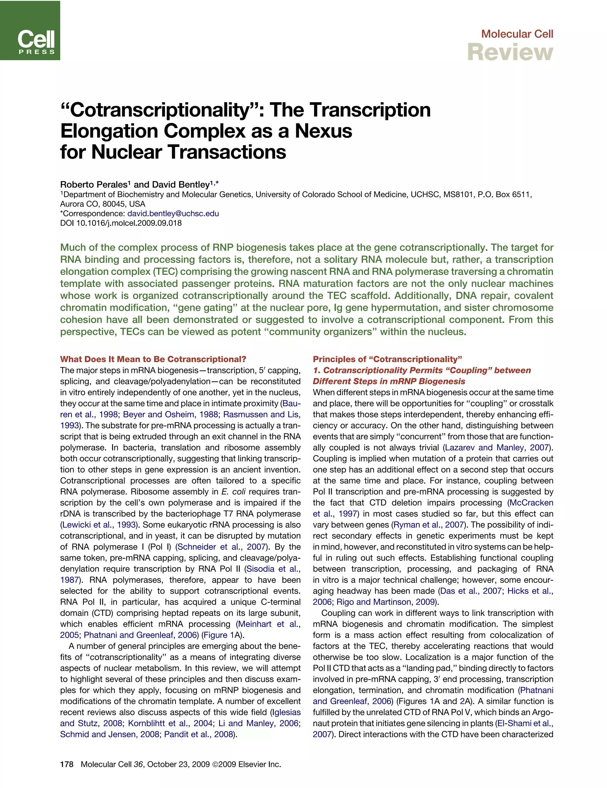
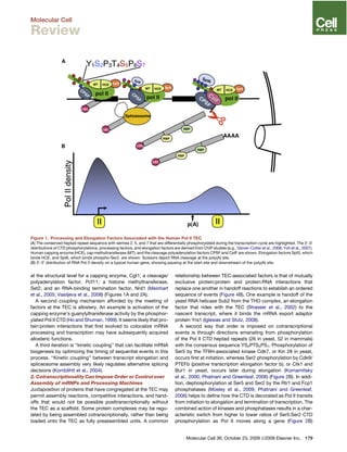
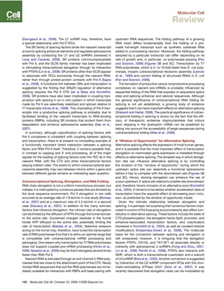
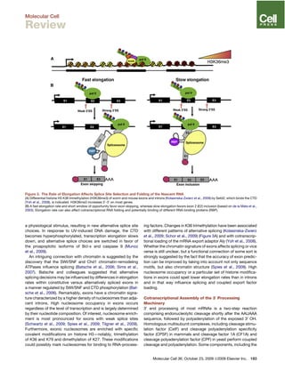
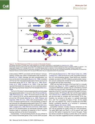
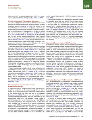
SlideShare a Scribd company logo
1 of 14
Download to read offline
Molecular Cell
Review
‘‘Cotranscriptionality’’: The Transcription
Elongation Complex as a Nexus
for Nuclear Transactions
Roberto Perales1 and David Bentley1,*
1Department of Biochemistry and Molecular Genetics, University of Colorado School of Medicine, UCHSC, MS8101, P.O. Box 6511,
Aurora CO, 80045, USA
*Correspondence: david.bentley@uchsc.edu
DOI 10.1016/j.molcel.2009.09.018
Much of the complex process of RNP biogenesis takes place at the gene cotranscriptionally. The target for
RNA binding and processing factors is, therefore, not a solitary RNA molecule but, rather, a transcription
elongation complex (TEC) comprising the growing nascent RNA and RNA polymerase traversing a chromatin
template with associated passenger proteins. RNA maturation factors are not the only nuclear machines
whose work is organized cotranscriptionally around the TEC scaffold. Additionally, DNA repair, covalent
chromatin modification, ‘‘gene gating’’ at the nuclear pore, Ig gene hypermutation, and sister chromosome
cohesion have all been demonstrated or suggested to involve a cotranscriptional component. From this
perspective, TECs can be viewed as potent ‘‘community organizers’’ within the nucleus.
What Does It Mean to Be Cotranscriptional?
The major steps in mRNA biogenesis—transcription, 50
capping,
splicing, and cleavage/polyadenylation—can be reconstituted
in vitro entirely independently of one another, yet in the nucleus,
they occur at the same time and place in intimate proximity (Bau-
ren et al., 1998; Beyer and Osheim, 1988; Rasmussen and Lis,
1993). The substrate for pre-mRNA processing is actually a tran-
script that is being extruded through an exit channel in the RNA
polymerase. In bacteria, translation and ribosome assembly
both occur cotranscriptionally, suggesting that linking transcrip-
tion to other steps in gene expression is an ancient invention.
Cotranscriptional processes are often tailored to a specific
RNA polymerase. Ribosome assembly in E. coli requires tran-
scription by the cell’s own polymerase and is impaired if the
rDNA is transcribed by the bacteriophage T7 RNA polymerase
(Lewicki et al., 1993). Some eukaryotic rRNA processing is also
cotranscriptional, and in yeast, it can be disrupted by mutation
of RNA polymerase I (Pol I) (Schneider et al., 2007). By the
same token, pre-mRNA capping, splicing, and cleavage/polya-
denylation require transcription by RNA Pol II (Sisodia et al.,
1987). RNA polymerases, therefore, appear to have been
selected for the ability to support cotranscriptional events.
RNA Pol II, in particular, has acquired a unique C-terminal
domain (CTD) comprising heptad repeats on its large subunit,
which enables efficient mRNA processing (Meinhart et al.,
2005; Phatnani and Greenleaf, 2006) (Figure 1A).
A number of general principles are emerging about the bene-
fits of ‘‘cotranscriptionality’’ as a means of integrating diverse
aspects of nuclear metabolism. In this review, we will attempt
to highlight several of these principles and then discuss exam-
ples for which they apply, focusing on mRNP biogenesis and
modifications of the chromatin template. A number of excellent
recent reviews also discuss aspects of this wide field (Iglesias
and Stutz, 2008; Kornblihtt et al., 2004; Li and Manley, 2006;
Schmid and Jensen, 2008; Pandit et al., 2008).
Principles of ‘‘Cotranscriptionality’’
1. Cotranscriptionality Permits ‘‘Coupling’’ between
Different Steps in mRNP Biogenesis
When different steps in mRNA biogenesis occur at the same time
and place, there will be opportunities for ‘‘coupling’’ or crosstalk
that makes those steps interdependent, thereby enhancing effi-
ciency or accuracy. On the other hand, distinguishing between
events that are simply ‘‘concurrent’’ from those that are function-
ally coupled is not always trivial (Lazarev and Manley, 2007).
Coupling is implied when mutation of a protein that carries out
one step has an additional effect on a second step that occurs
at the same time and place. For instance, coupling between
Pol II transcription and pre-mRNA processing is suggested by
the fact that CTD deletion impairs processing (McCracken
et al., 1997) in most cases studied so far, but this effect can
vary between genes (Ryman et al., 2007). The possibility of indi-
rect secondary effects in genetic experiments must be kept
in mind, however, and reconstituted in vitro systems can be help-
ful in ruling out such effects. Establishing functional coupling
between transcription, processing, and packaging of RNA
in vitro is a major technical challenge; however, some encour-
aging headway has been made (Das et al., 2007; Hicks et al.,
2006; Rigo and Martinson, 2009).
Coupling can work in different ways to link transcription with
mRNA biogenesis and chromatin modification. The simplest
form is a mass action effect resulting from colocalization of
factors at the TEC, thereby accelerating reactions that would
otherwise be too slow. Localization is a major function of the
Pol II CTD that acts as a ‘‘landing pad,’’ binding directly to factors
involved in pre-mRNA capping, 30
end processing, transcription
elongation, termination, and chromatin modification (Phatnani
and Greenleaf, 2006) (Figures 1A and 2A). A similar function is
fulfilled by the unrelated CTD of RNA Pol V, which binds an Argo-
naut protein that initiates gene silencing in plants (El-Shami et al.,
2007). Direct interactions with the CTD have been characterized
178 Molecular Cell 36, October 23, 2009 ª2009 Elsevier Inc.
at the structural level for a capping enzyme, Cgt1; a cleavage/
polyadenylation factor, Pcf11; a histone methyltransferase,
Set2; and an RNA-binding termination factor, Nrd1 (Meinhart
et al., 2005; Vasiljeva et al., 2008) (Figures 1A and 2A).
A second coupling mechanism afforded by the meeting of
factors at the TEC is allostery. An example is activation of the
capping enzyme’s guanylyltransferase activity by the phosphor-
ylated Pol II CTD (Ho and Shuman, 1999). It seems likely that pro-
tein:protein interactions that first evolved to colocalize mRNA
processing and transcription may have subsequently acquired
allosteric functions.
A third iteration is ‘‘kinetic coupling’’ that can facilitate mRNA
biogenesis by optimizing the timing of sequential events in this
process. ‘‘Kinetic coupling’’ between transcript elongation and
spliceosome assembly very likely regulates alternative splicing
decisions (Kornblihtt et al., 2004).
2. Cotranscriptionality Can Impose Order or Control over
Assembly of mRNPs and Processing Machines
Juxtaposition of proteins that have congregated at the TEC may
permit assembly reactions, competitive interactions, and hand-
offs that would not be possible posttranscriptionally without
the TEC as a scaffold. Some protein complexes may be regu-
lated by being assembled cotranscriptionally, rather than being
loaded onto the TEC as fully preassembled units. A common
relationship between TEC-associated factors is that of mutually
exclusive protein:protein and protein:RNA interactions that
replace one another in handoff reactions to establish an ordered
sequence of events (Figure 4B). One example is handoff of the
yeast RNA helicase Sub2 from the THO complex, an elongation
factor that rides with the TEC (Strasser et al., 2002) to the
nascent transcript, where it binds the mRNA export adaptor
protein Yra1 (Iglesias and Stutz, 2008).
A second way that order is imposed on cotranscriptional
events is through directions emanating from phosphorylation
of the Pol II CTD heptad repeats (26 in yeast, 52 in mammals)
with the consensus sequence YS2PTS5PS7. Phosphorylation of
Ser5 by the TFIIH-associated kinase Cdk7, or Kin 28 in yeast,
occurs first at initiation, whereas Ser2 phosphorylation by Cdk9/
PTEFb (positive transcription elongation factor b), or Ctk1 and
Bur1 in yeast, occurs later during elongation (Komarnitsky
et al., 2000; Phatnani and Greenleaf, 2006) (Figure 2B). In addi-
tion, dephosphorylation at Ser5 and Ser2 by the Rtr1 and Fcp1
phosphatases (Mosley et al., 2009; Phatnani and Greenleaf,
2006) helps to define how the CTD is decorated as Pol II transits
from initiation to elongation and termination of transcription. The
combined action of kinases and phosphatases results in a char-
acteristic switch from higher to lower ratios of Ser5:Ser2 CTD
phosphorylation as Pol II moves along a gene (Figure 2B)
Figure 1. Processing and Elongation Factors Associated with the Human Pol II TEC
(A) The conserved heptad repeat sequence with serines 2, 5, and 7 that are differentially phosphorylated during the transcription cycle are highlighted. The 50
-30
distributions of CTD phosphorylations, processing factors, and elongation factors are derived from ChIP studies (e.g., Glover-Cutter et al., 2008; Yoh et al., 2007).
Human capping enzyme (HCE), cap methyltransferase (MT), and the cleavage polyadenylation factors CPSF and CstF are shown. Elongation factors Spt5, which
binds HCE, and Spt6, which binds phospho-Ser2, are shown. Scissors depict RNA cleavage at the poly(A) site.
(B) 50
-30
distribution of RNA Pol II density on a typical human gene, showing pausing at the start site and downstream of the poly(A) site.
Molecular Cell 36, October 23, 2009 ª2009 Elsevier Inc. 179
Molecular Cell
Review
(Komarnitsky et al., 2000). Ser7 residues of the CTD heptads are
also phosphorylated within genes (Chapman et al., 2007) by
Kin28/Cdk7 in yeast and mammalian cells (Akhtar et al., 2009).
As a result of sequential phosphorylation, proteins that recognize
Ser 5 phosphorylated heptads, such as the yeast Nrd1 protein
(Vasiljeva et al., 2008), will be recruited to the TEC earlier than
those that recognize Ser2 phosphorylated heptads such as the
30
processing factor Pcf11 (Licatalosi et al., 2002).
3. The TEC Is a Locator for Nuclear Machines
The impact of cotranscriptionality is not limited to mRNA produc-
tion. The TEC is used to localize protein machines that carry out
DNA repair, covalent DNA modification, and gene silencing to
the places in the genome where they are required. An example
of this locator function is transcription-coupled DNA repair in
which the nucleotide excision repair machine recognizes a
stalled Pol II TEC and specifically removes DNA lesions on the
template strand (Lindsey-Boltz and Sancar, 2007). The TEC
may also be an active participant in relocating chromatin-associ-
ated proteins, including histones and cohesins (Lengronne et al.,
2004; Workman, 2006) (Figures 4A and 4C).
The Pol II CTD Organizes an ‘‘mRNA Factory’’
The CTD functions as a flexible landing pad for Pol II-interacting
proteins, including pre-mRNA-processing factors and chromatin
modifiers (Phatnani and Greenleaf, 2006). The length of the fully
extended CTD would be many times the diameter of core Pol II
(Meinhart et al., 2005), providing ample space, in principle, for
binding to multiple partners. The heptad repeats are phosphory-
lated and dephosphorylated by kinases and phosphatases at the
S2, S5, and S7 positions in a manner that is coordinated with the
initiation, elongation, and termination phases of the transcription
cycle (reviewed in Meinhart et al., 2005; Phatnani and Greenleaf,
2006). CTD phosphorylation is potentially astronomically com-
plex, and elucidating how it controls loading and unloading of
Pol II passenger proteins is an important problem.
In vitro, the CTD enhances capping, splicing, and 30
end
formation independently of ongoing transcription, and CTD dele-
tion disrupts these processing reactions in vivo (reviewed in Hir-
ose and Manley, 2000). Although the CTD is necessary for effi-
cient pre-mRNA processing, it is not sufficient. Simply pinning
the CTD onto T7 RNA polymerase or Pol I does not permit effi-
cient processing (Natalizio et al., 2009).
A dynamic ‘‘mRNA factory’’ complex of Pol II with associated
proteins is probably responsible for simultaneous synthesis, pro-
cessing, and packaging of the mRNP (Figure 1A). The poly-
merase with nascent RNA and numerous passenger proteins
would almost certainly exert too much viscous drag to move at
a high speed through nucleoplasm. The most likely solution to
Figure 2. CTD Phosphorylation and Processing Factor Recruitment at the Budding Yeast Pol II TEC
(A) 50
-30
distributions of factors are based on ChIP studies as in Figure 1A. Four factors that bind directly to the CTD are depicted: the cap methyltransferase (MT)
and guanylyltransferase (GT), the termination factor Nrd1, and the cleavage/polyadenylation factor Pcf11. Gene gating by putative interaction between the
nuclear pore and the TEC is depicted by a yellow arrow.
(B) ChIP-Chip analysis of the 50
-30
distribution of total Pol II density (left) and Ser2 and Ser5 phosphorylated Pol II (right) on a typical highly transcribed yeast gene
(RPL3). Results are from P. Megee and D.B., unpublished data.
180 Molecular Cell 36, October 23, 2009 ª2009 Elsevier Inc.
Molecular Cell
Review
this hydrodynamic problem is that the chromatin template is
threaded through a stationary mRNA factory (Jackson and
Cook, 1995).
First, Put Your Cap On
Pre-mRNAs are modified at their 50
ends by addition of a
7-methyl G50
ppp50
N cap (reviewed in Shuman, 2001) when the
transcript is only 25–50 bases long. Capping is a three-step
process that does not require specific sequences in the RNA.
RNA triphosphatase removes the g-phosphate of the first nucle-
otide, and then GMP is added by RNA guanylyltransferase.
Finally, the guanine is methylated at N7. In metazoans, the
capping enzyme is a bifunctional polypeptide with triphospha-
tase and guanylyltransferase activities. The GMP transfer reac-
tion is reversible, and capping is driven forward by the concerted
action of both guanylyltransferase and methyltransferase, yet
these two enzymes do not associate with one another (Shuman,
2001). The solution to this problem is that both the capping
enzyme and the methyltransferase bind directly and specifically
to the phosphorylated Pol II CTD (Shuman, 2001). When tran-
scription initiates, phosphorylation of the CTD on Ser5 residues
permits loading of capping enzyme onto the TEC and allosteric
activation of the guanylyltransferase (Ho and Shuman, 1999).
The overall capping reaction is, therefore, facilitated by both co-
localization of the capping enzymes on the phosphorylated CTD
and allosteric activation.
The relationship between capping enzymes and Pol II illus-
trates the two-way nature of communication between process-
ing factors and the transcription machinery. Not only is capping
enhanced by interaction of capping enzymes with the CTD, but
capping enzymes can also stimulate or inhibit transcription initi-
ation and/or early elongation (Mandal et al., 2004; Myers et al.,
2002; Schroeder et al., 2004). Polymerase complexes paused
at 50
ends are found on many metazoan genes, and they are
probably important for cotranscriptional capping. 50
pausing is
regulated by the Pol II-associated elongation factor Spt5, which
also allosterically activates the cap guanylyltransferase (Wen
and Shatkin, 1999). Another regulator of elongation, the HIV
Tat protein, also activates guanylyltransferase and enhances
capping of viral transcripts (Chiu et al., 2002). Capping could
be coupled to escape of paused Pol II, thereby ensuring that
polymerases that enter productive elongation will have an appro-
priately modified 50
end. Conversely pausing could promote
capping, perhaps by restricting the distance between the RNA
50
end and capping enzymes sitting on the CTD. Capping is
not usually regarded as a regulated step in gene expression;
however, capping factor recruitment could, in principle, be regu-
lated by Spt5 or CTD phosphorylation. A block to capping
enzyme recruitment has been reported at yeast silent mating-
type loci (Gao and Gross, 2008). In the future, it will be of interest
to investigate whether or not capping is affected by polymerase
pausing at the transcription start site.
The influence of capping enzymes on mRNA production may
not be limited to 50
ends. Human capping enzymes are found
at 50
ends and throughout genes, including 30
flanking regions
even more than a kilobase downstream of the poly(A) site
(Glover-Cutter et al., 2008) (Figure 1B). Capping factors, there-
fore, appear to linger on the Pol II ‘‘landing pad’’ long after addi-
tion of the cap, and they could, therefore, potentially influence
elongation, termination, and 30
end processing like the vaccinia
virus capping enzyme. They might also cap the pervasive non-
coding short transcripts detected within genes and at 50
and 30
ends (Kapranov et al., 2007). In addition, capping enzyme has
been suggested to promote R loop formation (see below) and
could thereby affect transcription elongation (Kaneko et al.,
2007).
Cotranscriptional Splicing.Some Assembly Required
Many, but not all, introns are removed cotranscriptionally rather
than posttranscriptionally, as vividly shown by EM studies (Beyer
and Osheim, 1988). Although not all splicing is completed
cotranscriptionally, it is probable that assembly of most spliceo-
somes, including those at alternative splice sites (Pandya-Jones
and Black, 2009), is initiated on the nascent transcript. The spli-
ceosome is one of the most elegant examples of ordered self-
assembly. In vitro, on a pre-made substrate without ongoing
transcription, U1 snRNP first base pairs to the 50
splice site,
and U2AF binds the 30
splice site, followed by U2 snRNP base
pairing the branch point. The tri-snRNP U4-U6/U5 then engages,
and the complex rearranges to assume the catalytically active
conformation as U1 and U4 are discarded. ChIP analysis of
cotranscriptional splicing on yeast genes supports a step-wise
assembly process similar to that which occurs in vitro (Gorne-
mann et al., 2005; Lacadie and Rosbash, 2005). On the other
hand, the possibility that preassembled higher-order snRNP
complexes are recruited cotranscriptionally cannot yet be
excluded, especially in mammalian cells (Listerman et al.,
2006). Little is known about spliceosome assembly at the TEC,
but there are reasons for thinking that it might differ from
assembly that is uncoupled from transcription. Splicing of
synthetic pre-mRNAs in injected Xenopus oocytes is less effi-
cient than splicing coupled to transcription in the same cells,
consistent with stimulation of splicing in vitro by the phosphory-
lated CTD (Bird et al., 2004; Hirose and Manley, 2000). It has
been suggested that cotranscriptional splicing might differ
from splicing uncoupled from transcription because, in the
former case, exons are held in place by tethering to the poly-
merase (Dye et al., 2006). The functional significance of exon
tethering has recently been questioned, however (Fong et al.,
2009). Unlike the other mRNA-processing steps, splicing is reit-
erated many times on most transcripts. How coupling with tran-
scription might affect the recycling of spliceosome components
for use on multiple introns within a transcript is an interesting
open question.
A number of intriguing connections have been uncovered
between the splicing machinery and TEC-associated proteins,
although the extent to which splicing is facilitated by direct pro-
tein:protein interactions between spliceosomes and Pol II
remains unresolved. The yeast U1 snRNP protein Prp40, which
bridges the 50
splice site and branch point, can bind directly to
the phospho-CTD (Phatnani and Greenleaf, 2006), but the func-
tional significance of this interaction remains to be established.
Human U1snRNP, but not other snRNPs, also coimmunoprecipi-
tates with Pol II (Das et al., 2007). Furthermore, U1snRNP at a 50
splice site can activate recruitment of Pol II and general tran-
scription factors to the promoter independently of splicing
Molecular Cell 36, October 23, 2009 ª2009 Elsevier Inc. 181
Molecular Cell
Review
(Damgaard et al., 2008). The U1 snRNP may, therefore, have
a special relationship with Pol II TECs.
The SR family of splicing factors binds the nascent transcript
at exonic splicing enhancer elements and regulates spliceosome
assembly by contacting the U1 and U2 snRNPs (reviewed in
Long and Caceres, 2009). SR proteins coimmunoprecipitate
with Pol II, and the SC35 family member has been implicated
in stimulating transcriptional elongation through its interaction
with PTEFb (Lin et al., 2008). SR proteins other than SC35 appear
to associate with TECs exclusively through the nascent RNA,
rather than through protein:protein contacts with Pol II (Sapra
et al., 2009). A functional link between SRs and transcription is
suggested by the finding that SRp20 regulation of alternative
splicing requires the Pol II CTD (de la Mata and Kornblihtt,
2006). SR proteins have also been implicated in coupling tran-
scription with splicing in an in vitro system in which transcripts
made by Pol II are selectively stabilized and spliced relative to
T7 transcripts (Hicks et al., 2006). This channeling of Pol II tran-
scripts into a productive splicing pathway is probably due to
facilitated binding of the nascent transcripts to RNA-binding
proteins (RBPs), including SR proteins that protect them from
degradation and enhance spliceosome assembly (Das et al.,
2007).
In summary, although copurification of splicing factors with
Pol II complexes is consistent with coupling between splicing
and transcription, there is at present no compelling example of
a functionally important direct interaction between a splicing
factor and RNA Pol II itself. Therefore, it remains possible that,
in contrast to capping and 30
end processing, all of the major
signals for the loading of splicing factors onto the TEC lie in the
nascent RNA, with the CTD and other transcriptional factors
playing indirect roles. The extent to which cotranscriptional spli-
ceosome assembly may vary between introns within a gene and
between different genes remains an interesting open question.
Cotranscriptional Splicing, Elongation, and RNA Folding
RNA chain elongation is not a uniform monotonous process, but
instead, it is interrupted by numerous pauses that are dictated by
the local sequence environment. Pol II elongation in live cells
occurred at an average rate of 1.9 kb/min in one study (Boireau
et al., 2007) and at a maximum rate of 4.3 kb/min in a second
case (Darzacq et al., 2007). In addition to the many extrinsic
factors that influence elongation, the intrinsic rate of elongation
can be limited by the diffusion of NTPs through the funnel domain
to the active site. Conserved charged residues in the funnel
hinder NTP diffusion to the active site, potentially limiting the
rate of transcription (Batada et al., 2004). Selective pressure
acting on the funnel may, therefore, have tuned the transcription
rate of RNA polymerase II so that it is within an optimal range that
is compatible with cotranscriptional mRNA processing and
packaging. One reason why transcription by T7 RNA polymerase
does not support coupled pre-mRNA processing (Hicks et al.,
2006; Natalizio et al., 2009) may be that it elongates several times
faster than RNA Pol II.
Nascent RNA is extruded through an exit channel in RNA poly-
merase that lies close to the attachment point of the CTD. Newly
minted RNA sequences that exit the RNA polymerase are imme-
diately available for interaction with RBPs and base pairing with
upstream RNA sequences. The folding pathway of a growing
RNA chain differs fundamentally from the folding of a pre-
made full-length transcript such as synthetic substrate RNA
added to a processing reaction. Moreover, the folding pathway
adopted by a particular transcript can differ depending on its
rate of growth and, in particular, on polymerase pausing (Pan
and Sosnick, 2006) (Figures 3B and 3C). Transcription by T7
RNA polymerase, which is 5- to 10-fold faster than E. coli poly-
merase, impairs cotranscriptional ribosome assembly (Lewicki
et al., 1993) and correct folding of structured RNAs in E. coli
(Pan and Sosnick, 2006).
The formation of productive versus nonproductive processing
complexes on nascent pre-mRNAs is probably influenced by
sequential folding of the RNA that exposes or sequesters splice
sites and splicing enhancer and silencer sequences. Although
the general significance of cotranscriptional RNA folding for
splicing is not yet established, a growing body of evidence
suggests that it can have important effects on alternative splicing
(Shepard and Hertel, 2008). The practical significance of cotran-
scriptional folding in splicing is shown by the fact that the effi-
cacy of therapeutic antisense oligonucleotides that induce
exon skipping in the dystrophin mRNA can be predicted by
taking into account the accessibility of target sequences during
cotranscriptional folding (Wee et al., 2008).
A ‘‘Window of Opportunity’’ for Alternative Splicing
Alternative splicing affects the expression of most human genes,
and it is possible that the most important effect of transcription
elongation on mammalian gene expression is mediated through
effects on alternative splicing. The simplest way in which elonga-
tion rate can influence alternative splicing is by controlling
the duration of the ‘‘window of opportunity’’ during which the
upstream splice site can assemble a functional spliceosome
before it has to compete with the downstream site (Figures 3B
and 3C). Hence, slowing elongation can enhance the use of
a poor upstream 30
splice site relative to a better site downstream
and, therefore, favors inclusion of an alternative exon (Kornblihtt
et al., 2004). It remains to be tested whether accelerated rates of
transcription have the opposite effect of decreasing exon inclu-
sion, as predicted by the window of opportunity model.
Given the intimate relationship between elongation and
splicing, it is perhaps not surprising that numerous factors impli-
cated in control of Pol II pausing and processivity also affect con-
stitutive or alternative splicing. These factors include the state of
CTD phosphorylation, the elongation factor Spt5, promoter- and
enhancer-associated transcription factors, and coactivators
(reviewed in Kornblihtt et al., 2004), as well as covalent histone
modifications (Kolasinska-Zwierz et al., 2009). The molecular
basis for the connection between splicing and elongation is
still unresolved; however, it is intriguing that the elongation
factors PTEFb, CA150, and TAT-SF1 all associate directly or
indirectly with spliceosomal U snRNPs (Fong and Zhou, 2001;
Lin et al., 2008; Pandit et al., 2008). PTEFb also interacts with
SKIP, which is both a transcriptional coactivator and a subunit
of U5 snRNP (Bres et al., 2005). Another connection is suggested
by interaction of the U2 snRNP component SF3a with the chro-
matin-remodeling ATPase Chd1 (Sims et al., 2007). It was
recently discovered that elongation rates can be modulated by
182 Molecular Cell 36, October 23, 2009 ª2009 Elsevier Inc.
Molecular Cell
Review
a physiological stimulus, resulting in new alternative splice site
choices. In response to UV-induced DNA damage, the CTD
becomes hyperphosphorylated, transcription elongation slows
down, and alternative splice choices are switched in favor of
the proapoptotic isoforms of Bcl-x and caspase 9 (Munoz
et al., 2009).
An intriguing connection with chromatin is suggested by the
discovery that the SWI/SNF and Chd1 chromatin-remodeling
ATPases influence splicing (Batsche et al., 2006; Sims et al.,
2007). Batsche and colleagues suggested that alternative
splicing decisions may be influenced by differences in elongation
rates within constitutive versus alternatively spliced exons in
a manner regulated by SWI/SNF and CTD phosphorylation (Bat-
sche et al., 2006). Remarkably, exons have a chromatin signa-
ture characterized by a higher density of nucleosomes than adja-
cent introns. High nucleosome occupancy in exons occurs
regardless of the level of transcription and is largely determined
by their nucleotide composition. Of interest, nucleosome enrich-
ment is most pronounced for exons with weak splice sites
(Schwartz et al., 2009; Spies et al., 2009; Tilgner et al., 2009).
Furthermore, exonic nucleosomes are enriched with specific
covalent modifications on histone H3—notably, trimethylation
of K36 and K79 and dimethylation of K27. These modifications
could possibly mark nucleosomes for binding to RNA-process-
ing factors. Changes in K36 trimethylation have been associated
with different patterns of alternative splicing (Kolasinska-Zwierz
et al., 2009; Schor et al., 2009) (Figure 3A) and with cotranscrip-
tional loading of the mRNA export adaptor Aly (Yoh et al., 2008).
Whether the chromatin signature of exons affects splicing or vice
versa is still unclear, but a functional connection of some sort is
strongly suggested by the fact that the accuracy of exon predic-
tion can be improved by taking into account not only sequence
motifs, but also chromatin structure (Spies et al., 2009). High
nucleosome occupancy or a particular set of histone modifica-
tions in exons could spell lower elongation rates than in introns
and in that way influence splicing and coupled export factor
loading.
Cotranscriptional Assembly of the 30
Processing
Machinery
30
end processing of most mRNAs is a two-step reaction
comprising endonucleolytic cleavage shortly after the AAUAAA
sequence, followed by polyadenylation of the exposed 30
OH.
Homologous multisubunit complexes, including cleavage stimu-
lation factor (CstF) and cleavage polyadenylation specificity
factor (CPSF) in mammals and cleavage factor 1A (CF1A) and
cleavage polyadenylation factor (CPF) in yeast perform coupled
cleavage and polyadenylation. Some components, including the
Figure 3. The Rate of Elongation Affects Splice Site Selection and Folding of the Nascent RNA
(A) Differential histone H3 K36 trimethylation (H3K36me3) of worm and mouse exons and introns (Kolasinska-Zwierz et al., 2009) by Setd2, which binds the CTD
(Yoh et al., 2008), is indicated. H3K36me3 increases 50
-30
on most genes.
(B) A fast elongation rate and short window of opportunity favor exon skipping, whereas slow elongation favors exon 2 (E2) inclusion (based on de la Mata et al.,
2003). Elongation rate can also affect cotranscriptional RNA folding and potentially binding of different RNA-binding proteins (RBP).
Molecular Cell 36, October 23, 2009 ª2009 Elsevier Inc. 183
Molecular Cell
Review
endonuclease, CPSF73, are shared with the histone 30
end-pro-
cessing complex that makes nonadenylated ends (reviewed in
Mandel et al., 2008). Cleavage and early polyadenylation can
occur at the site of transcription (Bauren et al., 1998), consistent
with the fact that cleavage/polyadenylation factors are found at
transcribed genes (Ahn et al., 2004; Gall et al., 1999; Glover-
Cutter et al., 2008; Licatalosi et al., 2002). It has also been
reported that poly(A) site cleavage can occur posttranscription-
ally following polymerase release from the template (West et al.,
2008).
The Pol II CTD binds 30
end-processing factors and stimulates
cleavage/polyadenylation in vivo and in vitro (Hirose and Manley,
1998; McCracken et al., 1997). The 50 kD subunit of CstF, the
Pcf11 subunit of CF1A, and the yeast termination factor Rtt103
all bind the CTD directly (Meinhart et al., 2005; Phatnani and
Greenleaf, 2006; Kim et al., 2004). Ser2 phosphorylation of the
CTD is of special significance for 30
end processing, at least in
part because the cleavage/polyadenylation factor Pcf11 prefer-
entially binds to heptad repeats with this modification (Ahn et al.,
2004; Licatalosi et al., 2002; Meinhart and Cramer, 2004). Modu-
lation of CTD phosphorylation as polymerase traverses a gene
therefore helps to coordinate the assembly of the 30
end-pro-
cessing machinery at the site of transcription.
Unexpectedly, 30
end-processing factors are not confined to
the 30
ends of transcribed genes. In fact, human CPSF and yeast
CF1A subunits (Dantonel et al., 1997; Glover-Cutter et al., 2008;
Licatalosi et al., 2002) are found at 50
ends, long before transcrip-
tion of 30
end-processing signals. These factors are, therefore,
probably recruited to the TEC initially by protein:protein interac-
tion and are subsequently handed off to the nascent RNA after
the poly(A) site has been transcribed.
CPSF can bind both the body of Pol II and CstF in a mutually
exclusive way (Nag et al., 2007), suggesting that formation of
a functional CPSF/CstF complex may be controlled by handoff
of CPSF from Pol II to CstF. A handoff reaction may also control
assembly of the Pcf11 and Clp1 subunits of the yeast 30
end-pro-
cessing complex, CF1A (Figure 4B). Clp1 and the export adaptor
Yra1 (Johnson et al., 2009) bind the same short region of Pcf11
and are thus likely to compete with one another. CF1A may,
therefore, be recruited to the gene in a partially assembled
form, with Yra1 occupying the place of Clp1. At the poly(A)
site, Clp1 may displace Yra1, which is handed off to the RNA,
thereby completing assembly of a functional 30
processing
complex (Saguez and Jensen, 2009).
Competition between mutually exclusive interactions at the
TEC may be exploited for quality control of 30
end processing.
The yeast RBPs Npl3 and Rna15, a CF1A subunit, both load
onto the TEC before it reaches the 30
end of the gene. These
two factors compete for binding to similar sites on the nascent
RNA, and it has been suggested that this competition enhances
Figure 4. The RNA Polymerase II TEC as a Locator of Chromatin Proteins
(A) Possible mechanism of cohesin (pink rings) localization by convergent transcription in yeast (based on Lengronne et al., 2004).
(B) Mutually exclusive protein:protein and protein:RNA interactions and handoff reactions facilitate assembly of export-competent mRNPs. In reaction 1, the
yeast export adaptor Yra1 is handed off from Pcf11 to the RNA helicase Sub2. In reaction 2, Yra1 is handed off to the export receptor Mex67/Mtr2.
(C) Mobilization of histones during Pol II transcription elongation. FACT and Swi/Snf travel with the TEC to facilitate disassembly and reassembly of nucleosomes.
Displaced histones could be handed off to the nascent RNA before they are redeposited behind the polymerase.
184 Molecular Cell 36, October 23, 2009 ª2009 Elsevier Inc.
Molecular Cell
Review
the accuracy of 30
processing by preventing Rna15 from recog-
nizing cryptic poly(A) sites within genes (Bucheli et al., 2007).
30
End Processing and Transcription Elongation
Like exons, poly(A) sites are associated with their own chromatin
signature: a localized nucleosome depletion over the AATAAA
consensus sequence (Spies et al., 2009). The function of this
chromatin feature is unknown, but it could cause a local increase
in transcriptional elongation rate that might influence 30
process-
ing. Further downstream, Pol II pausing 1–2 kb past the poly(A)
site is common to many genes (Boireau et al., 2007; Darzacq
et al., 2007; Glover-Cutter et al., 2008) (Figure 1B). At this pause
site, CTD Ser2 is highly phosphorylated (Figure 1), and maximal
levels of cleavage/polyadenylation factors are associated with
the TEC (Glover-Cutter et al., 2008). Although the precise rela-
tionship between poly(A) site processing and subsequent tran-
scription termination is unclear, it seems likely that both events
are coordinated with the downstream pause.
Alternative poly(A) site choice has recently been recognized as
a widespread phenomenon that shapes the repertoire of 30
UTR
sequences (Licatalosi et al., 2008; Sandberg et al., 2008) and
is perturbed in cancer cells (Mayr and Bartel, 2009). It is possible
that the rate of transcriptional elongation can affect the choice
between alternative poly(A) sites, and if this were the case,
then effects of chromatin structure on elongation at 30
ends could
influence these important RNA processing decisions. Although
early recruitment of 30
end-processing factors appears to be
a general phenomenon, it remains to be determined whether
RNA cleavage at the poly(A) site always precedes transcription
termination or whether the timing of cleavage and termination
differs between genes.
Poly(A) site cleavage at the 30
end may not be sufficient to cut
the mRNA loose from the TEC. In mammalian cells, an additional
release step requires the CTD (Custodio et al., 2007) and
completion of splicing (Rigo and Martinson, 2009). In yeast, the
THO complex, the RNA helicase Sub2, the export factor
Mex67, and the phosphatase Glc7 remodel the RNP at the 30
end of the gene and pry it away from the cleavage/polyadenyla-
tion apparatus (Gilbert and Guthrie, 2004; Qu et al., 2009; Rouge-
maille et al., 2008).
Cotranscriptional Recruitment of Factors
that Dismantle the TEC
A major advantage of cotranscriptional rather than posttran-
scriptional recognition of 30
end-processing sites is that it
permits the coupling of transcription termination with recognition
of the poly(A) signal that marks the end of the message
(Rosonina et al., 2006). There are two main models for how
cleavage/polyadenylation factors stimulate dissociation of the
extraordinarily stable TEC. The ‘‘allosteric’’ model invokes a
poly(A) site-dependent conformational change in the TEC that
reduces its processivity. The ‘‘torpedo’’ model, on the other
hand, proposes that the cut site in the nascent RNA permits
access to a 50
-30
RNA exonuclease that degrades the nascent
RNA tail and destabilizes the TEC. Evidence on these models
is divided, but in both the allosteric and torpedo scenarios, it is
clear that cotranscriptional loading of 30
end-processing factors,
including Pcf11 and the 50
-30
RNA exonuclease Xrn2/Rat1, ulti-
mately leads to dissociation of the TEC (reviewed in Rosonina
et al., 2006).
A second mechanism of Pol II termination is employed in yeast
at noncoding genes that lack poly(A) sites. At these genes,
a complex comprising Nrd1, Nab3, and the RNA helicase Sen1
is recruited to the TEC and terminates transcription indepen-
dently of a 50
-30
RNA exonuclease (Steinmetz et al., 2001; Kim
et al., 2006). The Nrd1 protein binds directly to Ser5-phosphory-
lated CTD heptads (Vasiljeva et al., 2008), as well as RNA. Posi-
tion-specific CTD phosphorylation on Ser5 at 50
ends of genes
ensures that this mechanism of termination only operates at
short distances from the transcription start site (Gudipati et al.,
2008). In summary, Pol II TECs, in a carefully regulated way,
recruit the factors that ultimately lead to their dissociation from
the template.
Specialized Cotranscriptional RNA Processing
In addition to capping, splicing, and cleavage/polyadenylation,
some transcripts are processed cotranscriptionally by A-I editing
or miRNA excision from introns. In the nascent GluR-B pre-
mRNA, an ADAR (adenosine deaminase acting on RNA) recog-
nizes RNA duplexes formed by intramolecular base pairing
between exon 11 and intron 11 and converts an A to I, thereby
switching a Q codon to R. Editing in this case must occur before
splicing, and the Pol II CTD is implicated in imposing this
sequence of events (Ryman et al., 2007).
miRNAs are released from Pol II-transcribed precursors by the
microprocessor complex that includes the RNase III family
member Drosha, which clips miRNA precursors at the base of
a hairpin. Drosha is found at genes harboring intronic miRNAs
(Morlando et al., 2008), strongly suggesting that it works cotran-
scriptionally. Furthermore, miRNA excision from introns is
completely compatible with splicing (Kim and Kim, 2007). The
microprocessor associates with spliceosomes (Kataoka et al.,
2009), but it is not known whether, like RNA editing, the timing
of miRNA excision relative to splicing is regulated by interactions
with Pol II.
Packaging, Export, and the Determination of RNA Fate
As it is extruded from the polymerase, a nascent transcript
encounters numerous RBPs, including cap binding complex
(CBC) (Listerman et al., 2006; Visa et al., 1996), hnRNPs (Dane-
holt, 2001), SR proteins (Long and Caceres, 2009), the exon-
junction complex (EJC) (Custodio et al., 2004), and zipcode-
binding proteins (ZBP) (Pan et al., 2007), some of which remain
attached for much of the transcript’s lifetime. In this way, cotran-
scriptional RBP loading governs the transport, translation, cyto-
plasmic localization, and life span of the mRNA (Daneholt, 2001;
Glisovic et al., 2008).
In addition to providing early protection from nucleases,
cotranscriptionality may impose order on RBP association with
the nascent transcript as the mature mRNP assembles. For
example, CBC association with the 50
cap early in transcript
synthesis (Listerman et al., 2006; Visa et al., 1996) may direct
subsequent interaction with the TREX complex (Cheng et al.,
2006). TREX comprises a subcomplex called THO plus the
export adaptor REF/Aly (Yra1 in yeast) and the RNA helicase
UAP56 (Sub2 in yeast) (Figure 4B). TREX associates with the
Molecular Cell 36, October 23, 2009 ª2009 Elsevier Inc. 185
Molecular Cell
Review
TEC and enhances elongation and mRNA export (Strasser et al.,
2002).
A particularly critical aspect of cotranscriptional mRNP
assembly is preparation for export. Nascent transcripts are
packaged for export by loading of adaptor proteins that allow
the mature mRNP to engage the export receptors Mex67/Mtr2
in yeast and TAP/p15 in metazoans (Iglesias and Stutz, 2008).
Recruitment of the export adaptors Yra1 and REF/Aly to the
nascent transcript is carefully monitored so that only perfectly
formed fully processed mRNPs become export competent (Lei
et al., 2001; Schmid and Jensen, 2008). Yra1 binds the RNA heli-
case Sub2 that also associates with the THO complex on the
TEC (Strasser et al., 2002). Yra1 recruitment requires interaction
with a 30
end-processing factor, Pcf11, that binds the phosphor-
ylated CTD (Johnson et al., 2009) (Figure 4B). This mechanism,
therefore, may ensure that export adaptor loading will only occur
if the machinery is in place to properly process the mRNA 30
end.
The mechanism of Yra1 loading also illustrates how the order of
assembly of mRNPs can be determined by the sequence of CTD
phosphorylation. Ser2 phosphorylation specifically facilitates
recruitment of Pcf11 and thereby indirectly specifies recruitment
of the export factor Yra1 at later times in the transcription cycle.
Following initial Yra1 loading by interaction with Pcf11, it is prob-
ably transferred to Sub2, whose helicase activity then facilitates
a second transfer to the mRNA/Mex67 complex (Figure 4B).
Both handoff reactions most likely occur at the TEC because
all players, Pcf11, Yra1, Sub2, and Mex67, localize to sites of
transcription (reviewed in Iglesias and Stutz, 2008).
In mammalian cells, the Yra1 homolog Aly is loaded cotran-
scriptionally by interaction with Iws1, a partner of the elongation
factor Spt6 that, like Pcf11, binds CTD heptads phosphorylated
on Ser2 (Yoh et al., 2007). In summary, the sequence of CTD
phosphorylations that accompanies the transcription cycle helps
to direct cotranscriptional mRNP assembly, as well as the pro-
cessing of the nascent transcript.
Cotranscriptional Quality Control
Quality control of mRNPs is facilitated by their cotranscriptional
assembly. This notion is suggested by the fact that slowing tran-
scription elongation suppresses the growth defects of mutants
that disrupt mRNP formation (Jensen et al., 2004). Yeast mRNPs
that are deemed not to be of ‘‘export quality’’ because they have
improperly formed 30
ends are detained close to the site of tran-
scription in a process that requires the exosome (Hilleren et al.,
2001), a complex of 30
-50
exonucleases that degrades defective
RNAs. The exosome can be recruited to transcribed genes
(Andrulis et al., 2002) and could, therefore, be positioned for
degradation of defective mRNPs when they are released from
the template with exposed 30
ends. A possible mechanism of
exosome loading in yeast is through Nrd1 and Npl3, which
both bind the TEC and the exosome (Burkard and Butler, 2000;
Vasiljeva and Buratowski, 2006).
Invasive Nascent Transcripts and R Loops
Another important reason for having RNPs assemble cotran-
scriptionally, rather than posttranscriptionally, is to protect the
DNA template from invasion by naked RNA. When normal
cotranscriptional handling of nascent RNA by RBPs is disrupted,
R loops can form by reannealing of the transcript with DNA
behind the polymerase. The displaced single-stranded nontem-
plate DNA strands are highly recombinogenic, causing genetic
instability (Li and Manley, 2006). Similarly, in E. coli, disruption
of cotranscriptional translation may induce nascent mRNA, no
longer engaged with ribosomes, to form R loops (Gowrishankar
and Harinarayanan, 2004). In yeast, R loops form in mutants of
the TREX complex (Huertas and Aguilera, 2003). R loop forma-
tion and genomic instability also occur in mammalian cells
when the SR proteins, ASF/SF2, or SC35 are depleted (Li and
Manley, 2006). R loops can obstruct elongating RNA polymer-
ases if they are not removed by RNaseH, and this mechanism
may help to explain the pile-ups of Pol II within transcribed genes
when SC35 is depleted (Lin et al., 2008).
Nuclear Pores and the TEC, a Role in ‘‘Gating’’?
In addition to processing and export factors, the TEC can also be
contacted by nuclear pore components, as predicted by the
‘‘gene gating’’ hypothesis, which proposes that export is facili-
tated by localizing active genes at the pore (Akhtar and Gasser,
2007) (Figure 2A). Coupling between transcription and mRNP
exit from the nucleus is suggested by the observation that
ongoing transcription facilitates interaction of Chironomus Bal-
biani Ring (BR) RNPs with nuclear pores (Kylberg et al., 2008).
Specific promoters, transcriptional activators, the coactivator
SAGA, 30
UTRs, and the exosome have all been implicated in
‘‘gene gating’’ (reviewed in Akhtar and Gasser, 2007). TEC inter-
action with the pore is supported by the fact that, when the THO
complex is compromised, pore proteins accumulate at the 30
ends of genes. This phenomenon may reflect a trapped interme-
diate in normal mRNP export (Rougemaille et al., 2008). TREX2 is
a four-subunit complex that bridges the pore with the Mex67/
Mtr2 and the THO complex (Fischer et al., 2002). Recent resolu-
tion of the TREX2 structure (Jani et al., 2009) gives cause for opti-
mism that the connection between transcription and mRNP
delivery to the nuclear pore will ultimately be elucidated at the
atomic level.
Cotranscriptional Looping?
Transcribed genes can adopt a looped conformation that was
first graphically revealed in the lampbrush chromosomes of
amphibian oocytes. Pol II and mRNA-processing factors are
distributed around lampbrush loops, and these structures criti-
cally depend on ongoing transcription (Gall et al., 1999). 3C
studies, which detect the spatial proximity of noncontiguous
DNA regions in the nucleus (Dekker et al., 2002), show that loops
also form between 50
and 30
ends of transcribed genes in somatic
cells (O’Sullivan et al., 2004). Insertion of the finger domain of
promoter-bound TFIIB into the RNA exit channel of Pol II at
both ends of the gene has been suggested as a mechanism for
tying together the base of a loop (Singh and Hampsey, 2007).
Cotranscriptional mRNA processing has also been proposed
to promote looping of the HIV provirus (Perkins et al., 2008).
Many questions remain about the role of cotranscriptional
events in gene looping, including: Are loops a result of threading
a gene through a stationary mRNA factory or a specific confor-
mation designed to facilitate communication and polymerase
recycling between the two ends of a gene? How often does
186 Molecular Cell 36, October 23, 2009 ª2009 Elsevier Inc.
Molecular Cell
Review
a transcribed gene adopt a looped conformation? One percent
of the time? Fifty percent of the time? And can a loop withstand
the torsional stress exerted by multiple polymerases on the same
gene?
The TEC Is a Locator for Chromatin Modifiers and
Silencers
The Pol II TEC is targeted by diverse proteins that modify the
chromatin template. One of the first cotranscriptional processes
identified in eukaryotes was transcription-coupled DNA repair in
which the TEC stalled at a DNA lesion attracts the nucleotide
excision repair machinery. Indeed, Pol II itself might be the
‘‘the most specific damage recognition protein’’ (Lindsey-Boltz
and Sancar, 2007). Another DNA modification that is probably
guided by the Pol II TEC is somatic hypermutation of immuno-
globulin genes by deamination of Cs in the displaced nontem-
plate strand of transcribed DNA. This reaction is catalyzed by
activation-induced deaminase (AID), which is probably associ-
ated with Pol II (Nambu et al., 2003).
Modification of histone N-terminal tails vastly enriches the
chromatin landscape, and accurate deposition of these covalent
marks is vital to many nuclear processes. The Pol II TEC serves
an important auxiliary function in guiding the placement of cova-
lent histone modifications at specific regions of the genome.
Some chromatin modifiers, including the H3K4 and K36 methyl-
transferases Set1 and Set2, bind to the Pol II CTD phosphory-
lated on Ser5 and Ser2 residues, respectively (Phatnani and
Greenleaf, 2006; Yoh et al., 2008). Transcription-dependent
H3K36 methylation by Set2 in yeast recruits a histone deacety-
lase that helps to shut down cryptic promoters (Workman,
2006). Other chromatin modifiers can latch onto the RNA
component of the TEC. A Chironomus histone acetyltransferase
is targeted to actively transcribed chromatin by interaction with
a complex of the RBP hrp65 and actin that binds to nascent
RNA (Sjolinder et al., 2005).
As a result of modifiers piggy-backing on the Pol II TEC, the
primary function of some transcription units is not to produce
an RNA transcript, but to establish a chromatin domain that is
marked by specific histone marks. In this way, one round of tran-
scription can exert a profound influence on the future expression
of that sequence. A recently identified antisense RNA of the
yeast GAL10 gene is present in only 1 cell in 14; however, the
transient presence of the Pol II TEC on the gene is sufficient to
establish a stable domain of H3K36 trimethylation and H3 de-
acetylation (Houseley et al., 2008).
Paradoxically, transcription by RNA Pol II plays a central role in
establishment of transcriptionally silent heterochromatin. This
new paradigm is based on silencing of centromeric chromatin
in fission yeast (reviewed in Moazed, 2009). Silencing is estab-
lished by the RITS complex (RNA-induced initiation of transcrip-
tional gene silencing) that recruits the methyltransferase CLRC,
which deposits the signature mark of heterochromatin, methyl-
ated H3K9. RITS is targeted to the TEC by binding of its Argonaut
1 subunit to short RNA duplexes formed between siRNAs and
the nascent chains made as Pol II transcribes the centromeric
repeats. Transcription of the repeats is limited to a brief interval
in S phase, but this is sufficient to establish a persistent hetero-
chromatin state that persists throughout the cell cycle.
Silencing at fission yeast centromeres is specifically disrupted
by mutations in the Rpb2 and Rpb7 subunits of Pol II (Djupedal
et al., 2005; Kato et al., 2005). Remarkably, splicing factors, likely
operating together with Pol II, help to maintain centromeric
silencing (Bayne et al., 2008). In plants, a separate nonessential
nuclear RNA polymerase, Pol V (IVb), forms the scaffold for co-
transcriptional gene silencing. The CTD of the Pol V large subunit
has GW/WG containing repeats unrelated to the Pol II CTD
heptads, but like the Pol II CTD, it is a landing pad for other
proteins, notably Argonaut 4 (El-Shami et al., 2007). Argonaut 4
situated on the CTD is thought to initiate silencing by binding
siRNAs that are duplexed with nascent transcripts (Wierzbicki
et al., 2008). In summary, likepre-mRNA processing, cotranscrip-
tional gene silencing is specific to particular RNA polymerases.
The TEC as a Machine that Moves Chromatin-
Associated Proteins
In addition to localizing histone modifiers, the Pol II TEC probably
also positions other chromatin proteins along the chromosome,
including histones. The act of transcription almost certainly influ-
ences nucleosome positioning by stimulating displacement in
front of the TEC and replacement in its wake (Workman, 2006).
This effect is mediated, in part, by remodelers and histone chap-
erones that travel with the TEC. In yeast, the remodeler SWI/SNF
and the H2A/H2B histone chaperone FACT travel with Pol II
(Workman, 2006) and facilitate transcription-coupled eviction
and redeposition of histones (Figure 4C). Whether these factors
bind directly to the TEC in addition to histones that are mobilized
by passage of the TEC is not known. It is also possible that
factor-independent effects of transcriptional elongation could
influence the localization of chromatin-associated proteins as
a result of torsional stress that accumulates within transcribed
DNA. When a restrained template is transcribed, twin domains
of supercoiling are established: positive in front and negative
behind the polymerase. Positive supercoiling ejects H2A/H2B
dimers from nucleosomes (Levchenko et al., 2005) in vitro,
consistent with their cotranscriptional displacement in vivo
(Workman, 2006). Histones displaced from chromatin by T7
RNA polymerase transcription in vitro bind more tightly to the
RNA transcript than to competitor DNA (Peng and Jackson,
1997). Displacement of histones by Pol II passage could, there-
fore, precipitate their binding to the nascent transcript unless
a mechanism is in place to avert this situation. Alternatively, tran-
sient interaction with nascent RNA could serve to localize a pool
of histones at the site of transcription ready for redeposition
(Figure 4C).
Another example of positioning by the Pol II TEC has been
proposed for cohesins that are thought to encircle pairs of sister
chromatids and maintain their cohesion during interphase.
Cohesins show a remarkable tendency to congregate between
convergent genes in yeast (Lengronne et al., 2004), where they
may facilitate transcriptional termination (Gullerova and Proud-
foot, 2008). Preventing transcription of one member of a pair of
convergent genes can abolish the build-up of cohesin in the in-
tergenic region, suggesting that the Pol II TEC pushes cohesins
into place (Lengronne et al., 2004) (Figure 4A). It remains to be
demonstrated directly, however, that the TEC can take on this
‘‘bulldozer’’-like function.
Molecular Cell 36, October 23, 2009 ª2009 Elsevier Inc. 187
Molecular Cell
Review
Future Directions
A detailed understanding of the role of the TEC as an integrator
and organizer of cotranscriptional activities will require answers
to a number of important questions, including:
How do the right factors associate with the Pol II TEC at the
right time on a particular gene? To what extent does a code of
CTD phosphorylation provide the necessary information as
opposed to specific signals in the sequence of the nascent tran-
script?
What is the relationship between cotranscriptional pre-mRNA
processing and chromatin? How does chromatin structure affect
cotranscriptional processing? Conversely, can processing,
particularly splicing, feed back on chromatin modification?
How does the rate of growth of the RNA chain affect mRNP
assembly and processing, particularly alternative splicing? How
is spliceosome assembly affected by being coupled to transcrip-
tion, and how does cotranscriptional spliceosome assembly
differ between different constitutive and alternative introns.
What is the structural basis for targeting of the TEC by pro-
cessing, packaging, and chromatin-modifying machines? In
a few cases, structures of these complexes have been eluci-
dated, but in many other cases, the nature of contacts with the
TEC is still undefined.
ACKNOWLEDGMENTS
We apologize to colleagues whose work could not be referenced because of
space limitations. Work in the authors’ lab is supported by National Institutes
of Health grants GM58163 and GM063873. We thank S. Johnson, S. Kim,
R. Allshire, A. Kornblihtt, M. Neuberger, D. Black, and P. Megee for helpful
comments and B. Erickson for help preparing the figures.
REFERENCES
Ahn, S.H., Kim, M., and Buratowski, S. (2004). Phosphorylation of serine 2
within the RNA polymerase II C-terminal domain couples transcription and 30
end processing. Mol. Cell 13, 67–76.
Akhtar, A., and Gasser, S.M. (2007). The nuclear envelope and transcriptional
control. Nat. Rev. Genet. 8, 507–517.
Akhtar, M.S., Heidemann, M., Tietjen, J.R., Zhang, D.W., Chapman, R.D., Eick,
D., and Ansari, A.Z. (2009). TFIIH kinase places bivalent marks on the carboxy-
terminal domain of RNA polymerase II. Mol. Cell 34, 387–393.
Andrulis, E.D., Werner, J., Nazarian, A., Erdjument-Bromage, H., Tempst, P.,
and Lis, J.T. (2002). The RNA processing exosome is linked to elongating
RNA polymerase II in Drosophila. Nature 420, 837–841.
Batada, N.N., Westover, K.D., Bushnell, D.A., Levitt, M., and Kornberg, R.D.
(2004). Diffusion of nucleoside triphosphates and role of the entry site to
the RNA polymerase II active center. Proc. Natl. Acad. Sci. USA 101,
17361–17364.
Batsche, E., Yaniv, M., and Muchardt, C. (2006). The human SWI/SNF subunit
Brm is a regulator of alternative splicing. Nat. Struct. Mol. Biol. 13, 22–29.
Bauren, G., Belikov, S., and Wieslander, L. (1998). Transcriptional termination
in the Balbiani ring 1 gene is closely coupled to 30
-end formation and excision
of the 30
-terminal intron. Genes Dev. 12, 2759–2769.
Bayne, E.H., Portoso, M., Kagansky, A., Kos-Braun, I.C., Urano, T., Ekwall, K.,
Alves, F., Rappsilber, J., and Allshire, R.C. (2008). Splicing factors facilitate
RNAi-directed silencing in fission yeast. Science 322, 602–606.
Beyer, A.L., and Osheim, Y.N. (1988). Splice site selection, rate of splicing, and
alternative splicing on nascent transcripts. Genes Dev. 2, 754–765.
Bird, G., Zorio, D.A., and Bentley, D.L. (2004). RNA polymerase II carboxy-
terminal domain phosphorylation is required for cotranscriptional pre-mRNA
splicing and 30
-end formation. Mol. Cell. Biol. 24, 8963–8969.
Boireau, S., Maiuri, P., Basyuk, E., de la Mata, M., Knezevich, A., Pradet-
Balade, B., Backer, V., Kornblihtt, A., Marcello, A., and Bertrand, E. (2007).
The transcriptional cycle of HIV-1 in real-time and live cells. J. Cell Biol. 179,
291–304.
Bres, V., Gomes, N., Pickle, L., and Jones, K.A. (2005). A human splicing
factor, SKIP, associates with P-TEFb and enhances transcription elongation
by HIV-1 Tat. Genes Dev. 19, 1211–1226.
Bucheli, M.E., He, X., Kaplan, C.D., Moore, C.L., and Buratowski, S. (2007).
Polyadenylation site choice in yeast is affected by competition between
Npl3 and polyadenylation factor CFI. RNA 13, 1756–1764.
Burkard, K.T., and Butler, J.S. (2000). A nuclear 30
-50
exonuclease involved in
mRNA degradation interacts with Poly(A) polymerase and the hnRNA protein
Npl3p. Mol. Cell. Biol. 20, 604–616.
Chapman, R.D., Heidemann, M., Albert, T.K., Mailhammer, R., Flatley, A.,
Meisterernst, M., Kremmer, E., and Eick, D. (2007). Transcribing RNA poly-
merase II is phosphorylated at CTD residue serine-7. Science 318, 1780–1782.
Cheng, H., Dufu, K., Lee, C.S., Hsu, J.L., Dias, A., and Reed, R. (2006). Human
mRNA export machinery recruited to the 50
end of mRNA. Cell 127, 1389–1400.
Chiu, Y.L., Ho, C.K., Saha, N., Schwer, B., Shuman, S., and Rana, T.M. (2002).
Tat stimulates cotranscriptional capping of HIV mRNA. Mol. Cell 10, 585–597.
Custodio, N., Carvalho, C., Condado, I., Antoniou, M., Blencowe, B.J., and
Carmo-Fonseca, M. (2004). In vivo recruitment of exon junction complex
proteins to transcription sites in mammalian cell nuclei. RNA 10, 622–633.
Custodio, N., Vivo, M., Antoniou, M., and Carmo-Fonseca, M. (2007). Splicing-
and cleavage-independent requirement of RNA polymerase II CTD for mRNA
release from the transcription site. J. Cell Biol. 179, 199–207.
Damgaard, C.K., Kahns, S., Lykke-Andersen, S., Nielsen, A.L., Jensen, T.H.,
and Kjems, J. (2008). A 50
splice site enhances the recruitment of basal tran-
scription initiation factors in vivo. Mol. Cell 29, 271–278.
Daneholt, B. (2001). Assembly and transport of a premessenger RNP particle.
Proc. Natl. Acad. Sci. USA 98, 7012–7017.
Dantonel, J.C., Murthy, K.G., Manley, J.L., and Tora, L. (1997). Transcription
factor TFIID recruits factor CPSF for formation of 30
end of mRNA. Nature
389, 399–402.
Darzacq, X., Shav-Tal, Y., de Turris, V., Brody, Y., Shenoy, S.M., Phair, R.D.,
and Singer, R.H. (2007). In vivo dynamics of RNA polymerase II transcription.
Nat. Struct. Mol. Biol. 14, 796–806.
Das, R., Yu, J., Zhang, Z., Gygi, M.P., Krainer, A.R., Gygi, S.P., and Reed, R.
(2007). SR proteins function in coupling RNAP II transcription to pre-mRNA
splicing. Mol. Cell 26, 867–881.
de la Mata, M., and Kornblihtt, A.R. (2006). RNA polymerase II C-terminal
domain mediates regulation of alternative splicing by SRp20. Nat. Struct.
Mol. Biol. 13, 973–980.
de la Mata, M., Alonso, C.R., Kadener, S., Fededa, J.P., Blaustein, M., Pelisch,
F., Cramer, P., Bentley, D., and Kornblihtt, A.R. (2003). A slow RNA polymerase
II affects alternative splicing in vivo. Mol. Cell 12, 525–532.
Dekker, J., Rippe, K., Dekker, M., and Kleckner, N. (2002). Capturing chromo-
some conformation. Science 295, 1306–1311.
Djupedal, I., Portoso, M., Spahr, H., Bonilla, C., Gustafsson, C.M., Allshire,
R.C., and Ekwall, K. (2005). RNA Pol II subunit Rpb7 promotes centro-
meric transcription and RNAi-directed chromatin silencing. Genes Dev. 19,
2301–2306.
Dye, M.J., Gromak, N., and Proudfoot, N.J. (2006). Exon tethering in transcrip-
tion by RNA polymerase II. Mol. Cell 21, 849–859.
El-Shami, M., Pontier, D., Lahmy, S., Braun, L., Picart, C., Vega, D., Hakimi,
M.A., Jacobsen, S.E., Cooke, R., and Lagrange, T. (2007). Reiterated WG/
GW motifs form functionally and evolutionarily conserved ARGONAUTE-
binding platforms in RNAi-related components. Genes Dev. 21, 2539–2544.
188 Molecular Cell 36, October 23, 2009 ª2009 Elsevier Inc.
Molecular Cell
Review
Fischer, T., Strasser, K., Racz, A., Rodriguez-Navarro, S., Oppizzi, M., Ihrig, P.,
Lechner, J., and Hurt, E. (2002). The mRNA export machinery requires the
novel Sac3p-Thp1p complex to dock at the nucleoplasmic entrance of the
nuclear pores. EMBO J. 21, 5843–5852.
Fong, Y.W., and Zhou, Q. (2001). Stimulatory effect of splicing factors on tran-
scriptional elongation. Nature 414, 929–932.
Fong, N., Ohman, M., and Bentley, D.L. (2009). Fast ribozyme cleavage
releases transcripts from RNA polymerase II and aborts co-transcriptional
pre-mRNA processing. Nat. Struct. Mol. Biol. 16, 916–922.
Gall, J.G., Bellini, M., Wu, Z., and Murphy, C. (1999). Assembly of the nuclear
transcription and processing machinery: Cajal bodies (coiled bodies) and tran-
scriptosomes. Mol. Biol. Cell 10, 4385–4402.
Gao, L., and Gross, D.S. (2008). Sir2 silences gene transcription by targeting
the transition between RNA polymerase II initiation and elongation. Mol. Cell.
Biol. 28, 3979–3994.
Gilbert, W., and Guthrie, C. (2004). The Glc7p nuclear phosphatase promotes
mRNA export by facilitating association of Mex67p with mRNA. Mol. Cell 13,
201–212.
Glisovic, T., Bachorik, J.L., Yong, J., and Dreyfuss, G. (2008). RNA-binding
proteins and post-transcriptional gene regulation. FEBS Lett. 582, 1977–1986.
Glover-Cutter, K., Kim, S., Espinosa, J., and Bentley, D.L. (2008). RNA poly-
merase II pauses and associates with pre-mRNA processing factors at both
ends of genes. Nat. Struct. Mol. Biol. 15, 71–78.
Gornemann, J., Kotovic, K.M., Hujer, K., and Neugebauer, K.M. (2005).
Cotranscriptional spliceosome assembly occurs in a stepwise fashion and
requires the cap binding complex. Mol. Cell 19, 53–63.
Gowrishankar, J., and Harinarayanan, R. (2004). Why is transcription coupled
to translation in bacteria? Mol. Microbiol. 54, 598–603.
Gudipati, R.K., Villa, T., Boulay, J., and Libri, D. (2008). Phosphorylation of the
RNA polymerase II C-terminal domain dictates transcription termination
choice. Nat. Struct. Mol. Biol. 15, 786–794.
Gullerova, M., and Proudfoot, N.J. (2008). Cohesin complex promotes tran-
scriptional termination between convergent genes in S. pombe. Cell 132,
983–995.
Hicks, M.J., Yang, C.R., Kotlajich, M.V., and Hertel, K.J. (2006). Linking
splicing to Pol II transcription stabilizes pre-mRNAs and influences splicing
patterns. PLoS Biol. 4, e147.
Hilleren, P., McCarthy, T., Rosbash, M., Parker, R., and Jensen, T.H. (2001).
Quality control of mRNA 30
-end processing is linked to the nuclear exosome.
Nature 413, 538–542.
Hirose, Y., and Manley, J.L. (1998). RNA polymerase II is an essential mRNA
polyadenylation factor. Nature 395, 93–96.
Hirose, Y., and Manley, J.L. (2000). RNA polymerase II and the integration of
nuclear events. Genes Dev. 14, 1415–1429.
Ho, C.K., and Shuman, S. (1999). Distinct roles for CTD Ser-2 and Ser-5 phos-
phorylation in the recruitment and allosteric activation of mammalian mRNA
capping enzyme. Mol. Cell 3, 405–411.
Houseley, J., Rubbi, L., Grunstein, M., Tollervey, D., and Vogelauer, M. (2008).
A ncRNA modulates histone modification and mRNA induction in the yeast
GAL gene cluster. Mol. Cell 32, 685–695.
Huertas, P., and Aguilera, A. (2003). Cotranscriptionally formed DNA:RNA
hybrids mediate transcription elongation impairment and transcription-associ-
ated recombination. Mol. Cell 12, 711–721.
Iglesias, N., and Stutz, F. (2008). Regulation of mRNP dynamics along the
export pathway. FEBS Lett. 582, 1987–1996.
Jackson, D.A., and Cook, P.R. (1995). The structural basis of nuclear function.
Int. Rev. Cytol. 162a, 125–149.
Jani, D., Lutz, S., Marshall, N., Fischer, T., Kohler, A., Ellisdon, A., Hurt, E., and
Stewart, M. (2009). Sus1, Cdc31, and the Sac3 CID region form a conserved
interaction platform that promotes nuclear pore association and mRNA export.
Mol. Cell 33, 727–737.
Jensen, T.H., Boulay, J., Olesen, J.R., Colin, J., Weyler, M., and Libri, D. (2004).
Modulation of transcription affects mRNP quality. Mol. Cell 16, 235–244.
Johnson, S.A., Cubberley, G., and Bentley, D.L. (2009). Cotranscriptional
recruitment of the mRNA export factor Yra1 by direct interaction with the 30
end processing factor Pcf11. Mol. Cell 33, 215–226.
Kaneko, S., Chu, C., Shatkin, A.J., and Manley, J.L. (2007). Human capping
enzyme promotes formation of transcriptional R loops in vitro. Proc. Natl.
Acad. Sci. USA 104, 17620–17625.
Kapranov, P., Cheng, J., Dike, S., Nix, D.A., Duttagupta, R., Willingham, A.T.,
Stadler, P.F., Hertel, J., Hackermuller, J., Hofacker, I.L., et al. (2007). RNA
maps reveal new RNA classes and a possible function for pervasive transcrip-
tion. Science 316, 1484–1488.
Kataoka, N., Fujita, M., and Ohno, M. (2009). Functional association of the
Microprocessor complex with the spliceosome. Mol. Cell. Biol. 29, 3243–3254.
Kato, H., Goto, D.B., Martienssen, R.A., Urano, T., Furukawa, K., and Mura-
kami, Y. (2005). RNA polymerase II is required for RNAi-dependent hetero-
chromatin assembly. Science 309, 467–469.
Kim, Y.K., and Kim, V.N. (2007). Processing of intronic microRNAs. EMBO J.
26, 775–783.
Kim, M., Krogan, N.J., Vasiljeva, L., Rando, O.J., Nedea, E., Greenblatt, J.F.,
and Buratowski, S. (2004). The yeast Rat1 exonuclease promotes transcription
termination by RNA polymerase II. Nature 432, 517–522.
Kim, M., Vasiljeva, L., Rando, O.J., Zhelkovsky, A., Moore, C., and Buratowski,
S. (2006). Distinct pathways for snoRNA and mRNA termination. Mol. Cell 24,
723–734.
Kolasinska-Zwierz, P., Down, T., Latorre, I., Liu, T., Liu, X.S., and Ahringer, J.
(2009). Differential chromatin marking of introns and expressed exons by
H3K36me3. Nat. Genet. 41, 376–381.
Komarnitsky, P., Cho, E.J., and Buratowski, S. (2000). Different phosphory-
lated forms of RNA polymerase II and associated mRNA processing factors
during transcription. Genes Dev. 14, 2452–2460.
Kornblihtt, A.R., de la Mata, M., Fededa, J.P., Munoz, M.J., and Nogues, G.
(2004). Multiple links between transcription and splicing. RNA 10, 1489–1498.
Kylberg, K., Bjorkroth, B., Ivarsson, B., Fomproix, N., and Daneholt, B. (2008).
Close coupling between transcription and exit of mRNP from the cell nucleus.
Exp. Cell Res. 314, 1708–1720.
Lacadie, S.A., and Rosbash, M. (2005). Cotranscriptional spliceosome
assembly dynamics and the role of U1 snRNA:50
ss base pairing in yeast.
Mol. Cell 19, 65–75.
Lazarev, D., and Manley, J.L. (2007). Concurrent splicing and transcription are
not sufficient to enhance splicing efficiency. RNA 13, 1546–1557.
Lei, E.P., Krebber, H., and Silver, P.A. (2001). Messenger RNAs are recruited
for nuclear export during transcription. Genes Dev. 15, 1771–1782.
Lengronne, A., Katou, Y., Mori, S., Yokobayashi, S., Kelly, G.P., Itoh, T., Wata-
nabe, Y., Shirahige, K., and Uhlmann, F. (2004). Cohesin relocation from sites
of chromosomal loading to places of convergent transcription. Nature 430,
573–578.
Levchenko, V., Jackson, B., and Jackson, V. (2005). Histone release during
transcription: displacement of the two H2A-H2B dimers in the nucleosome
is dependent on different levels of transcription-induced positive stress.
Biochemistry 44, 5357–5372.
Lewicki, B.T., Margus, T., Remme, J., and Nierhaus, K.H. (1993). Coupling of
rRNA transcription and ribosomal assembly in vivo. Formation of active ribo-
somal subunits in Escherichia coli requires transcription of rRNA genes by
host RNA polymerase which cannot be replaced by bacteriophage T7 RNA
polymerase. J. Mol. Biol. 231, 581–593.
Li, X., and Manley, J.L. (2006). Cotranscriptional processes and their influence
on genome stability. Genes Dev. 20, 1838–1847.
Licatalosi, D.D., Geiger, G., Minet, M., Schroeder, S., Cilli, K., McNeil, J.B., and
Bentley, D.L. (2002). Functional interaction of yeast pre-mRNA 30
end process-
ing factors with RNA polymerase II. Mol. Cell 9, 1101–1111.
Molecular Cell 36, October 23, 2009 ª2009 Elsevier Inc. 189
Molecular Cell
Review
Licatalosi, D.D., Mele, A., Fak, J.J., Ule, J., Kayikci, M., Chi, S.W., Clark, T.A.,
Schweitzer, A.C., Blume, J.E., Wang, X., et al. (2008). HITS-CLIP yields
genome-wide insights into brain alternative RNA processing. Nature 456,
464–469.
Lin, S., Coutinho-Mansfield, G., Wang, D., Pandit, S., and Fu, X.D. (2008). The
splicing factor SC35 has an active role in transcriptional elongation. Nat.
Struct. Mol. Biol. 15, 819–826.
Lindsey-Boltz, L.A., and Sancar, A. (2007). RNA polymerase: the most specific
damage recognition protein in cellular responses to DNA damage? Proc. Natl.
Acad. Sci. USA 104, 13213–13214.
Listerman, I., Sapra, A.K., and Neugebauer, K.M. (2006). Cotranscriptional
coupling of splicing factor recruitment and precursor messenger RNA splicing
in mammalian cells. Nat. Struct. Mol. Biol. 13, 815–822.
Long, J.C., and Caceres, J.F. (2009). The SR protein family of splicing factors:
master regulators of gene expression. Biochem. J. 417, 15–27.
Mandal, S.S., Chu, C., Wada, T., Handa, H., Shatkin, A.J., and Reinberg, D.
(2004). Functional interactions of RNA-capping enzyme with factors that posi-
tively and negatively regulate promoter escape by RNA polymerase II. Proc.
Natl. Acad. Sci. USA 101, 7572–7577.
Mandel, C.R., Bai, Y., and Tong, L. (2008). Protein factors in pre-mRNA 30
-end
processing. Cell. Mol. Life Sci. 65, 1099–1122.
Mayr, C., and Bartel, D.P. (2009). Widespread shortening of 30
UTRs by alterna-
tive cleavage and polyadenylation activates oncogenes in cancer cells. Cell
138, 673–684.
McCracken, S., Fong, N., Yankulov, K., Ballantyne, S., Pan, G.H., Greenblatt,
J., Patterson, S.D., Wickens, M., and Bentley, D.L. (1997). The C-terminal
domain of RNA polymerase II couples messenger RNA processing to tran-
scription. Nature 385, 357–361.
Meinhart, A., and Cramer, P. (2004). Recognition of RNA polymerase II car-
boxy-terminal domain by 30
-RNA-processing factors. Nature 430, 223–226.
Meinhart, A., Kamenski, T., Hoeppner, S., Baumli, S., and Cramer, P. (2005).
A structural perspective of CTD function. Genes Dev. 19, 1401–1415.
Moazed, D. (2009). Small RNAs in transcriptional gene silencing and genome
defence. Nature 457, 413–420.
Morlando, M., Ballarino, M., Gromak, N., Pagano, F., Bozzoni, I., and Proud-
foot, N.J. (2008). Primary microRNA transcripts are processed co-transcrip-
tionally. Nat. Struct. Mol. Biol. 15, 902–909.
Mosley, A.L., Pattenden, S.G., Carey, M., Venkatesh, S., Gilmore, J.M., Flo-
rens, L., Workman, J.L., and Washburn, M.P. (2009). Rtr1 is a CTD phospha-
tase that regulates RNA polymerase II during the transition from serine 5 to
serine 2 phosphorylation. Mol. Cell 34, 168–178.
Munoz, M.J., Perez Santangelo, M.S., Paronetto, M.P., de la Mata, M., Pelisch,
F., Boireau, S., Glover-Cutter, K., Ben-Dov, C., Blaustein, M., Lozano, J.J.,
et al. (2009). DNA damage regulates alternative splicing through inhibition of
RNA polymerase II elongation. Cell 137, 708–720.
Myers, L.C., Lacomis, L., Erdjument-Bromage, H., and Tempst, P. (2002). The
yeast capping enzyme represses RNA polymerase II transcription. Mol. Cell
10, 883–894.
Nag, A., Narsinh, K., and Martinson, H.G. (2007). The poly(A)-dependent tran-
scriptional pause is mediated by CPSF acting on the body of the polymerase.
Nat. Struct. Mol. Biol. 14, 662–669.
Nambu, Y., Sugai, M., Gonda, H., Lee, C.G., Katakai, T., Agata, Y., Yokota, Y.,
and Shimizu, A. (2003). Transcription-coupled events associating with immu-
noglobulin switch region chromatin. Science 302, 2137–2140.
Natalizio, B.J., Robson-Dixon, N.D., and Garcia-Blanco, M.A. (2009). The
carboxyl-terminal domain of RNA polymerase II is not sufficient to enhance
the efficiency of pre-mRNA capping or splicing in the context of a different
polymerase. J. Biol. Chem. 284, 8692–8702.
O’Sullivan, J.M., Tan-Wong, S.M., Morillon, A., Lee, B., Coles, J., Mellor, J.,
and Proudfoot, N.J. (2004). Gene loops juxtapose promoters and terminators
in yeast. Nat. Genet. 36, 1014–1018.
Pan, T., and Sosnick, T. (2006). RNA folding during transcription. Annu. Rev.
Biophys. Biomol. Struct. 35, 161–175.
Pan, F., Huttelmaier, S., Singer, R.H., and Gu, W. (2007). ZBP2 facilitates
binding of ZBP1 to beta-actin mRNA during transcription. Mol. Cell. Biol. 27,
8340–8351.
Pandit, S., Wang, D., and Fu, X.D. (2008). Functional integration of transcrip-
tional and RNA processing machineries. Curr. Opin. Cell Biol. 20, 260–265.
Pandya-Jones, A., and Black, D. (2009). Co-transcriptional splicing of consti-
tutive and alternative exons. RNA 15, 1896–1908.
Peng, H.F., and Jackson, V. (1997). Measurement of the frequency of histone
displacement during the in vitro transcription of nucleosomes: RNA is
a competitor for these histones. Biochemistry 36, 12371–12382.
Perkins, K.J., Lusic, M., Mitar, I., Giacca, M., and Proudfoot, N.J. (2008). Tran-
scription-dependent gene looping of the HIV-1 provirus is dictated by recog-
nition of pre-mRNA processing signals. Mol. Cell 29, 56–68.
Phatnani, H.P., and Greenleaf, A.L. (2006). Phosphorylation and functions of
the RNA polymerase II CTD. Genes Dev. 20, 2922–2936.
Qu, X., Lykke-Andersen, S., Nasser, T., Saguez, C., Bertrand, E., Jensen, T.H.,
and Moore, C. (2009). Assembly of an export-component mRNP is needed for
efficient release of the 30
-end processing complex after polyadenylation. Mol.
Cell. Biol. 29, 5327–5338.
Rasmussen, E.B., and Lis, J.T. (1993). In-vivo transcriptional pausing and cap
formation on 3 drosophila heat-shock genes. Proc. Natl. Acad. Sci. USA 90,
7923–7927.
Rigo, F., and Martinson, H.G. (2009). Polyadenylation releases mRNA from
RNA polymerase II in a process that is licensed by splicing. RNA 15, 823–836.
Rosonina, E., Kaneko, S., and Manley, J.L. (2006). Terminating the transcript:
breaking up is hard to do. Genes Dev. 20, 1050–1056.
Rougemaille, M., Dieppois, G., Kisseleva-Romanova, E., Gudipati, R.K.,
Lemoine, S., Blugeon, C., Boulay, J., Jensen, T.H., Stutz, F., Devaux, F., and
Libri, D. (2008). THO/Sub2p functions to coordinate 30
-end processing with
gene-nuclear pore association. Cell 135, 308–321.
Ryman, K., Fong, N., Bratt, E., Bentley, D.L., and Ohman, M. (2007). The
C-terminal domain of RNA Pol II helps ensure that editing precedes splicing
of the GluR-B transcript. RNA 13, 1071–1078.
Saguez, C., and Jensen, T.H. (2009). Assembly of export-competent mRNP:
it’s all about being connected. Mol. Cell 33, 139–140.
Sandberg, R., Neilson, J.R., Sarma, A., Sharp, P.A., and Burge, C.B. (2008).
Proliferating cells express mRNAs with shortened 30
untranslated regions
and fewer microRNA target sites. Science 320, 1643–1647.
Sapra, A.K., Anko, M.L., Grishina, I., Lorenz, M., Pabis, M., Poser, I., Rollins, J.,
Weiland, E.M., and Neugebauer, K.M. (2009). SR protein family members
display diverse activities in the formation of nascent and mature mRNPs
in vivo. Mol. Cell 34, 179–190.
Schmid, M., and Jensen, T.H. (2008). Quality control of mRNP in the nucleus.
Chromosoma 117, 419–429.
Schneider, D.A., Michel, A., Sikes, M.L., Vu, L., Dodd, J.A., Salgia, S., Osheim,
Y.N., Beyer, A.L., and Nomura, M. (2007). Transcription elongation by RNA
polymerase I is linked to efficient rRNA processing and ribosome assembly.
Mol. Cell 26, 217–229.
Schor, I.E., Rascovan, N., Pelisch, F., Allo, M., and Kornblihtt, A.R. (2009).
Neuronal cell depolarization induces intragenic chromatin modifica-
tions affecting NCAM alternative splicing. Proc. Natl. Acad. Sci. USA 106,
4325–4330.
Schroeder, S.C., Zorio, D.A., Schwer, B., Shuman, S., and Bentley, D. (2004).
A function of yeast mRNA cap methyltransferase, Abd1, in transcription by
RNA polymerase II. Mol. Cell 13, 377–387.
Schwartz, S., Meshorer, E., and Ast, G. (2009). Chromatin organization marks
exon-intron structure. Nat. Struct. Mol. Biol. 16, 990–995.
Shepard, P.J., and Hertel, K.J. (2008). Conserved RNA secondary structures
promote alternative splicing. RNA 14, 1463–1469.
190 Molecular Cell 36, October 23, 2009 ª2009 Elsevier Inc.
Molecular Cell
Review
Shuman, S. (2001). Structure, mechanism, and evolution of the mRNA capping
apparatus. Prog. Nucleic Acid Res. Mol. Biol. 66, 1–40.
Sims, R.J., III, Millhouse, S., Chen, C.F., Lewis, B.A., Erdjument-Bromage, H.,
Tempst, P., Manley, J.L., and Reinberg, D. (2007). Recognition of trimethylated
histone H3 lysine 4 facilitates the recruitment of transcription postinitiation
factors and pre-mRNA splicing. Mol. Cell 28, 665–676.
Singh, B.N., and Hampsey, M. (2007). A transcription-independent role for
TFIIB in gene looping. Mol. Cell 27, 806–816.
Sisodia, S.S., Sollner, W.B., and Cleveland, D.W. (1987). Specificity of RNA
maturation pathways: RNAs transcribed by RNA polymerase III are not
substrates for splicing or polyadenylation. Mol. Cell. Biol. 7, 3602–3612.
Sjolinder, M., Bjork, P., Soderberg, E., Sabri, N., Farrants, A.K., and Visa, N.
(2005). The growing pre-mRNA recruits actin and chromatin-modifying factors
to transcriptionally active genes. Genes Dev. 19, 1871–1884.
Spies, N., Nielsen, C., Padgett, R., and Burge, C.B. (2009). Biased chromatin
signatures around polyadenylation sites and exons. Mol. Cell 36, this issue,
245–254.
Steinmetz, E.J., Conrad, N.K., Brow, D.A., and Corden, J.L. (2001). RNA-
binding protein Nrd1 directs poly(A)-independent 30
-end formation of RNA
polymerase II transcripts. Nature 413, 327–331.
Strasser, K., Masuda, S., Mason, P., Pfannstiel, J., Oppizzi, M., Rodriguez-
Navarro, S., Rondon, A.G., Aguilera, A., Struhl, K., Reed, R., and Hurt, E.
(2002). TREX is a conserved complex coupling transcription with messenger
RNA export. Nature 417, 304–308.
Tilgner, H., Nikolaou, C., Althammer, S., Sammeth, M., Beato, M., Valcarcel, J.,
and Guigo, R. (2009). Nucleosome positioning as a determinant of exon recog-
nition. Nat. Struct. Mol. Biol. 16, 996–1001.
Vasiljeva, L., and Buratowski, S. (2006). Nrd1 interacts with the nuclear exo-
some for 30
processing of RNA polymerase II transcripts. Mol. Cell 21,
239–248.
Vasiljeva, L., Kim, M., Mutschler, H., Buratowski, S., and Meinhart, A. (2008).
The Nrd1-Nab3-Sen1 termination complex interacts with the Ser5-phosphor-
ylated RNA polymerase II C-terminal domain. Nat. Struct. Mol. Biol. 15,
795–804.
Visa, N., Izaurralde, E., Ferreira, J., Daneholt, B., and Mattaj, I.W. (1996).
A nuclear cap-binding complex binds Balbiani ring pre-mRNA cotranscription-
ally and accompanies the ribonucleoprotein particle during nuclear export. J.
Cell Biol. 133, 5–14.
Wee, K.B., Pramono, Z.A., Wang, J.L., MacDorman, K.F., Lai, P.S., and Yee,
W.C. (2008). Dynamics of co-transcriptional pre-mRNA folding influences
the induction of dystrophin exon skipping by antisense oligonucleotides.
PLoS ONE 3, e1844.
Wen, Y., and Shatkin, A.J. (1999). Transcription elongation factor hSPT5 stim-
ulates mRNA capping. Genes Dev. 13, 1774–1779.
West, S., Proudfoot, N.J., and Dye, M.J. (2008). Molecular dissection of
mammalian RNA polymerase II transcriptional termination. Mol. Cell 29,
600–610.
Wierzbicki, A.T., Haag, J.R., and Pikaard, C.S. (2008). Noncoding transcription
by RNA polymerase Pol IVb/Pol V mediates transcriptional silencing of over-
lapping and adjacent genes. Cell 135, 635–648.
Workman, J.L. (2006). Nucleosome displacement in transcription. Genes Dev.
20, 2009–2017.
Yoh, S.M., Cho, H., Pickle, L., Evans, R.M., and Jones, K.A. (2007). The Spt6
SH2 domain binds Ser2-P RNAPII to direct Iws1-dependent mRNA splicing
and export. Genes Dev. 21, 160–174.
Yoh, S.M., Lucas, J.S., and Jones, K.A. (2008). The Iws1:Spt6:CTD complex
controls cotranscriptional mRNA biosynthesis and HYPB/Setd2-mediated
histone H3K36 methylation. Genes Dev. 22, 3422–3434.
Molecular Cell 36, October 23, 2009 ª2009 Elsevier Inc. 191
Molecular Cell
Review

More Related Content

What's hot

Chromatin remodeling and plant stress
Chromatin remodeling and plant stressChromatin remodeling and plant stress
Chromatin remodeling and plant stressAbinash Kar
 
Horrix et al. - 2010
Horrix et al. - 2010Horrix et al. - 2010
Horrix et al. - 2010Cristina Voss
 
Transcription associated mutation
Transcription associated mutationTranscription associated mutation
Transcription associated mutationsonam mahawar
 
DTwohig-LiteratureReview-Prions_TheUPR_&MetalIons
DTwohig-LiteratureReview-Prions_TheUPR_&MetalIonsDTwohig-LiteratureReview-Prions_TheUPR_&MetalIons
DTwohig-LiteratureReview-Prions_TheUPR_&MetalIonsDaniel Twohig
 
Taxol and 10-deacetylbaccatinIII induce distinct changes
Taxol and 10-deacetylbaccatinIII induce distinct changesTaxol and 10-deacetylbaccatinIII induce distinct changes
Taxol and 10-deacetylbaccatinIII induce distinct changesNeesar Ahmed
 
Science-2013-Vannier-239-42
Science-2013-Vannier-239-42Science-2013-Vannier-239-42
Science-2013-Vannier-239-42Sumit Sandhu
 
Retinoblastoma family proteins New players in DNA repair by non-homologous
Retinoblastoma family proteins New players in DNA repair by non-homologousRetinoblastoma family proteins New players in DNA repair by non-homologous
Retinoblastoma family proteins New players in DNA repair by non-homologousMaciej Luczynski
 
Poster of Experimental Biology 2014, San Diego, CA
Poster of Experimental Biology 2014, San Diego, CAPoster of Experimental Biology 2014, San Diego, CA
Poster of Experimental Biology 2014, San Diego, CAStefano Martellucci
 
mBio-2015-Melnyk-
mBio-2015-Melnyk-mBio-2015-Melnyk-
mBio-2015-Melnyk-Ryan Melnyk
 
Suppressor mutation
Suppressor mutationSuppressor mutation
Suppressor mutationDeepika Rana
 
Wojciechowski stevens 2021_a_dynamic_duo
Wojciechowski stevens 2021_a_dynamic_duoWojciechowski stevens 2021_a_dynamic_duo
Wojciechowski stevens 2021_a_dynamic_duoFranProfesor1
 
Alvarez kolberg edelbrock-sasselli_ortega_qiu_syrgiannis_mirau_chen_chin_weig...
Alvarez kolberg edelbrock-sasselli_ortega_qiu_syrgiannis_mirau_chen_chin_weig...Alvarez kolberg edelbrock-sasselli_ortega_qiu_syrgiannis_mirau_chen_chin_weig...
Alvarez kolberg edelbrock-sasselli_ortega_qiu_syrgiannis_mirau_chen_chin_weig...FranProfesor1
 
Regulation of pten activity by its carboxyl terminal autoinhibitory
Regulation of pten activity by its carboxyl terminal autoinhibitoryRegulation of pten activity by its carboxyl terminal autoinhibitory
Regulation of pten activity by its carboxyl terminal autoinhibitoryChau Chan Lao
 

What's hot (20)

Sarah POSTER-final
Sarah POSTER-finalSarah POSTER-final
Sarah POSTER-final
 
Chromatin remodeling and plant stress
Chromatin remodeling and plant stressChromatin remodeling and plant stress
Chromatin remodeling and plant stress
 
Horrix et al. - 2010
Horrix et al. - 2010Horrix et al. - 2010
Horrix et al. - 2010
 
Transcription associated mutation
Transcription associated mutationTranscription associated mutation
Transcription associated mutation
 
DTwohig-LiteratureReview-Prions_TheUPR_&MetalIons
DTwohig-LiteratureReview-Prions_TheUPR_&MetalIonsDTwohig-LiteratureReview-Prions_TheUPR_&MetalIons
DTwohig-LiteratureReview-Prions_TheUPR_&MetalIons
 
IME 1
IME 1IME 1
IME 1
 
Taxol and 10-deacetylbaccatinIII induce distinct changes
Taxol and 10-deacetylbaccatinIII induce distinct changesTaxol and 10-deacetylbaccatinIII induce distinct changes
Taxol and 10-deacetylbaccatinIII induce distinct changes
 
Science-2013-Vannier-239-42
Science-2013-Vannier-239-42Science-2013-Vannier-239-42
Science-2013-Vannier-239-42
 
Retinoblastoma family proteins New players in DNA repair by non-homologous
Retinoblastoma family proteins New players in DNA repair by non-homologousRetinoblastoma family proteins New players in DNA repair by non-homologous
Retinoblastoma family proteins New players in DNA repair by non-homologous
 
Poster of Experimental Biology 2014, San Diego, CA
Poster of Experimental Biology 2014, San Diego, CAPoster of Experimental Biology 2014, San Diego, CA
Poster of Experimental Biology 2014, San Diego, CA
 
mBio-2015-Melnyk-
mBio-2015-Melnyk-mBio-2015-Melnyk-
mBio-2015-Melnyk-
 
Mmp9 GLIA
Mmp9 GLIAMmp9 GLIA
Mmp9 GLIA
 
769.full
769.full769.full
769.full
 
Suppressor mutation
Suppressor mutationSuppressor mutation
Suppressor mutation
 
Wojciechowski stevens 2021_a_dynamic_duo
Wojciechowski stevens 2021_a_dynamic_duoWojciechowski stevens 2021_a_dynamic_duo
Wojciechowski stevens 2021_a_dynamic_duo
 
Alvarez kolberg edelbrock-sasselli_ortega_qiu_syrgiannis_mirau_chen_chin_weig...
Alvarez kolberg edelbrock-sasselli_ortega_qiu_syrgiannis_mirau_chen_chin_weig...Alvarez kolberg edelbrock-sasselli_ortega_qiu_syrgiannis_mirau_chen_chin_weig...
Alvarez kolberg edelbrock-sasselli_ortega_qiu_syrgiannis_mirau_chen_chin_weig...
 
Regulation of pten activity by its carboxyl terminal autoinhibitory
Regulation of pten activity by its carboxyl terminal autoinhibitoryRegulation of pten activity by its carboxyl terminal autoinhibitory
Regulation of pten activity by its carboxyl terminal autoinhibitory
 
jbc1998
jbc1998jbc1998
jbc1998
 
Epigenomics gyanika
Epigenomics   gyanikaEpigenomics   gyanika
Epigenomics gyanika
 
3.wall.transcription
3.wall.transcription3.wall.transcription
3.wall.transcription
 

Viewers also liked

Global Entrepreneurship Institute lecture oct 2012 v3
Global Entrepreneurship Institute lecture oct 2012 v3Global Entrepreneurship Institute lecture oct 2012 v3
Global Entrepreneurship Institute lecture oct 2012 v3Nic Brisbourne
 
Jankowski, Presentation, Scholarly Publishing And The Web, Final Version, Sin...
Jankowski, Presentation, Scholarly Publishing And The Web, Final Version, Sin...Jankowski, Presentation, Scholarly Publishing And The Web, Final Version, Sin...
Jankowski, Presentation, Scholarly Publishing And The Web, Final Version, Sin...Nick Jankowski
 
DFJ Esprit - why european vc evca study
DFJ Esprit -  why european vc evca studyDFJ Esprit -  why european vc evca study
DFJ Esprit - why european vc evca studyNic Brisbourne
 
Intro to Forward Partners for JLAB - July 2014
Intro to Forward Partners for JLAB - July 2014Intro to Forward Partners for JLAB - July 2014
Intro to Forward Partners for JLAB - July 2014Nic Brisbourne
 
Forward tv event slides v4
Forward tv event slides v4Forward tv event slides v4
Forward tv event slides v4Nic Brisbourne
 
CompTIA Certifications Approved for Directive 8570.1
CompTIA Certifications Approved for Directive 8570.1CompTIA Certifications Approved for Directive 8570.1
CompTIA Certifications Approved for Directive 8570.1GTS Learning, Inc.
 
Presentation to UCL Nov 2013
Presentation to UCL Nov 2013Presentation to UCL Nov 2013
Presentation to UCL Nov 2013Nic Brisbourne
 
Drug Resistant Tuberculosis. A Survival Guide For Clinicians. Ntc.08
Drug Resistant Tuberculosis. A Survival Guide For Clinicians. Ntc.08Drug Resistant Tuberculosis. A Survival Guide For Clinicians. Ntc.08
Drug Resistant Tuberculosis. A Survival Guide For Clinicians. Ntc.08Raul Rojas
 

Viewers also liked (8)

Global Entrepreneurship Institute lecture oct 2012 v3
Global Entrepreneurship Institute lecture oct 2012 v3Global Entrepreneurship Institute lecture oct 2012 v3
Global Entrepreneurship Institute lecture oct 2012 v3
 
Jankowski, Presentation, Scholarly Publishing And The Web, Final Version, Sin...
Jankowski, Presentation, Scholarly Publishing And The Web, Final Version, Sin...Jankowski, Presentation, Scholarly Publishing And The Web, Final Version, Sin...
Jankowski, Presentation, Scholarly Publishing And The Web, Final Version, Sin...
 
DFJ Esprit - why european vc evca study
DFJ Esprit -  why european vc evca studyDFJ Esprit -  why european vc evca study
DFJ Esprit - why european vc evca study
 
Intro to Forward Partners for JLAB - July 2014
Intro to Forward Partners for JLAB - July 2014Intro to Forward Partners for JLAB - July 2014
Intro to Forward Partners for JLAB - July 2014
 
Forward tv event slides v4
Forward tv event slides v4Forward tv event slides v4
Forward tv event slides v4
 
CompTIA Certifications Approved for Directive 8570.1
CompTIA Certifications Approved for Directive 8570.1CompTIA Certifications Approved for Directive 8570.1
CompTIA Certifications Approved for Directive 8570.1
 
Presentation to UCL Nov 2013
Presentation to UCL Nov 2013Presentation to UCL Nov 2013
Presentation to UCL Nov 2013
 
Drug Resistant Tuberculosis. A Survival Guide For Clinicians. Ntc.08
Drug Resistant Tuberculosis. A Survival Guide For Clinicians. Ntc.08Drug Resistant Tuberculosis. A Survival Guide For Clinicians. Ntc.08
Drug Resistant Tuberculosis. A Survival Guide For Clinicians. Ntc.08
 

Similar to Perales and Bentley MolCell 2009

Senior Thesis-Analyzing the interactions between MYOGEF and a component of er...
Senior Thesis-Analyzing the interactions between MYOGEF and a component of er...Senior Thesis-Analyzing the interactions between MYOGEF and a component of er...
Senior Thesis-Analyzing the interactions between MYOGEF and a component of er...Dougan McGrath
 
Akhtar et al. Stem Cell Reports (2015)
Akhtar et al. Stem Cell Reports (2015)Akhtar et al. Stem Cell Reports (2015)
Akhtar et al. Stem Cell Reports (2015)Aslam Akhtar, MS
 
Kou Molecular and Cellular Biology 2003 3186-3201
Kou Molecular and Cellular Biology 2003 3186-3201Kou Molecular and Cellular Biology 2003 3186-3201
Kou Molecular and Cellular Biology 2003 3186-3201Jordan Irvin
 
13-miller-chap-7b-lecture.ppt
13-miller-chap-7b-lecture.ppt13-miller-chap-7b-lecture.ppt
13-miller-chap-7b-lecture.pptBalakumaran779282
 
Transcription in eucaryotes
Transcription in eucaryotes Transcription in eucaryotes
Transcription in eucaryotes KhetnaMantaw
 
13-miller-chap-7b-lecture.ppt
13-miller-chap-7b-lecture.ppt13-miller-chap-7b-lecture.ppt
13-miller-chap-7b-lecture.pptSrishtiVerma95
 
CELLULAR REPROGRAMMING: Current Technology, Perspectives and Generation of iP...
CELLULAR REPROGRAMMING: Current Technology, Perspectives and Generation of iP...CELLULAR REPROGRAMMING: Current Technology, Perspectives and Generation of iP...
CELLULAR REPROGRAMMING: Current Technology, Perspectives and Generation of iP...Munna Yadav
 
Walmacq J Biol Chem 2009 19601-12
Walmacq J Biol Chem 2009 19601-12Walmacq J Biol Chem 2009 19601-12
Walmacq J Biol Chem 2009 19601-12Jordan Irvin
 
PNAS-2013-Arambula-8212-7
PNAS-2013-Arambula-8212-7PNAS-2013-Arambula-8212-7
PNAS-2013-Arambula-8212-7Wenge Wong
 
Molecular cell biology 2 transcription
Molecular cell biology 2 transcriptionMolecular cell biology 2 transcription
Molecular cell biology 2 transcriptionDr. sreeremya S
 
Klf2 is an essential factor that sustains ground state pluripotency cell st...
Klf2 is an essential factor that sustains ground state pluripotency   cell st...Klf2 is an essential factor that sustains ground state pluripotency   cell st...
Klf2 is an essential factor that sustains ground state pluripotency cell st...Jia-Chi Yeo, PhD
 
transciption powerpoint
transciption powerpointtransciption powerpoint
transciption powerpointNikka Bañez
 
Rho Kinase Promotes Alloimmune Responses by Regulating the Proliferation and ...
Rho Kinase Promotes Alloimmune Responses by Regulating the Proliferation and ...Rho Kinase Promotes Alloimmune Responses by Regulating the Proliferation and ...
Rho Kinase Promotes Alloimmune Responses by Regulating the Proliferation and ...Federal University of Bahia
 
The Thiazide-sensitive NaCl
The Thiazide-sensitive NaClThe Thiazide-sensitive NaCl
The Thiazide-sensitive NaClAvin Snyder
 
DANSTAN MOGIRE senior paper
DANSTAN MOGIRE senior paperDANSTAN MOGIRE senior paper
DANSTAN MOGIRE senior paperDanstan Arasa
 

Similar to Perales and Bentley MolCell 2009 (20)

Senior Thesis-Analyzing the interactions between MYOGEF and a component of er...
Senior Thesis-Analyzing the interactions between MYOGEF and a component of er...Senior Thesis-Analyzing the interactions between MYOGEF and a component of er...
Senior Thesis-Analyzing the interactions between MYOGEF and a component of er...
 
Akhtar et al. Stem Cell Reports (2015)
Akhtar et al. Stem Cell Reports (2015)Akhtar et al. Stem Cell Reports (2015)
Akhtar et al. Stem Cell Reports (2015)
 
Kou Molecular and Cellular Biology 2003 3186-3201
Kou Molecular and Cellular Biology 2003 3186-3201Kou Molecular and Cellular Biology 2003 3186-3201
Kou Molecular and Cellular Biology 2003 3186-3201
 
13-miller-chap-7b-lecture.ppt
13-miller-chap-7b-lecture.ppt13-miller-chap-7b-lecture.ppt
13-miller-chap-7b-lecture.ppt
 
Transcription in eucaryotes
Transcription in eucaryotes Transcription in eucaryotes
Transcription in eucaryotes
 
13-miller-chap-7b-lecture.ppt
13-miller-chap-7b-lecture.ppt13-miller-chap-7b-lecture.ppt
13-miller-chap-7b-lecture.ppt
 
Choo et al., 2004
Choo et al., 2004Choo et al., 2004
Choo et al., 2004
 
CELLULAR REPROGRAMMING: Current Technology, Perspectives and Generation of iP...
CELLULAR REPROGRAMMING: Current Technology, Perspectives and Generation of iP...CELLULAR REPROGRAMMING: Current Technology, Perspectives and Generation of iP...
CELLULAR REPROGRAMMING: Current Technology, Perspectives and Generation of iP...
 
Walmacq J Biol Chem 2009 19601-12
Walmacq J Biol Chem 2009 19601-12Walmacq J Biol Chem 2009 19601-12
Walmacq J Biol Chem 2009 19601-12
 
PNAS-2013-Arambula-8212-7
PNAS-2013-Arambula-8212-7PNAS-2013-Arambula-8212-7
PNAS-2013-Arambula-8212-7
 
1
11
1
 
Molecular cell biology 2 transcription
Molecular cell biology 2 transcriptionMolecular cell biology 2 transcription
Molecular cell biology 2 transcription
 
Klf2 is an essential factor that sustains ground state pluripotency cell st...
Klf2 is an essential factor that sustains ground state pluripotency   cell st...Klf2 is an essential factor that sustains ground state pluripotency   cell st...
Klf2 is an essential factor that sustains ground state pluripotency cell st...
 
transciption powerpoint
transciption powerpointtransciption powerpoint
transciption powerpoint
 
2015 GPR126
2015 GPR1262015 GPR126
2015 GPR126
 
Rho Kinase Promotes Alloimmune Responses by Regulating the Proliferation and ...
Rho Kinase Promotes Alloimmune Responses by Regulating the Proliferation and ...Rho Kinase Promotes Alloimmune Responses by Regulating the Proliferation and ...
Rho Kinase Promotes Alloimmune Responses by Regulating the Proliferation and ...
 
Structure and function of the ribosome
Structure and function of the ribosomeStructure and function of the ribosome
Structure and function of the ribosome
 
Master
MasterMaster
Master
 
The Thiazide-sensitive NaCl
The Thiazide-sensitive NaClThe Thiazide-sensitive NaCl
The Thiazide-sensitive NaCl
 
DANSTAN MOGIRE senior paper
DANSTAN MOGIRE senior paperDANSTAN MOGIRE senior paper
DANSTAN MOGIRE senior paper
 

Perales and Bentley MolCell 2009

  • 1. Molecular Cell Review ‘‘Cotranscriptionality’’: The Transcription Elongation Complex as a Nexus for Nuclear Transactions Roberto Perales1 and David Bentley1,* 1Department of Biochemistry and Molecular Genetics, University of Colorado School of Medicine, UCHSC, MS8101, P.O. Box 6511, Aurora CO, 80045, USA *Correspondence: david.bentley@uchsc.edu DOI 10.1016/j.molcel.2009.09.018 Much of the complex process of RNP biogenesis takes place at the gene cotranscriptionally. The target for RNA binding and processing factors is, therefore, not a solitary RNA molecule but, rather, a transcription elongation complex (TEC) comprising the growing nascent RNA and RNA polymerase traversing a chromatin template with associated passenger proteins. RNA maturation factors are not the only nuclear machines whose work is organized cotranscriptionally around the TEC scaffold. Additionally, DNA repair, covalent chromatin modification, ‘‘gene gating’’ at the nuclear pore, Ig gene hypermutation, and sister chromosome cohesion have all been demonstrated or suggested to involve a cotranscriptional component. From this perspective, TECs can be viewed as potent ‘‘community organizers’’ within the nucleus. What Does It Mean to Be Cotranscriptional? The major steps in mRNA biogenesis—transcription, 50 capping, splicing, and cleavage/polyadenylation—can be reconstituted in vitro entirely independently of one another, yet in the nucleus, they occur at the same time and place in intimate proximity (Bau- ren et al., 1998; Beyer and Osheim, 1988; Rasmussen and Lis, 1993). The substrate for pre-mRNA processing is actually a tran- script that is being extruded through an exit channel in the RNA polymerase. In bacteria, translation and ribosome assembly both occur cotranscriptionally, suggesting that linking transcrip- tion to other steps in gene expression is an ancient invention. Cotranscriptional processes are often tailored to a specific RNA polymerase. Ribosome assembly in E. coli requires tran- scription by the cell’s own polymerase and is impaired if the rDNA is transcribed by the bacteriophage T7 RNA polymerase (Lewicki et al., 1993). Some eukaryotic rRNA processing is also cotranscriptional, and in yeast, it can be disrupted by mutation of RNA polymerase I (Pol I) (Schneider et al., 2007). By the same token, pre-mRNA capping, splicing, and cleavage/polya- denylation require transcription by RNA Pol II (Sisodia et al., 1987). RNA polymerases, therefore, appear to have been selected for the ability to support cotranscriptional events. RNA Pol II, in particular, has acquired a unique C-terminal domain (CTD) comprising heptad repeats on its large subunit, which enables efficient mRNA processing (Meinhart et al., 2005; Phatnani and Greenleaf, 2006) (Figure 1A). A number of general principles are emerging about the bene- fits of ‘‘cotranscriptionality’’ as a means of integrating diverse aspects of nuclear metabolism. In this review, we will attempt to highlight several of these principles and then discuss exam- ples for which they apply, focusing on mRNP biogenesis and modifications of the chromatin template. A number of excellent recent reviews also discuss aspects of this wide field (Iglesias and Stutz, 2008; Kornblihtt et al., 2004; Li and Manley, 2006; Schmid and Jensen, 2008; Pandit et al., 2008). Principles of ‘‘Cotranscriptionality’’ 1. Cotranscriptionality Permits ‘‘Coupling’’ between Different Steps in mRNP Biogenesis When different steps in mRNA biogenesis occur at the same time and place, there will be opportunities for ‘‘coupling’’ or crosstalk that makes those steps interdependent, thereby enhancing effi- ciency or accuracy. On the other hand, distinguishing between events that are simply ‘‘concurrent’’ from those that are function- ally coupled is not always trivial (Lazarev and Manley, 2007). Coupling is implied when mutation of a protein that carries out one step has an additional effect on a second step that occurs at the same time and place. For instance, coupling between Pol II transcription and pre-mRNA processing is suggested by the fact that CTD deletion impairs processing (McCracken et al., 1997) in most cases studied so far, but this effect can vary between genes (Ryman et al., 2007). The possibility of indi- rect secondary effects in genetic experiments must be kept in mind, however, and reconstituted in vitro systems can be help- ful in ruling out such effects. Establishing functional coupling between transcription, processing, and packaging of RNA in vitro is a major technical challenge; however, some encour- aging headway has been made (Das et al., 2007; Hicks et al., 2006; Rigo and Martinson, 2009). Coupling can work in different ways to link transcription with mRNA biogenesis and chromatin modification. The simplest form is a mass action effect resulting from colocalization of factors at the TEC, thereby accelerating reactions that would otherwise be too slow. Localization is a major function of the Pol II CTD that acts as a ‘‘landing pad,’’ binding directly to factors involved in pre-mRNA capping, 30 end processing, transcription elongation, termination, and chromatin modification (Phatnani and Greenleaf, 2006) (Figures 1A and 2A). A similar function is fulfilled by the unrelated CTD of RNA Pol V, which binds an Argo- naut protein that initiates gene silencing in plants (El-Shami et al., 2007). Direct interactions with the CTD have been characterized 178 Molecular Cell 36, October 23, 2009 ª2009 Elsevier Inc.
  • 2. at the structural level for a capping enzyme, Cgt1; a cleavage/ polyadenylation factor, Pcf11; a histone methyltransferase, Set2; and an RNA-binding termination factor, Nrd1 (Meinhart et al., 2005; Vasiljeva et al., 2008) (Figures 1A and 2A). A second coupling mechanism afforded by the meeting of factors at the TEC is allostery. An example is activation of the capping enzyme’s guanylyltransferase activity by the phosphor- ylated Pol II CTD (Ho and Shuman, 1999). It seems likely that pro- tein:protein interactions that first evolved to colocalize mRNA processing and transcription may have subsequently acquired allosteric functions. A third iteration is ‘‘kinetic coupling’’ that can facilitate mRNA biogenesis by optimizing the timing of sequential events in this process. ‘‘Kinetic coupling’’ between transcript elongation and spliceosome assembly very likely regulates alternative splicing decisions (Kornblihtt et al., 2004). 2. Cotranscriptionality Can Impose Order or Control over Assembly of mRNPs and Processing Machines Juxtaposition of proteins that have congregated at the TEC may permit assembly reactions, competitive interactions, and hand- offs that would not be possible posttranscriptionally without the TEC as a scaffold. Some protein complexes may be regu- lated by being assembled cotranscriptionally, rather than being loaded onto the TEC as fully preassembled units. A common relationship between TEC-associated factors is that of mutually exclusive protein:protein and protein:RNA interactions that replace one another in handoff reactions to establish an ordered sequence of events (Figure 4B). One example is handoff of the yeast RNA helicase Sub2 from the THO complex, an elongation factor that rides with the TEC (Strasser et al., 2002) to the nascent transcript, where it binds the mRNA export adaptor protein Yra1 (Iglesias and Stutz, 2008). A second way that order is imposed on cotranscriptional events is through directions emanating from phosphorylation of the Pol II CTD heptad repeats (26 in yeast, 52 in mammals) with the consensus sequence YS2PTS5PS7. Phosphorylation of Ser5 by the TFIIH-associated kinase Cdk7, or Kin 28 in yeast, occurs first at initiation, whereas Ser2 phosphorylation by Cdk9/ PTEFb (positive transcription elongation factor b), or Ctk1 and Bur1 in yeast, occurs later during elongation (Komarnitsky et al., 2000; Phatnani and Greenleaf, 2006) (Figure 2B). In addi- tion, dephosphorylation at Ser5 and Ser2 by the Rtr1 and Fcp1 phosphatases (Mosley et al., 2009; Phatnani and Greenleaf, 2006) helps to define how the CTD is decorated as Pol II transits from initiation to elongation and termination of transcription. The combined action of kinases and phosphatases results in a char- acteristic switch from higher to lower ratios of Ser5:Ser2 CTD phosphorylation as Pol II moves along a gene (Figure 2B) Figure 1. Processing and Elongation Factors Associated with the Human Pol II TEC (A) The conserved heptad repeat sequence with serines 2, 5, and 7 that are differentially phosphorylated during the transcription cycle are highlighted. The 50 -30 distributions of CTD phosphorylations, processing factors, and elongation factors are derived from ChIP studies (e.g., Glover-Cutter et al., 2008; Yoh et al., 2007). Human capping enzyme (HCE), cap methyltransferase (MT), and the cleavage polyadenylation factors CPSF and CstF are shown. Elongation factors Spt5, which binds HCE, and Spt6, which binds phospho-Ser2, are shown. Scissors depict RNA cleavage at the poly(A) site. (B) 50 -30 distribution of RNA Pol II density on a typical human gene, showing pausing at the start site and downstream of the poly(A) site. Molecular Cell 36, October 23, 2009 ª2009 Elsevier Inc. 179 Molecular Cell Review
  • 3. (Komarnitsky et al., 2000). Ser7 residues of the CTD heptads are also phosphorylated within genes (Chapman et al., 2007) by Kin28/Cdk7 in yeast and mammalian cells (Akhtar et al., 2009). As a result of sequential phosphorylation, proteins that recognize Ser 5 phosphorylated heptads, such as the yeast Nrd1 protein (Vasiljeva et al., 2008), will be recruited to the TEC earlier than those that recognize Ser2 phosphorylated heptads such as the 30 processing factor Pcf11 (Licatalosi et al., 2002). 3. The TEC Is a Locator for Nuclear Machines The impact of cotranscriptionality is not limited to mRNA produc- tion. The TEC is used to localize protein machines that carry out DNA repair, covalent DNA modification, and gene silencing to the places in the genome where they are required. An example of this locator function is transcription-coupled DNA repair in which the nucleotide excision repair machine recognizes a stalled Pol II TEC and specifically removes DNA lesions on the template strand (Lindsey-Boltz and Sancar, 2007). The TEC may also be an active participant in relocating chromatin-associ- ated proteins, including histones and cohesins (Lengronne et al., 2004; Workman, 2006) (Figures 4A and 4C). The Pol II CTD Organizes an ‘‘mRNA Factory’’ The CTD functions as a flexible landing pad for Pol II-interacting proteins, including pre-mRNA-processing factors and chromatin modifiers (Phatnani and Greenleaf, 2006). The length of the fully extended CTD would be many times the diameter of core Pol II (Meinhart et al., 2005), providing ample space, in principle, for binding to multiple partners. The heptad repeats are phosphory- lated and dephosphorylated by kinases and phosphatases at the S2, S5, and S7 positions in a manner that is coordinated with the initiation, elongation, and termination phases of the transcription cycle (reviewed in Meinhart et al., 2005; Phatnani and Greenleaf, 2006). CTD phosphorylation is potentially astronomically com- plex, and elucidating how it controls loading and unloading of Pol II passenger proteins is an important problem. In vitro, the CTD enhances capping, splicing, and 30 end formation independently of ongoing transcription, and CTD dele- tion disrupts these processing reactions in vivo (reviewed in Hir- ose and Manley, 2000). Although the CTD is necessary for effi- cient pre-mRNA processing, it is not sufficient. Simply pinning the CTD onto T7 RNA polymerase or Pol I does not permit effi- cient processing (Natalizio et al., 2009). A dynamic ‘‘mRNA factory’’ complex of Pol II with associated proteins is probably responsible for simultaneous synthesis, pro- cessing, and packaging of the mRNP (Figure 1A). The poly- merase with nascent RNA and numerous passenger proteins would almost certainly exert too much viscous drag to move at a high speed through nucleoplasm. The most likely solution to Figure 2. CTD Phosphorylation and Processing Factor Recruitment at the Budding Yeast Pol II TEC (A) 50 -30 distributions of factors are based on ChIP studies as in Figure 1A. Four factors that bind directly to the CTD are depicted: the cap methyltransferase (MT) and guanylyltransferase (GT), the termination factor Nrd1, and the cleavage/polyadenylation factor Pcf11. Gene gating by putative interaction between the nuclear pore and the TEC is depicted by a yellow arrow. (B) ChIP-Chip analysis of the 50 -30 distribution of total Pol II density (left) and Ser2 and Ser5 phosphorylated Pol II (right) on a typical highly transcribed yeast gene (RPL3). Results are from P. Megee and D.B., unpublished data. 180 Molecular Cell 36, October 23, 2009 ª2009 Elsevier Inc. Molecular Cell Review
  • 4. this hydrodynamic problem is that the chromatin template is threaded through a stationary mRNA factory (Jackson and Cook, 1995). First, Put Your Cap On Pre-mRNAs are modified at their 50 ends by addition of a 7-methyl G50 ppp50 N cap (reviewed in Shuman, 2001) when the transcript is only 25–50 bases long. Capping is a three-step process that does not require specific sequences in the RNA. RNA triphosphatase removes the g-phosphate of the first nucle- otide, and then GMP is added by RNA guanylyltransferase. Finally, the guanine is methylated at N7. In metazoans, the capping enzyme is a bifunctional polypeptide with triphospha- tase and guanylyltransferase activities. The GMP transfer reac- tion is reversible, and capping is driven forward by the concerted action of both guanylyltransferase and methyltransferase, yet these two enzymes do not associate with one another (Shuman, 2001). The solution to this problem is that both the capping enzyme and the methyltransferase bind directly and specifically to the phosphorylated Pol II CTD (Shuman, 2001). When tran- scription initiates, phosphorylation of the CTD on Ser5 residues permits loading of capping enzyme onto the TEC and allosteric activation of the guanylyltransferase (Ho and Shuman, 1999). The overall capping reaction is, therefore, facilitated by both co- localization of the capping enzymes on the phosphorylated CTD and allosteric activation. The relationship between capping enzymes and Pol II illus- trates the two-way nature of communication between process- ing factors and the transcription machinery. Not only is capping enhanced by interaction of capping enzymes with the CTD, but capping enzymes can also stimulate or inhibit transcription initi- ation and/or early elongation (Mandal et al., 2004; Myers et al., 2002; Schroeder et al., 2004). Polymerase complexes paused at 50 ends are found on many metazoan genes, and they are probably important for cotranscriptional capping. 50 pausing is regulated by the Pol II-associated elongation factor Spt5, which also allosterically activates the cap guanylyltransferase (Wen and Shatkin, 1999). Another regulator of elongation, the HIV Tat protein, also activates guanylyltransferase and enhances capping of viral transcripts (Chiu et al., 2002). Capping could be coupled to escape of paused Pol II, thereby ensuring that polymerases that enter productive elongation will have an appro- priately modified 50 end. Conversely pausing could promote capping, perhaps by restricting the distance between the RNA 50 end and capping enzymes sitting on the CTD. Capping is not usually regarded as a regulated step in gene expression; however, capping factor recruitment could, in principle, be regu- lated by Spt5 or CTD phosphorylation. A block to capping enzyme recruitment has been reported at yeast silent mating- type loci (Gao and Gross, 2008). In the future, it will be of interest to investigate whether or not capping is affected by polymerase pausing at the transcription start site. The influence of capping enzymes on mRNA production may not be limited to 50 ends. Human capping enzymes are found at 50 ends and throughout genes, including 30 flanking regions even more than a kilobase downstream of the poly(A) site (Glover-Cutter et al., 2008) (Figure 1B). Capping factors, there- fore, appear to linger on the Pol II ‘‘landing pad’’ long after addi- tion of the cap, and they could, therefore, potentially influence elongation, termination, and 30 end processing like the vaccinia virus capping enzyme. They might also cap the pervasive non- coding short transcripts detected within genes and at 50 and 30 ends (Kapranov et al., 2007). In addition, capping enzyme has been suggested to promote R loop formation (see below) and could thereby affect transcription elongation (Kaneko et al., 2007). Cotranscriptional Splicing.Some Assembly Required Many, but not all, introns are removed cotranscriptionally rather than posttranscriptionally, as vividly shown by EM studies (Beyer and Osheim, 1988). Although not all splicing is completed cotranscriptionally, it is probable that assembly of most spliceo- somes, including those at alternative splice sites (Pandya-Jones and Black, 2009), is initiated on the nascent transcript. The spli- ceosome is one of the most elegant examples of ordered self- assembly. In vitro, on a pre-made substrate without ongoing transcription, U1 snRNP first base pairs to the 50 splice site, and U2AF binds the 30 splice site, followed by U2 snRNP base pairing the branch point. The tri-snRNP U4-U6/U5 then engages, and the complex rearranges to assume the catalytically active conformation as U1 and U4 are discarded. ChIP analysis of cotranscriptional splicing on yeast genes supports a step-wise assembly process similar to that which occurs in vitro (Gorne- mann et al., 2005; Lacadie and Rosbash, 2005). On the other hand, the possibility that preassembled higher-order snRNP complexes are recruited cotranscriptionally cannot yet be excluded, especially in mammalian cells (Listerman et al., 2006). Little is known about spliceosome assembly at the TEC, but there are reasons for thinking that it might differ from assembly that is uncoupled from transcription. Splicing of synthetic pre-mRNAs in injected Xenopus oocytes is less effi- cient than splicing coupled to transcription in the same cells, consistent with stimulation of splicing in vitro by the phosphory- lated CTD (Bird et al., 2004; Hirose and Manley, 2000). It has been suggested that cotranscriptional splicing might differ from splicing uncoupled from transcription because, in the former case, exons are held in place by tethering to the poly- merase (Dye et al., 2006). The functional significance of exon tethering has recently been questioned, however (Fong et al., 2009). Unlike the other mRNA-processing steps, splicing is reit- erated many times on most transcripts. How coupling with tran- scription might affect the recycling of spliceosome components for use on multiple introns within a transcript is an interesting open question. A number of intriguing connections have been uncovered between the splicing machinery and TEC-associated proteins, although the extent to which splicing is facilitated by direct pro- tein:protein interactions between spliceosomes and Pol II remains unresolved. The yeast U1 snRNP protein Prp40, which bridges the 50 splice site and branch point, can bind directly to the phospho-CTD (Phatnani and Greenleaf, 2006), but the func- tional significance of this interaction remains to be established. Human U1snRNP, but not other snRNPs, also coimmunoprecipi- tates with Pol II (Das et al., 2007). Furthermore, U1snRNP at a 50 splice site can activate recruitment of Pol II and general tran- scription factors to the promoter independently of splicing Molecular Cell 36, October 23, 2009 ª2009 Elsevier Inc. 181 Molecular Cell Review
  • 5. (Damgaard et al., 2008). The U1 snRNP may, therefore, have a special relationship with Pol II TECs. The SR family of splicing factors binds the nascent transcript at exonic splicing enhancer elements and regulates spliceosome assembly by contacting the U1 and U2 snRNPs (reviewed in Long and Caceres, 2009). SR proteins coimmunoprecipitate with Pol II, and the SC35 family member has been implicated in stimulating transcriptional elongation through its interaction with PTEFb (Lin et al., 2008). SR proteins other than SC35 appear to associate with TECs exclusively through the nascent RNA, rather than through protein:protein contacts with Pol II (Sapra et al., 2009). A functional link between SRs and transcription is suggested by the finding that SRp20 regulation of alternative splicing requires the Pol II CTD (de la Mata and Kornblihtt, 2006). SR proteins have also been implicated in coupling tran- scription with splicing in an in vitro system in which transcripts made by Pol II are selectively stabilized and spliced relative to T7 transcripts (Hicks et al., 2006). This channeling of Pol II tran- scripts into a productive splicing pathway is probably due to facilitated binding of the nascent transcripts to RNA-binding proteins (RBPs), including SR proteins that protect them from degradation and enhance spliceosome assembly (Das et al., 2007). In summary, although copurification of splicing factors with Pol II complexes is consistent with coupling between splicing and transcription, there is at present no compelling example of a functionally important direct interaction between a splicing factor and RNA Pol II itself. Therefore, it remains possible that, in contrast to capping and 30 end processing, all of the major signals for the loading of splicing factors onto the TEC lie in the nascent RNA, with the CTD and other transcriptional factors playing indirect roles. The extent to which cotranscriptional spli- ceosome assembly may vary between introns within a gene and between different genes remains an interesting open question. Cotranscriptional Splicing, Elongation, and RNA Folding RNA chain elongation is not a uniform monotonous process, but instead, it is interrupted by numerous pauses that are dictated by the local sequence environment. Pol II elongation in live cells occurred at an average rate of 1.9 kb/min in one study (Boireau et al., 2007) and at a maximum rate of 4.3 kb/min in a second case (Darzacq et al., 2007). In addition to the many extrinsic factors that influence elongation, the intrinsic rate of elongation can be limited by the diffusion of NTPs through the funnel domain to the active site. Conserved charged residues in the funnel hinder NTP diffusion to the active site, potentially limiting the rate of transcription (Batada et al., 2004). Selective pressure acting on the funnel may, therefore, have tuned the transcription rate of RNA polymerase II so that it is within an optimal range that is compatible with cotranscriptional mRNA processing and packaging. One reason why transcription by T7 RNA polymerase does not support coupled pre-mRNA processing (Hicks et al., 2006; Natalizio et al., 2009) may be that it elongates several times faster than RNA Pol II. Nascent RNA is extruded through an exit channel in RNA poly- merase that lies close to the attachment point of the CTD. Newly minted RNA sequences that exit the RNA polymerase are imme- diately available for interaction with RBPs and base pairing with upstream RNA sequences. The folding pathway of a growing RNA chain differs fundamentally from the folding of a pre- made full-length transcript such as synthetic substrate RNA added to a processing reaction. Moreover, the folding pathway adopted by a particular transcript can differ depending on its rate of growth and, in particular, on polymerase pausing (Pan and Sosnick, 2006) (Figures 3B and 3C). Transcription by T7 RNA polymerase, which is 5- to 10-fold faster than E. coli poly- merase, impairs cotranscriptional ribosome assembly (Lewicki et al., 1993) and correct folding of structured RNAs in E. coli (Pan and Sosnick, 2006). The formation of productive versus nonproductive processing complexes on nascent pre-mRNAs is probably influenced by sequential folding of the RNA that exposes or sequesters splice sites and splicing enhancer and silencer sequences. Although the general significance of cotranscriptional RNA folding for splicing is not yet established, a growing body of evidence suggests that it can have important effects on alternative splicing (Shepard and Hertel, 2008). The practical significance of cotran- scriptional folding in splicing is shown by the fact that the effi- cacy of therapeutic antisense oligonucleotides that induce exon skipping in the dystrophin mRNA can be predicted by taking into account the accessibility of target sequences during cotranscriptional folding (Wee et al., 2008). A ‘‘Window of Opportunity’’ for Alternative Splicing Alternative splicing affects the expression of most human genes, and it is possible that the most important effect of transcription elongation on mammalian gene expression is mediated through effects on alternative splicing. The simplest way in which elonga- tion rate can influence alternative splicing is by controlling the duration of the ‘‘window of opportunity’’ during which the upstream splice site can assemble a functional spliceosome before it has to compete with the downstream site (Figures 3B and 3C). Hence, slowing elongation can enhance the use of a poor upstream 30 splice site relative to a better site downstream and, therefore, favors inclusion of an alternative exon (Kornblihtt et al., 2004). It remains to be tested whether accelerated rates of transcription have the opposite effect of decreasing exon inclu- sion, as predicted by the window of opportunity model. Given the intimate relationship between elongation and splicing, it is perhaps not surprising that numerous factors impli- cated in control of Pol II pausing and processivity also affect con- stitutive or alternative splicing. These factors include the state of CTD phosphorylation, the elongation factor Spt5, promoter- and enhancer-associated transcription factors, and coactivators (reviewed in Kornblihtt et al., 2004), as well as covalent histone modifications (Kolasinska-Zwierz et al., 2009). The molecular basis for the connection between splicing and elongation is still unresolved; however, it is intriguing that the elongation factors PTEFb, CA150, and TAT-SF1 all associate directly or indirectly with spliceosomal U snRNPs (Fong and Zhou, 2001; Lin et al., 2008; Pandit et al., 2008). PTEFb also interacts with SKIP, which is both a transcriptional coactivator and a subunit of U5 snRNP (Bres et al., 2005). Another connection is suggested by interaction of the U2 snRNP component SF3a with the chro- matin-remodeling ATPase Chd1 (Sims et al., 2007). It was recently discovered that elongation rates can be modulated by 182 Molecular Cell 36, October 23, 2009 ª2009 Elsevier Inc. Molecular Cell Review
  • 6. a physiological stimulus, resulting in new alternative splice site choices. In response to UV-induced DNA damage, the CTD becomes hyperphosphorylated, transcription elongation slows down, and alternative splice choices are switched in favor of the proapoptotic isoforms of Bcl-x and caspase 9 (Munoz et al., 2009). An intriguing connection with chromatin is suggested by the discovery that the SWI/SNF and Chd1 chromatin-remodeling ATPases influence splicing (Batsche et al., 2006; Sims et al., 2007). Batsche and colleagues suggested that alternative splicing decisions may be influenced by differences in elongation rates within constitutive versus alternatively spliced exons in a manner regulated by SWI/SNF and CTD phosphorylation (Bat- sche et al., 2006). Remarkably, exons have a chromatin signa- ture characterized by a higher density of nucleosomes than adja- cent introns. High nucleosome occupancy in exons occurs regardless of the level of transcription and is largely determined by their nucleotide composition. Of interest, nucleosome enrich- ment is most pronounced for exons with weak splice sites (Schwartz et al., 2009; Spies et al., 2009; Tilgner et al., 2009). Furthermore, exonic nucleosomes are enriched with specific covalent modifications on histone H3—notably, trimethylation of K36 and K79 and dimethylation of K27. These modifications could possibly mark nucleosomes for binding to RNA-process- ing factors. Changes in K36 trimethylation have been associated with different patterns of alternative splicing (Kolasinska-Zwierz et al., 2009; Schor et al., 2009) (Figure 3A) and with cotranscrip- tional loading of the mRNA export adaptor Aly (Yoh et al., 2008). Whether the chromatin signature of exons affects splicing or vice versa is still unclear, but a functional connection of some sort is strongly suggested by the fact that the accuracy of exon predic- tion can be improved by taking into account not only sequence motifs, but also chromatin structure (Spies et al., 2009). High nucleosome occupancy or a particular set of histone modifica- tions in exons could spell lower elongation rates than in introns and in that way influence splicing and coupled export factor loading. Cotranscriptional Assembly of the 30 Processing Machinery 30 end processing of most mRNAs is a two-step reaction comprising endonucleolytic cleavage shortly after the AAUAAA sequence, followed by polyadenylation of the exposed 30 OH. Homologous multisubunit complexes, including cleavage stimu- lation factor (CstF) and cleavage polyadenylation specificity factor (CPSF) in mammals and cleavage factor 1A (CF1A) and cleavage polyadenylation factor (CPF) in yeast perform coupled cleavage and polyadenylation. Some components, including the Figure 3. The Rate of Elongation Affects Splice Site Selection and Folding of the Nascent RNA (A) Differential histone H3 K36 trimethylation (H3K36me3) of worm and mouse exons and introns (Kolasinska-Zwierz et al., 2009) by Setd2, which binds the CTD (Yoh et al., 2008), is indicated. H3K36me3 increases 50 -30 on most genes. (B) A fast elongation rate and short window of opportunity favor exon skipping, whereas slow elongation favors exon 2 (E2) inclusion (based on de la Mata et al., 2003). Elongation rate can also affect cotranscriptional RNA folding and potentially binding of different RNA-binding proteins (RBP). Molecular Cell 36, October 23, 2009 ª2009 Elsevier Inc. 183 Molecular Cell Review
  • 7. endonuclease, CPSF73, are shared with the histone 30 end-pro- cessing complex that makes nonadenylated ends (reviewed in Mandel et al., 2008). Cleavage and early polyadenylation can occur at the site of transcription (Bauren et al., 1998), consistent with the fact that cleavage/polyadenylation factors are found at transcribed genes (Ahn et al., 2004; Gall et al., 1999; Glover- Cutter et al., 2008; Licatalosi et al., 2002). It has also been reported that poly(A) site cleavage can occur posttranscription- ally following polymerase release from the template (West et al., 2008). The Pol II CTD binds 30 end-processing factors and stimulates cleavage/polyadenylation in vivo and in vitro (Hirose and Manley, 1998; McCracken et al., 1997). The 50 kD subunit of CstF, the Pcf11 subunit of CF1A, and the yeast termination factor Rtt103 all bind the CTD directly (Meinhart et al., 2005; Phatnani and Greenleaf, 2006; Kim et al., 2004). Ser2 phosphorylation of the CTD is of special significance for 30 end processing, at least in part because the cleavage/polyadenylation factor Pcf11 prefer- entially binds to heptad repeats with this modification (Ahn et al., 2004; Licatalosi et al., 2002; Meinhart and Cramer, 2004). Modu- lation of CTD phosphorylation as polymerase traverses a gene therefore helps to coordinate the assembly of the 30 end-pro- cessing machinery at the site of transcription. Unexpectedly, 30 end-processing factors are not confined to the 30 ends of transcribed genes. In fact, human CPSF and yeast CF1A subunits (Dantonel et al., 1997; Glover-Cutter et al., 2008; Licatalosi et al., 2002) are found at 50 ends, long before transcrip- tion of 30 end-processing signals. These factors are, therefore, probably recruited to the TEC initially by protein:protein interac- tion and are subsequently handed off to the nascent RNA after the poly(A) site has been transcribed. CPSF can bind both the body of Pol II and CstF in a mutually exclusive way (Nag et al., 2007), suggesting that formation of a functional CPSF/CstF complex may be controlled by handoff of CPSF from Pol II to CstF. A handoff reaction may also control assembly of the Pcf11 and Clp1 subunits of the yeast 30 end-pro- cessing complex, CF1A (Figure 4B). Clp1 and the export adaptor Yra1 (Johnson et al., 2009) bind the same short region of Pcf11 and are thus likely to compete with one another. CF1A may, therefore, be recruited to the gene in a partially assembled form, with Yra1 occupying the place of Clp1. At the poly(A) site, Clp1 may displace Yra1, which is handed off to the RNA, thereby completing assembly of a functional 30 processing complex (Saguez and Jensen, 2009). Competition between mutually exclusive interactions at the TEC may be exploited for quality control of 30 end processing. The yeast RBPs Npl3 and Rna15, a CF1A subunit, both load onto the TEC before it reaches the 30 end of the gene. These two factors compete for binding to similar sites on the nascent RNA, and it has been suggested that this competition enhances Figure 4. The RNA Polymerase II TEC as a Locator of Chromatin Proteins (A) Possible mechanism of cohesin (pink rings) localization by convergent transcription in yeast (based on Lengronne et al., 2004). (B) Mutually exclusive protein:protein and protein:RNA interactions and handoff reactions facilitate assembly of export-competent mRNPs. In reaction 1, the yeast export adaptor Yra1 is handed off from Pcf11 to the RNA helicase Sub2. In reaction 2, Yra1 is handed off to the export receptor Mex67/Mtr2. (C) Mobilization of histones during Pol II transcription elongation. FACT and Swi/Snf travel with the TEC to facilitate disassembly and reassembly of nucleosomes. Displaced histones could be handed off to the nascent RNA before they are redeposited behind the polymerase. 184 Molecular Cell 36, October 23, 2009 ª2009 Elsevier Inc. Molecular Cell Review
  • 8. the accuracy of 30 processing by preventing Rna15 from recog- nizing cryptic poly(A) sites within genes (Bucheli et al., 2007). 30 End Processing and Transcription Elongation Like exons, poly(A) sites are associated with their own chromatin signature: a localized nucleosome depletion over the AATAAA consensus sequence (Spies et al., 2009). The function of this chromatin feature is unknown, but it could cause a local increase in transcriptional elongation rate that might influence 30 process- ing. Further downstream, Pol II pausing 1–2 kb past the poly(A) site is common to many genes (Boireau et al., 2007; Darzacq et al., 2007; Glover-Cutter et al., 2008) (Figure 1B). At this pause site, CTD Ser2 is highly phosphorylated (Figure 1), and maximal levels of cleavage/polyadenylation factors are associated with the TEC (Glover-Cutter et al., 2008). Although the precise rela- tionship between poly(A) site processing and subsequent tran- scription termination is unclear, it seems likely that both events are coordinated with the downstream pause. Alternative poly(A) site choice has recently been recognized as a widespread phenomenon that shapes the repertoire of 30 UTR sequences (Licatalosi et al., 2008; Sandberg et al., 2008) and is perturbed in cancer cells (Mayr and Bartel, 2009). It is possible that the rate of transcriptional elongation can affect the choice between alternative poly(A) sites, and if this were the case, then effects of chromatin structure on elongation at 30 ends could influence these important RNA processing decisions. Although early recruitment of 30 end-processing factors appears to be a general phenomenon, it remains to be determined whether RNA cleavage at the poly(A) site always precedes transcription termination or whether the timing of cleavage and termination differs between genes. Poly(A) site cleavage at the 30 end may not be sufficient to cut the mRNA loose from the TEC. In mammalian cells, an additional release step requires the CTD (Custodio et al., 2007) and completion of splicing (Rigo and Martinson, 2009). In yeast, the THO complex, the RNA helicase Sub2, the export factor Mex67, and the phosphatase Glc7 remodel the RNP at the 30 end of the gene and pry it away from the cleavage/polyadenyla- tion apparatus (Gilbert and Guthrie, 2004; Qu et al., 2009; Rouge- maille et al., 2008). Cotranscriptional Recruitment of Factors that Dismantle the TEC A major advantage of cotranscriptional rather than posttran- scriptional recognition of 30 end-processing sites is that it permits the coupling of transcription termination with recognition of the poly(A) signal that marks the end of the message (Rosonina et al., 2006). There are two main models for how cleavage/polyadenylation factors stimulate dissociation of the extraordinarily stable TEC. The ‘‘allosteric’’ model invokes a poly(A) site-dependent conformational change in the TEC that reduces its processivity. The ‘‘torpedo’’ model, on the other hand, proposes that the cut site in the nascent RNA permits access to a 50 -30 RNA exonuclease that degrades the nascent RNA tail and destabilizes the TEC. Evidence on these models is divided, but in both the allosteric and torpedo scenarios, it is clear that cotranscriptional loading of 30 end-processing factors, including Pcf11 and the 50 -30 RNA exonuclease Xrn2/Rat1, ulti- mately leads to dissociation of the TEC (reviewed in Rosonina et al., 2006). A second mechanism of Pol II termination is employed in yeast at noncoding genes that lack poly(A) sites. At these genes, a complex comprising Nrd1, Nab3, and the RNA helicase Sen1 is recruited to the TEC and terminates transcription indepen- dently of a 50 -30 RNA exonuclease (Steinmetz et al., 2001; Kim et al., 2006). The Nrd1 protein binds directly to Ser5-phosphory- lated CTD heptads (Vasiljeva et al., 2008), as well as RNA. Posi- tion-specific CTD phosphorylation on Ser5 at 50 ends of genes ensures that this mechanism of termination only operates at short distances from the transcription start site (Gudipati et al., 2008). In summary, Pol II TECs, in a carefully regulated way, recruit the factors that ultimately lead to their dissociation from the template. Specialized Cotranscriptional RNA Processing In addition to capping, splicing, and cleavage/polyadenylation, some transcripts are processed cotranscriptionally by A-I editing or miRNA excision from introns. In the nascent GluR-B pre- mRNA, an ADAR (adenosine deaminase acting on RNA) recog- nizes RNA duplexes formed by intramolecular base pairing between exon 11 and intron 11 and converts an A to I, thereby switching a Q codon to R. Editing in this case must occur before splicing, and the Pol II CTD is implicated in imposing this sequence of events (Ryman et al., 2007). miRNAs are released from Pol II-transcribed precursors by the microprocessor complex that includes the RNase III family member Drosha, which clips miRNA precursors at the base of a hairpin. Drosha is found at genes harboring intronic miRNAs (Morlando et al., 2008), strongly suggesting that it works cotran- scriptionally. Furthermore, miRNA excision from introns is completely compatible with splicing (Kim and Kim, 2007). The microprocessor associates with spliceosomes (Kataoka et al., 2009), but it is not known whether, like RNA editing, the timing of miRNA excision relative to splicing is regulated by interactions with Pol II. Packaging, Export, and the Determination of RNA Fate As it is extruded from the polymerase, a nascent transcript encounters numerous RBPs, including cap binding complex (CBC) (Listerman et al., 2006; Visa et al., 1996), hnRNPs (Dane- holt, 2001), SR proteins (Long and Caceres, 2009), the exon- junction complex (EJC) (Custodio et al., 2004), and zipcode- binding proteins (ZBP) (Pan et al., 2007), some of which remain attached for much of the transcript’s lifetime. In this way, cotran- scriptional RBP loading governs the transport, translation, cyto- plasmic localization, and life span of the mRNA (Daneholt, 2001; Glisovic et al., 2008). In addition to providing early protection from nucleases, cotranscriptionality may impose order on RBP association with the nascent transcript as the mature mRNP assembles. For example, CBC association with the 50 cap early in transcript synthesis (Listerman et al., 2006; Visa et al., 1996) may direct subsequent interaction with the TREX complex (Cheng et al., 2006). TREX comprises a subcomplex called THO plus the export adaptor REF/Aly (Yra1 in yeast) and the RNA helicase UAP56 (Sub2 in yeast) (Figure 4B). TREX associates with the Molecular Cell 36, October 23, 2009 ª2009 Elsevier Inc. 185 Molecular Cell Review
  • 9. TEC and enhances elongation and mRNA export (Strasser et al., 2002). A particularly critical aspect of cotranscriptional mRNP assembly is preparation for export. Nascent transcripts are packaged for export by loading of adaptor proteins that allow the mature mRNP to engage the export receptors Mex67/Mtr2 in yeast and TAP/p15 in metazoans (Iglesias and Stutz, 2008). Recruitment of the export adaptors Yra1 and REF/Aly to the nascent transcript is carefully monitored so that only perfectly formed fully processed mRNPs become export competent (Lei et al., 2001; Schmid and Jensen, 2008). Yra1 binds the RNA heli- case Sub2 that also associates with the THO complex on the TEC (Strasser et al., 2002). Yra1 recruitment requires interaction with a 30 end-processing factor, Pcf11, that binds the phosphor- ylated CTD (Johnson et al., 2009) (Figure 4B). This mechanism, therefore, may ensure that export adaptor loading will only occur if the machinery is in place to properly process the mRNA 30 end. The mechanism of Yra1 loading also illustrates how the order of assembly of mRNPs can be determined by the sequence of CTD phosphorylation. Ser2 phosphorylation specifically facilitates recruitment of Pcf11 and thereby indirectly specifies recruitment of the export factor Yra1 at later times in the transcription cycle. Following initial Yra1 loading by interaction with Pcf11, it is prob- ably transferred to Sub2, whose helicase activity then facilitates a second transfer to the mRNA/Mex67 complex (Figure 4B). Both handoff reactions most likely occur at the TEC because all players, Pcf11, Yra1, Sub2, and Mex67, localize to sites of transcription (reviewed in Iglesias and Stutz, 2008). In mammalian cells, the Yra1 homolog Aly is loaded cotran- scriptionally by interaction with Iws1, a partner of the elongation factor Spt6 that, like Pcf11, binds CTD heptads phosphorylated on Ser2 (Yoh et al., 2007). In summary, the sequence of CTD phosphorylations that accompanies the transcription cycle helps to direct cotranscriptional mRNP assembly, as well as the pro- cessing of the nascent transcript. Cotranscriptional Quality Control Quality control of mRNPs is facilitated by their cotranscriptional assembly. This notion is suggested by the fact that slowing tran- scription elongation suppresses the growth defects of mutants that disrupt mRNP formation (Jensen et al., 2004). Yeast mRNPs that are deemed not to be of ‘‘export quality’’ because they have improperly formed 30 ends are detained close to the site of tran- scription in a process that requires the exosome (Hilleren et al., 2001), a complex of 30 -50 exonucleases that degrades defective RNAs. The exosome can be recruited to transcribed genes (Andrulis et al., 2002) and could, therefore, be positioned for degradation of defective mRNPs when they are released from the template with exposed 30 ends. A possible mechanism of exosome loading in yeast is through Nrd1 and Npl3, which both bind the TEC and the exosome (Burkard and Butler, 2000; Vasiljeva and Buratowski, 2006). Invasive Nascent Transcripts and R Loops Another important reason for having RNPs assemble cotran- scriptionally, rather than posttranscriptionally, is to protect the DNA template from invasion by naked RNA. When normal cotranscriptional handling of nascent RNA by RBPs is disrupted, R loops can form by reannealing of the transcript with DNA behind the polymerase. The displaced single-stranded nontem- plate DNA strands are highly recombinogenic, causing genetic instability (Li and Manley, 2006). Similarly, in E. coli, disruption of cotranscriptional translation may induce nascent mRNA, no longer engaged with ribosomes, to form R loops (Gowrishankar and Harinarayanan, 2004). In yeast, R loops form in mutants of the TREX complex (Huertas and Aguilera, 2003). R loop forma- tion and genomic instability also occur in mammalian cells when the SR proteins, ASF/SF2, or SC35 are depleted (Li and Manley, 2006). R loops can obstruct elongating RNA polymer- ases if they are not removed by RNaseH, and this mechanism may help to explain the pile-ups of Pol II within transcribed genes when SC35 is depleted (Lin et al., 2008). Nuclear Pores and the TEC, a Role in ‘‘Gating’’? In addition to processing and export factors, the TEC can also be contacted by nuclear pore components, as predicted by the ‘‘gene gating’’ hypothesis, which proposes that export is facili- tated by localizing active genes at the pore (Akhtar and Gasser, 2007) (Figure 2A). Coupling between transcription and mRNP exit from the nucleus is suggested by the observation that ongoing transcription facilitates interaction of Chironomus Bal- biani Ring (BR) RNPs with nuclear pores (Kylberg et al., 2008). Specific promoters, transcriptional activators, the coactivator SAGA, 30 UTRs, and the exosome have all been implicated in ‘‘gene gating’’ (reviewed in Akhtar and Gasser, 2007). TEC inter- action with the pore is supported by the fact that, when the THO complex is compromised, pore proteins accumulate at the 30 ends of genes. This phenomenon may reflect a trapped interme- diate in normal mRNP export (Rougemaille et al., 2008). TREX2 is a four-subunit complex that bridges the pore with the Mex67/ Mtr2 and the THO complex (Fischer et al., 2002). Recent resolu- tion of the TREX2 structure (Jani et al., 2009) gives cause for opti- mism that the connection between transcription and mRNP delivery to the nuclear pore will ultimately be elucidated at the atomic level. Cotranscriptional Looping? Transcribed genes can adopt a looped conformation that was first graphically revealed in the lampbrush chromosomes of amphibian oocytes. Pol II and mRNA-processing factors are distributed around lampbrush loops, and these structures criti- cally depend on ongoing transcription (Gall et al., 1999). 3C studies, which detect the spatial proximity of noncontiguous DNA regions in the nucleus (Dekker et al., 2002), show that loops also form between 50 and 30 ends of transcribed genes in somatic cells (O’Sullivan et al., 2004). Insertion of the finger domain of promoter-bound TFIIB into the RNA exit channel of Pol II at both ends of the gene has been suggested as a mechanism for tying together the base of a loop (Singh and Hampsey, 2007). Cotranscriptional mRNA processing has also been proposed to promote looping of the HIV provirus (Perkins et al., 2008). Many questions remain about the role of cotranscriptional events in gene looping, including: Are loops a result of threading a gene through a stationary mRNA factory or a specific confor- mation designed to facilitate communication and polymerase recycling between the two ends of a gene? How often does 186 Molecular Cell 36, October 23, 2009 ª2009 Elsevier Inc. Molecular Cell Review
  • 10. a transcribed gene adopt a looped conformation? One percent of the time? Fifty percent of the time? And can a loop withstand the torsional stress exerted by multiple polymerases on the same gene? The TEC Is a Locator for Chromatin Modifiers and Silencers The Pol II TEC is targeted by diverse proteins that modify the chromatin template. One of the first cotranscriptional processes identified in eukaryotes was transcription-coupled DNA repair in which the TEC stalled at a DNA lesion attracts the nucleotide excision repair machinery. Indeed, Pol II itself might be the ‘‘the most specific damage recognition protein’’ (Lindsey-Boltz and Sancar, 2007). Another DNA modification that is probably guided by the Pol II TEC is somatic hypermutation of immuno- globulin genes by deamination of Cs in the displaced nontem- plate strand of transcribed DNA. This reaction is catalyzed by activation-induced deaminase (AID), which is probably associ- ated with Pol II (Nambu et al., 2003). Modification of histone N-terminal tails vastly enriches the chromatin landscape, and accurate deposition of these covalent marks is vital to many nuclear processes. The Pol II TEC serves an important auxiliary function in guiding the placement of cova- lent histone modifications at specific regions of the genome. Some chromatin modifiers, including the H3K4 and K36 methyl- transferases Set1 and Set2, bind to the Pol II CTD phosphory- lated on Ser5 and Ser2 residues, respectively (Phatnani and Greenleaf, 2006; Yoh et al., 2008). Transcription-dependent H3K36 methylation by Set2 in yeast recruits a histone deacety- lase that helps to shut down cryptic promoters (Workman, 2006). Other chromatin modifiers can latch onto the RNA component of the TEC. A Chironomus histone acetyltransferase is targeted to actively transcribed chromatin by interaction with a complex of the RBP hrp65 and actin that binds to nascent RNA (Sjolinder et al., 2005). As a result of modifiers piggy-backing on the Pol II TEC, the primary function of some transcription units is not to produce an RNA transcript, but to establish a chromatin domain that is marked by specific histone marks. In this way, one round of tran- scription can exert a profound influence on the future expression of that sequence. A recently identified antisense RNA of the yeast GAL10 gene is present in only 1 cell in 14; however, the transient presence of the Pol II TEC on the gene is sufficient to establish a stable domain of H3K36 trimethylation and H3 de- acetylation (Houseley et al., 2008). Paradoxically, transcription by RNA Pol II plays a central role in establishment of transcriptionally silent heterochromatin. This new paradigm is based on silencing of centromeric chromatin in fission yeast (reviewed in Moazed, 2009). Silencing is estab- lished by the RITS complex (RNA-induced initiation of transcrip- tional gene silencing) that recruits the methyltransferase CLRC, which deposits the signature mark of heterochromatin, methyl- ated H3K9. RITS is targeted to the TEC by binding of its Argonaut 1 subunit to short RNA duplexes formed between siRNAs and the nascent chains made as Pol II transcribes the centromeric repeats. Transcription of the repeats is limited to a brief interval in S phase, but this is sufficient to establish a persistent hetero- chromatin state that persists throughout the cell cycle. Silencing at fission yeast centromeres is specifically disrupted by mutations in the Rpb2 and Rpb7 subunits of Pol II (Djupedal et al., 2005; Kato et al., 2005). Remarkably, splicing factors, likely operating together with Pol II, help to maintain centromeric silencing (Bayne et al., 2008). In plants, a separate nonessential nuclear RNA polymerase, Pol V (IVb), forms the scaffold for co- transcriptional gene silencing. The CTD of the Pol V large subunit has GW/WG containing repeats unrelated to the Pol II CTD heptads, but like the Pol II CTD, it is a landing pad for other proteins, notably Argonaut 4 (El-Shami et al., 2007). Argonaut 4 situated on the CTD is thought to initiate silencing by binding siRNAs that are duplexed with nascent transcripts (Wierzbicki et al., 2008). In summary, likepre-mRNA processing, cotranscrip- tional gene silencing is specific to particular RNA polymerases. The TEC as a Machine that Moves Chromatin- Associated Proteins In addition to localizing histone modifiers, the Pol II TEC probably also positions other chromatin proteins along the chromosome, including histones. The act of transcription almost certainly influ- ences nucleosome positioning by stimulating displacement in front of the TEC and replacement in its wake (Workman, 2006). This effect is mediated, in part, by remodelers and histone chap- erones that travel with the TEC. In yeast, the remodeler SWI/SNF and the H2A/H2B histone chaperone FACT travel with Pol II (Workman, 2006) and facilitate transcription-coupled eviction and redeposition of histones (Figure 4C). Whether these factors bind directly to the TEC in addition to histones that are mobilized by passage of the TEC is not known. It is also possible that factor-independent effects of transcriptional elongation could influence the localization of chromatin-associated proteins as a result of torsional stress that accumulates within transcribed DNA. When a restrained template is transcribed, twin domains of supercoiling are established: positive in front and negative behind the polymerase. Positive supercoiling ejects H2A/H2B dimers from nucleosomes (Levchenko et al., 2005) in vitro, consistent with their cotranscriptional displacement in vivo (Workman, 2006). Histones displaced from chromatin by T7 RNA polymerase transcription in vitro bind more tightly to the RNA transcript than to competitor DNA (Peng and Jackson, 1997). Displacement of histones by Pol II passage could, there- fore, precipitate their binding to the nascent transcript unless a mechanism is in place to avert this situation. Alternatively, tran- sient interaction with nascent RNA could serve to localize a pool of histones at the site of transcription ready for redeposition (Figure 4C). Another example of positioning by the Pol II TEC has been proposed for cohesins that are thought to encircle pairs of sister chromatids and maintain their cohesion during interphase. Cohesins show a remarkable tendency to congregate between convergent genes in yeast (Lengronne et al., 2004), where they may facilitate transcriptional termination (Gullerova and Proud- foot, 2008). Preventing transcription of one member of a pair of convergent genes can abolish the build-up of cohesin in the in- tergenic region, suggesting that the Pol II TEC pushes cohesins into place (Lengronne et al., 2004) (Figure 4A). It remains to be demonstrated directly, however, that the TEC can take on this ‘‘bulldozer’’-like function. Molecular Cell 36, October 23, 2009 ª2009 Elsevier Inc. 187 Molecular Cell Review
  • 11. Future Directions A detailed understanding of the role of the TEC as an integrator and organizer of cotranscriptional activities will require answers to a number of important questions, including: How do the right factors associate with the Pol II TEC at the right time on a particular gene? To what extent does a code of CTD phosphorylation provide the necessary information as opposed to specific signals in the sequence of the nascent tran- script? What is the relationship between cotranscriptional pre-mRNA processing and chromatin? How does chromatin structure affect cotranscriptional processing? Conversely, can processing, particularly splicing, feed back on chromatin modification? How does the rate of growth of the RNA chain affect mRNP assembly and processing, particularly alternative splicing? How is spliceosome assembly affected by being coupled to transcrip- tion, and how does cotranscriptional spliceosome assembly differ between different constitutive and alternative introns. What is the structural basis for targeting of the TEC by pro- cessing, packaging, and chromatin-modifying machines? In a few cases, structures of these complexes have been eluci- dated, but in many other cases, the nature of contacts with the TEC is still undefined. ACKNOWLEDGMENTS We apologize to colleagues whose work could not be referenced because of space limitations. Work in the authors’ lab is supported by National Institutes of Health grants GM58163 and GM063873. We thank S. Johnson, S. Kim, R. Allshire, A. Kornblihtt, M. Neuberger, D. Black, and P. Megee for helpful comments and B. Erickson for help preparing the figures. REFERENCES Ahn, S.H., Kim, M., and Buratowski, S. (2004). Phosphorylation of serine 2 within the RNA polymerase II C-terminal domain couples transcription and 30 end processing. Mol. Cell 13, 67–76. Akhtar, A., and Gasser, S.M. (2007). The nuclear envelope and transcriptional control. Nat. Rev. Genet. 8, 507–517. Akhtar, M.S., Heidemann, M., Tietjen, J.R., Zhang, D.W., Chapman, R.D., Eick, D., and Ansari, A.Z. (2009). TFIIH kinase places bivalent marks on the carboxy- terminal domain of RNA polymerase II. Mol. Cell 34, 387–393. Andrulis, E.D., Werner, J., Nazarian, A., Erdjument-Bromage, H., Tempst, P., and Lis, J.T. (2002). The RNA processing exosome is linked to elongating RNA polymerase II in Drosophila. Nature 420, 837–841. Batada, N.N., Westover, K.D., Bushnell, D.A., Levitt, M., and Kornberg, R.D. (2004). Diffusion of nucleoside triphosphates and role of the entry site to the RNA polymerase II active center. Proc. Natl. Acad. Sci. USA 101, 17361–17364. Batsche, E., Yaniv, M., and Muchardt, C. (2006). The human SWI/SNF subunit Brm is a regulator of alternative splicing. Nat. Struct. Mol. Biol. 13, 22–29. Bauren, G., Belikov, S., and Wieslander, L. (1998). Transcriptional termination in the Balbiani ring 1 gene is closely coupled to 30 -end formation and excision of the 30 -terminal intron. Genes Dev. 12, 2759–2769. Bayne, E.H., Portoso, M., Kagansky, A., Kos-Braun, I.C., Urano, T., Ekwall, K., Alves, F., Rappsilber, J., and Allshire, R.C. (2008). Splicing factors facilitate RNAi-directed silencing in fission yeast. Science 322, 602–606. Beyer, A.L., and Osheim, Y.N. (1988). Splice site selection, rate of splicing, and alternative splicing on nascent transcripts. Genes Dev. 2, 754–765. Bird, G., Zorio, D.A., and Bentley, D.L. (2004). RNA polymerase II carboxy- terminal domain phosphorylation is required for cotranscriptional pre-mRNA splicing and 30 -end formation. Mol. Cell. Biol. 24, 8963–8969. Boireau, S., Maiuri, P., Basyuk, E., de la Mata, M., Knezevich, A., Pradet- Balade, B., Backer, V., Kornblihtt, A., Marcello, A., and Bertrand, E. (2007). The transcriptional cycle of HIV-1 in real-time and live cells. J. Cell Biol. 179, 291–304. Bres, V., Gomes, N., Pickle, L., and Jones, K.A. (2005). A human splicing factor, SKIP, associates with P-TEFb and enhances transcription elongation by HIV-1 Tat. Genes Dev. 19, 1211–1226. Bucheli, M.E., He, X., Kaplan, C.D., Moore, C.L., and Buratowski, S. (2007). Polyadenylation site choice in yeast is affected by competition between Npl3 and polyadenylation factor CFI. RNA 13, 1756–1764. Burkard, K.T., and Butler, J.S. (2000). A nuclear 30 -50 exonuclease involved in mRNA degradation interacts with Poly(A) polymerase and the hnRNA protein Npl3p. Mol. Cell. Biol. 20, 604–616. Chapman, R.D., Heidemann, M., Albert, T.K., Mailhammer, R., Flatley, A., Meisterernst, M., Kremmer, E., and Eick, D. (2007). Transcribing RNA poly- merase II is phosphorylated at CTD residue serine-7. Science 318, 1780–1782. Cheng, H., Dufu, K., Lee, C.S., Hsu, J.L., Dias, A., and Reed, R. (2006). Human mRNA export machinery recruited to the 50 end of mRNA. Cell 127, 1389–1400. Chiu, Y.L., Ho, C.K., Saha, N., Schwer, B., Shuman, S., and Rana, T.M. (2002). Tat stimulates cotranscriptional capping of HIV mRNA. Mol. Cell 10, 585–597. Custodio, N., Carvalho, C., Condado, I., Antoniou, M., Blencowe, B.J., and Carmo-Fonseca, M. (2004). In vivo recruitment of exon junction complex proteins to transcription sites in mammalian cell nuclei. RNA 10, 622–633. Custodio, N., Vivo, M., Antoniou, M., and Carmo-Fonseca, M. (2007). Splicing- and cleavage-independent requirement of RNA polymerase II CTD for mRNA release from the transcription site. J. Cell Biol. 179, 199–207. Damgaard, C.K., Kahns, S., Lykke-Andersen, S., Nielsen, A.L., Jensen, T.H., and Kjems, J. (2008). A 50 splice site enhances the recruitment of basal tran- scription initiation factors in vivo. Mol. Cell 29, 271–278. Daneholt, B. (2001). Assembly and transport of a premessenger RNP particle. Proc. Natl. Acad. Sci. USA 98, 7012–7017. Dantonel, J.C., Murthy, K.G., Manley, J.L., and Tora, L. (1997). Transcription factor TFIID recruits factor CPSF for formation of 30 end of mRNA. Nature 389, 399–402. Darzacq, X., Shav-Tal, Y., de Turris, V., Brody, Y., Shenoy, S.M., Phair, R.D., and Singer, R.H. (2007). In vivo dynamics of RNA polymerase II transcription. Nat. Struct. Mol. Biol. 14, 796–806. Das, R., Yu, J., Zhang, Z., Gygi, M.P., Krainer, A.R., Gygi, S.P., and Reed, R. (2007). SR proteins function in coupling RNAP II transcription to pre-mRNA splicing. Mol. Cell 26, 867–881. de la Mata, M., and Kornblihtt, A.R. (2006). RNA polymerase II C-terminal domain mediates regulation of alternative splicing by SRp20. Nat. Struct. Mol. Biol. 13, 973–980. de la Mata, M., Alonso, C.R., Kadener, S., Fededa, J.P., Blaustein, M., Pelisch, F., Cramer, P., Bentley, D., and Kornblihtt, A.R. (2003). A slow RNA polymerase II affects alternative splicing in vivo. Mol. Cell 12, 525–532. Dekker, J., Rippe, K., Dekker, M., and Kleckner, N. (2002). Capturing chromo- some conformation. Science 295, 1306–1311. Djupedal, I., Portoso, M., Spahr, H., Bonilla, C., Gustafsson, C.M., Allshire, R.C., and Ekwall, K. (2005). RNA Pol II subunit Rpb7 promotes centro- meric transcription and RNAi-directed chromatin silencing. Genes Dev. 19, 2301–2306. Dye, M.J., Gromak, N., and Proudfoot, N.J. (2006). Exon tethering in transcrip- tion by RNA polymerase II. Mol. Cell 21, 849–859. El-Shami, M., Pontier, D., Lahmy, S., Braun, L., Picart, C., Vega, D., Hakimi, M.A., Jacobsen, S.E., Cooke, R., and Lagrange, T. (2007). Reiterated WG/ GW motifs form functionally and evolutionarily conserved ARGONAUTE- binding platforms in RNAi-related components. Genes Dev. 21, 2539–2544. 188 Molecular Cell 36, October 23, 2009 ª2009 Elsevier Inc. Molecular Cell Review
  • 12. Fischer, T., Strasser, K., Racz, A., Rodriguez-Navarro, S., Oppizzi, M., Ihrig, P., Lechner, J., and Hurt, E. (2002). The mRNA export machinery requires the novel Sac3p-Thp1p complex to dock at the nucleoplasmic entrance of the nuclear pores. EMBO J. 21, 5843–5852. Fong, Y.W., and Zhou, Q. (2001). Stimulatory effect of splicing factors on tran- scriptional elongation. Nature 414, 929–932. Fong, N., Ohman, M., and Bentley, D.L. (2009). Fast ribozyme cleavage releases transcripts from RNA polymerase II and aborts co-transcriptional pre-mRNA processing. Nat. Struct. Mol. Biol. 16, 916–922. Gall, J.G., Bellini, M., Wu, Z., and Murphy, C. (1999). Assembly of the nuclear transcription and processing machinery: Cajal bodies (coiled bodies) and tran- scriptosomes. Mol. Biol. Cell 10, 4385–4402. Gao, L., and Gross, D.S. (2008). Sir2 silences gene transcription by targeting the transition between RNA polymerase II initiation and elongation. Mol. Cell. Biol. 28, 3979–3994. Gilbert, W., and Guthrie, C. (2004). The Glc7p nuclear phosphatase promotes mRNA export by facilitating association of Mex67p with mRNA. Mol. Cell 13, 201–212. Glisovic, T., Bachorik, J.L., Yong, J., and Dreyfuss, G. (2008). RNA-binding proteins and post-transcriptional gene regulation. FEBS Lett. 582, 1977–1986. Glover-Cutter, K., Kim, S., Espinosa, J., and Bentley, D.L. (2008). RNA poly- merase II pauses and associates with pre-mRNA processing factors at both ends of genes. Nat. Struct. Mol. Biol. 15, 71–78. Gornemann, J., Kotovic, K.M., Hujer, K., and Neugebauer, K.M. (2005). Cotranscriptional spliceosome assembly occurs in a stepwise fashion and requires the cap binding complex. Mol. Cell 19, 53–63. Gowrishankar, J., and Harinarayanan, R. (2004). Why is transcription coupled to translation in bacteria? Mol. Microbiol. 54, 598–603. Gudipati, R.K., Villa, T., Boulay, J., and Libri, D. (2008). Phosphorylation of the RNA polymerase II C-terminal domain dictates transcription termination choice. Nat. Struct. Mol. Biol. 15, 786–794. Gullerova, M., and Proudfoot, N.J. (2008). Cohesin complex promotes tran- scriptional termination between convergent genes in S. pombe. Cell 132, 983–995. Hicks, M.J., Yang, C.R., Kotlajich, M.V., and Hertel, K.J. (2006). Linking splicing to Pol II transcription stabilizes pre-mRNAs and influences splicing patterns. PLoS Biol. 4, e147. Hilleren, P., McCarthy, T., Rosbash, M., Parker, R., and Jensen, T.H. (2001). Quality control of mRNA 30 -end processing is linked to the nuclear exosome. Nature 413, 538–542. Hirose, Y., and Manley, J.L. (1998). RNA polymerase II is an essential mRNA polyadenylation factor. Nature 395, 93–96. Hirose, Y., and Manley, J.L. (2000). RNA polymerase II and the integration of nuclear events. Genes Dev. 14, 1415–1429. Ho, C.K., and Shuman, S. (1999). Distinct roles for CTD Ser-2 and Ser-5 phos- phorylation in the recruitment and allosteric activation of mammalian mRNA capping enzyme. Mol. Cell 3, 405–411. Houseley, J., Rubbi, L., Grunstein, M., Tollervey, D., and Vogelauer, M. (2008). A ncRNA modulates histone modification and mRNA induction in the yeast GAL gene cluster. Mol. Cell 32, 685–695. Huertas, P., and Aguilera, A. (2003). Cotranscriptionally formed DNA:RNA hybrids mediate transcription elongation impairment and transcription-associ- ated recombination. Mol. Cell 12, 711–721. Iglesias, N., and Stutz, F. (2008). Regulation of mRNP dynamics along the export pathway. FEBS Lett. 582, 1987–1996. Jackson, D.A., and Cook, P.R. (1995). The structural basis of nuclear function. Int. Rev. Cytol. 162a, 125–149. Jani, D., Lutz, S., Marshall, N., Fischer, T., Kohler, A., Ellisdon, A., Hurt, E., and Stewart, M. (2009). Sus1, Cdc31, and the Sac3 CID region form a conserved interaction platform that promotes nuclear pore association and mRNA export. Mol. Cell 33, 727–737. Jensen, T.H., Boulay, J., Olesen, J.R., Colin, J., Weyler, M., and Libri, D. (2004). Modulation of transcription affects mRNP quality. Mol. Cell 16, 235–244. Johnson, S.A., Cubberley, G., and Bentley, D.L. (2009). Cotranscriptional recruitment of the mRNA export factor Yra1 by direct interaction with the 30 end processing factor Pcf11. Mol. Cell 33, 215–226. Kaneko, S., Chu, C., Shatkin, A.J., and Manley, J.L. (2007). Human capping enzyme promotes formation of transcriptional R loops in vitro. Proc. Natl. Acad. Sci. USA 104, 17620–17625. Kapranov, P., Cheng, J., Dike, S., Nix, D.A., Duttagupta, R., Willingham, A.T., Stadler, P.F., Hertel, J., Hackermuller, J., Hofacker, I.L., et al. (2007). RNA maps reveal new RNA classes and a possible function for pervasive transcrip- tion. Science 316, 1484–1488. Kataoka, N., Fujita, M., and Ohno, M. (2009). Functional association of the Microprocessor complex with the spliceosome. Mol. Cell. Biol. 29, 3243–3254. Kato, H., Goto, D.B., Martienssen, R.A., Urano, T., Furukawa, K., and Mura- kami, Y. (2005). RNA polymerase II is required for RNAi-dependent hetero- chromatin assembly. Science 309, 467–469. Kim, Y.K., and Kim, V.N. (2007). Processing of intronic microRNAs. EMBO J. 26, 775–783. Kim, M., Krogan, N.J., Vasiljeva, L., Rando, O.J., Nedea, E., Greenblatt, J.F., and Buratowski, S. (2004). The yeast Rat1 exonuclease promotes transcription termination by RNA polymerase II. Nature 432, 517–522. Kim, M., Vasiljeva, L., Rando, O.J., Zhelkovsky, A., Moore, C., and Buratowski, S. (2006). Distinct pathways for snoRNA and mRNA termination. Mol. Cell 24, 723–734. Kolasinska-Zwierz, P., Down, T., Latorre, I., Liu, T., Liu, X.S., and Ahringer, J. (2009). Differential chromatin marking of introns and expressed exons by H3K36me3. Nat. Genet. 41, 376–381. Komarnitsky, P., Cho, E.J., and Buratowski, S. (2000). Different phosphory- lated forms of RNA polymerase II and associated mRNA processing factors during transcription. Genes Dev. 14, 2452–2460. Kornblihtt, A.R., de la Mata, M., Fededa, J.P., Munoz, M.J., and Nogues, G. (2004). Multiple links between transcription and splicing. RNA 10, 1489–1498. Kylberg, K., Bjorkroth, B., Ivarsson, B., Fomproix, N., and Daneholt, B. (2008). Close coupling between transcription and exit of mRNP from the cell nucleus. Exp. Cell Res. 314, 1708–1720. Lacadie, S.A., and Rosbash, M. (2005). Cotranscriptional spliceosome assembly dynamics and the role of U1 snRNA:50 ss base pairing in yeast. Mol. Cell 19, 65–75. Lazarev, D., and Manley, J.L. (2007). Concurrent splicing and transcription are not sufficient to enhance splicing efficiency. RNA 13, 1546–1557. Lei, E.P., Krebber, H., and Silver, P.A. (2001). Messenger RNAs are recruited for nuclear export during transcription. Genes Dev. 15, 1771–1782. Lengronne, A., Katou, Y., Mori, S., Yokobayashi, S., Kelly, G.P., Itoh, T., Wata- nabe, Y., Shirahige, K., and Uhlmann, F. (2004). Cohesin relocation from sites of chromosomal loading to places of convergent transcription. Nature 430, 573–578. Levchenko, V., Jackson, B., and Jackson, V. (2005). Histone release during transcription: displacement of the two H2A-H2B dimers in the nucleosome is dependent on different levels of transcription-induced positive stress. Biochemistry 44, 5357–5372. Lewicki, B.T., Margus, T., Remme, J., and Nierhaus, K.H. (1993). Coupling of rRNA transcription and ribosomal assembly in vivo. Formation of active ribo- somal subunits in Escherichia coli requires transcription of rRNA genes by host RNA polymerase which cannot be replaced by bacteriophage T7 RNA polymerase. J. Mol. Biol. 231, 581–593. Li, X., and Manley, J.L. (2006). Cotranscriptional processes and their influence on genome stability. Genes Dev. 20, 1838–1847. Licatalosi, D.D., Geiger, G., Minet, M., Schroeder, S., Cilli, K., McNeil, J.B., and Bentley, D.L. (2002). Functional interaction of yeast pre-mRNA 30 end process- ing factors with RNA polymerase II. Mol. Cell 9, 1101–1111. Molecular Cell 36, October 23, 2009 ª2009 Elsevier Inc. 189 Molecular Cell Review
  • 13. Licatalosi, D.D., Mele, A., Fak, J.J., Ule, J., Kayikci, M., Chi, S.W., Clark, T.A., Schweitzer, A.C., Blume, J.E., Wang, X., et al. (2008). HITS-CLIP yields genome-wide insights into brain alternative RNA processing. Nature 456, 464–469. Lin, S., Coutinho-Mansfield, G., Wang, D., Pandit, S., and Fu, X.D. (2008). The splicing factor SC35 has an active role in transcriptional elongation. Nat. Struct. Mol. Biol. 15, 819–826. Lindsey-Boltz, L.A., and Sancar, A. (2007). RNA polymerase: the most specific damage recognition protein in cellular responses to DNA damage? Proc. Natl. Acad. Sci. USA 104, 13213–13214. Listerman, I., Sapra, A.K., and Neugebauer, K.M. (2006). Cotranscriptional coupling of splicing factor recruitment and precursor messenger RNA splicing in mammalian cells. Nat. Struct. Mol. Biol. 13, 815–822. Long, J.C., and Caceres, J.F. (2009). The SR protein family of splicing factors: master regulators of gene expression. Biochem. J. 417, 15–27. Mandal, S.S., Chu, C., Wada, T., Handa, H., Shatkin, A.J., and Reinberg, D. (2004). Functional interactions of RNA-capping enzyme with factors that posi- tively and negatively regulate promoter escape by RNA polymerase II. Proc. Natl. Acad. Sci. USA 101, 7572–7577. Mandel, C.R., Bai, Y., and Tong, L. (2008). Protein factors in pre-mRNA 30 -end processing. Cell. Mol. Life Sci. 65, 1099–1122. Mayr, C., and Bartel, D.P. (2009). Widespread shortening of 30 UTRs by alterna- tive cleavage and polyadenylation activates oncogenes in cancer cells. Cell 138, 673–684. McCracken, S., Fong, N., Yankulov, K., Ballantyne, S., Pan, G.H., Greenblatt, J., Patterson, S.D., Wickens, M., and Bentley, D.L. (1997). The C-terminal domain of RNA polymerase II couples messenger RNA processing to tran- scription. Nature 385, 357–361. Meinhart, A., and Cramer, P. (2004). Recognition of RNA polymerase II car- boxy-terminal domain by 30 -RNA-processing factors. Nature 430, 223–226. Meinhart, A., Kamenski, T., Hoeppner, S., Baumli, S., and Cramer, P. (2005). A structural perspective of CTD function. Genes Dev. 19, 1401–1415. Moazed, D. (2009). Small RNAs in transcriptional gene silencing and genome defence. Nature 457, 413–420. Morlando, M., Ballarino, M., Gromak, N., Pagano, F., Bozzoni, I., and Proud- foot, N.J. (2008). Primary microRNA transcripts are processed co-transcrip- tionally. Nat. Struct. Mol. Biol. 15, 902–909. Mosley, A.L., Pattenden, S.G., Carey, M., Venkatesh, S., Gilmore, J.M., Flo- rens, L., Workman, J.L., and Washburn, M.P. (2009). Rtr1 is a CTD phospha- tase that regulates RNA polymerase II during the transition from serine 5 to serine 2 phosphorylation. Mol. Cell 34, 168–178. Munoz, M.J., Perez Santangelo, M.S., Paronetto, M.P., de la Mata, M., Pelisch, F., Boireau, S., Glover-Cutter, K., Ben-Dov, C., Blaustein, M., Lozano, J.J., et al. (2009). DNA damage regulates alternative splicing through inhibition of RNA polymerase II elongation. Cell 137, 708–720. Myers, L.C., Lacomis, L., Erdjument-Bromage, H., and Tempst, P. (2002). The yeast capping enzyme represses RNA polymerase II transcription. Mol. Cell 10, 883–894. Nag, A., Narsinh, K., and Martinson, H.G. (2007). The poly(A)-dependent tran- scriptional pause is mediated by CPSF acting on the body of the polymerase. Nat. Struct. Mol. Biol. 14, 662–669. Nambu, Y., Sugai, M., Gonda, H., Lee, C.G., Katakai, T., Agata, Y., Yokota, Y., and Shimizu, A. (2003). Transcription-coupled events associating with immu- noglobulin switch region chromatin. Science 302, 2137–2140. Natalizio, B.J., Robson-Dixon, N.D., and Garcia-Blanco, M.A. (2009). The carboxyl-terminal domain of RNA polymerase II is not sufficient to enhance the efficiency of pre-mRNA capping or splicing in the context of a different polymerase. J. Biol. Chem. 284, 8692–8702. O’Sullivan, J.M., Tan-Wong, S.M., Morillon, A., Lee, B., Coles, J., Mellor, J., and Proudfoot, N.J. (2004). Gene loops juxtapose promoters and terminators in yeast. Nat. Genet. 36, 1014–1018. Pan, T., and Sosnick, T. (2006). RNA folding during transcription. Annu. Rev. Biophys. Biomol. Struct. 35, 161–175. Pan, F., Huttelmaier, S., Singer, R.H., and Gu, W. (2007). ZBP2 facilitates binding of ZBP1 to beta-actin mRNA during transcription. Mol. Cell. Biol. 27, 8340–8351. Pandit, S., Wang, D., and Fu, X.D. (2008). Functional integration of transcrip- tional and RNA processing machineries. Curr. Opin. Cell Biol. 20, 260–265. Pandya-Jones, A., and Black, D. (2009). Co-transcriptional splicing of consti- tutive and alternative exons. RNA 15, 1896–1908. Peng, H.F., and Jackson, V. (1997). Measurement of the frequency of histone displacement during the in vitro transcription of nucleosomes: RNA is a competitor for these histones. Biochemistry 36, 12371–12382. Perkins, K.J., Lusic, M., Mitar, I., Giacca, M., and Proudfoot, N.J. (2008). Tran- scription-dependent gene looping of the HIV-1 provirus is dictated by recog- nition of pre-mRNA processing signals. Mol. Cell 29, 56–68. Phatnani, H.P., and Greenleaf, A.L. (2006). Phosphorylation and functions of the RNA polymerase II CTD. Genes Dev. 20, 2922–2936. Qu, X., Lykke-Andersen, S., Nasser, T., Saguez, C., Bertrand, E., Jensen, T.H., and Moore, C. (2009). Assembly of an export-component mRNP is needed for efficient release of the 30 -end processing complex after polyadenylation. Mol. Cell. Biol. 29, 5327–5338. Rasmussen, E.B., and Lis, J.T. (1993). In-vivo transcriptional pausing and cap formation on 3 drosophila heat-shock genes. Proc. Natl. Acad. Sci. USA 90, 7923–7927. Rigo, F., and Martinson, H.G. (2009). Polyadenylation releases mRNA from RNA polymerase II in a process that is licensed by splicing. RNA 15, 823–836. Rosonina, E., Kaneko, S., and Manley, J.L. (2006). Terminating the transcript: breaking up is hard to do. Genes Dev. 20, 1050–1056. Rougemaille, M., Dieppois, G., Kisseleva-Romanova, E., Gudipati, R.K., Lemoine, S., Blugeon, C., Boulay, J., Jensen, T.H., Stutz, F., Devaux, F., and Libri, D. (2008). THO/Sub2p functions to coordinate 30 -end processing with gene-nuclear pore association. Cell 135, 308–321. Ryman, K., Fong, N., Bratt, E., Bentley, D.L., and Ohman, M. (2007). The C-terminal domain of RNA Pol II helps ensure that editing precedes splicing of the GluR-B transcript. RNA 13, 1071–1078. Saguez, C., and Jensen, T.H. (2009). Assembly of export-competent mRNP: it’s all about being connected. Mol. Cell 33, 139–140. Sandberg, R., Neilson, J.R., Sarma, A., Sharp, P.A., and Burge, C.B. (2008). Proliferating cells express mRNAs with shortened 30 untranslated regions and fewer microRNA target sites. Science 320, 1643–1647. Sapra, A.K., Anko, M.L., Grishina, I., Lorenz, M., Pabis, M., Poser, I., Rollins, J., Weiland, E.M., and Neugebauer, K.M. (2009). SR protein family members display diverse activities in the formation of nascent and mature mRNPs in vivo. Mol. Cell 34, 179–190. Schmid, M., and Jensen, T.H. (2008). Quality control of mRNP in the nucleus. Chromosoma 117, 419–429. Schneider, D.A., Michel, A., Sikes, M.L., Vu, L., Dodd, J.A., Salgia, S., Osheim, Y.N., Beyer, A.L., and Nomura, M. (2007). Transcription elongation by RNA polymerase I is linked to efficient rRNA processing and ribosome assembly. Mol. Cell 26, 217–229. Schor, I.E., Rascovan, N., Pelisch, F., Allo, M., and Kornblihtt, A.R. (2009). Neuronal cell depolarization induces intragenic chromatin modifica- tions affecting NCAM alternative splicing. Proc. Natl. Acad. Sci. USA 106, 4325–4330. Schroeder, S.C., Zorio, D.A., Schwer, B., Shuman, S., and Bentley, D. (2004). A function of yeast mRNA cap methyltransferase, Abd1, in transcription by RNA polymerase II. Mol. Cell 13, 377–387. Schwartz, S., Meshorer, E., and Ast, G. (2009). Chromatin organization marks exon-intron structure. Nat. Struct. Mol. Biol. 16, 990–995. Shepard, P.J., and Hertel, K.J. (2008). Conserved RNA secondary structures promote alternative splicing. RNA 14, 1463–1469. 190 Molecular Cell 36, October 23, 2009 ª2009 Elsevier Inc. Molecular Cell Review
  • 14. Shuman, S. (2001). Structure, mechanism, and evolution of the mRNA capping apparatus. Prog. Nucleic Acid Res. Mol. Biol. 66, 1–40. Sims, R.J., III, Millhouse, S., Chen, C.F., Lewis, B.A., Erdjument-Bromage, H., Tempst, P., Manley, J.L., and Reinberg, D. (2007). Recognition of trimethylated histone H3 lysine 4 facilitates the recruitment of transcription postinitiation factors and pre-mRNA splicing. Mol. Cell 28, 665–676. Singh, B.N., and Hampsey, M. (2007). A transcription-independent role for TFIIB in gene looping. Mol. Cell 27, 806–816. Sisodia, S.S., Sollner, W.B., and Cleveland, D.W. (1987). Specificity of RNA maturation pathways: RNAs transcribed by RNA polymerase III are not substrates for splicing or polyadenylation. Mol. Cell. Biol. 7, 3602–3612. Sjolinder, M., Bjork, P., Soderberg, E., Sabri, N., Farrants, A.K., and Visa, N. (2005). The growing pre-mRNA recruits actin and chromatin-modifying factors to transcriptionally active genes. Genes Dev. 19, 1871–1884. Spies, N., Nielsen, C., Padgett, R., and Burge, C.B. (2009). Biased chromatin signatures around polyadenylation sites and exons. Mol. Cell 36, this issue, 245–254. Steinmetz, E.J., Conrad, N.K., Brow, D.A., and Corden, J.L. (2001). RNA- binding protein Nrd1 directs poly(A)-independent 30 -end formation of RNA polymerase II transcripts. Nature 413, 327–331. Strasser, K., Masuda, S., Mason, P., Pfannstiel, J., Oppizzi, M., Rodriguez- Navarro, S., Rondon, A.G., Aguilera, A., Struhl, K., Reed, R., and Hurt, E. (2002). TREX is a conserved complex coupling transcription with messenger RNA export. Nature 417, 304–308. Tilgner, H., Nikolaou, C., Althammer, S., Sammeth, M., Beato, M., Valcarcel, J., and Guigo, R. (2009). Nucleosome positioning as a determinant of exon recog- nition. Nat. Struct. Mol. Biol. 16, 996–1001. Vasiljeva, L., and Buratowski, S. (2006). Nrd1 interacts with the nuclear exo- some for 30 processing of RNA polymerase II transcripts. Mol. Cell 21, 239–248. Vasiljeva, L., Kim, M., Mutschler, H., Buratowski, S., and Meinhart, A. (2008). The Nrd1-Nab3-Sen1 termination complex interacts with the Ser5-phosphor- ylated RNA polymerase II C-terminal domain. Nat. Struct. Mol. Biol. 15, 795–804. Visa, N., Izaurralde, E., Ferreira, J., Daneholt, B., and Mattaj, I.W. (1996). A nuclear cap-binding complex binds Balbiani ring pre-mRNA cotranscription- ally and accompanies the ribonucleoprotein particle during nuclear export. J. Cell Biol. 133, 5–14. Wee, K.B., Pramono, Z.A., Wang, J.L., MacDorman, K.F., Lai, P.S., and Yee, W.C. (2008). Dynamics of co-transcriptional pre-mRNA folding influences the induction of dystrophin exon skipping by antisense oligonucleotides. PLoS ONE 3, e1844. Wen, Y., and Shatkin, A.J. (1999). Transcription elongation factor hSPT5 stim- ulates mRNA capping. Genes Dev. 13, 1774–1779. West, S., Proudfoot, N.J., and Dye, M.J. (2008). Molecular dissection of mammalian RNA polymerase II transcriptional termination. Mol. Cell 29, 600–610. Wierzbicki, A.T., Haag, J.R., and Pikaard, C.S. (2008). Noncoding transcription by RNA polymerase Pol IVb/Pol V mediates transcriptional silencing of over- lapping and adjacent genes. Cell 135, 635–648. Workman, J.L. (2006). Nucleosome displacement in transcription. Genes Dev. 20, 2009–2017. Yoh, S.M., Cho, H., Pickle, L., Evans, R.M., and Jones, K.A. (2007). The Spt6 SH2 domain binds Ser2-P RNAPII to direct Iws1-dependent mRNA splicing and export. Genes Dev. 21, 160–174. Yoh, S.M., Lucas, J.S., and Jones, K.A. (2008). The Iws1:Spt6:CTD complex controls cotranscriptional mRNA biosynthesis and HYPB/Setd2-mediated histone H3K36 methylation. Genes Dev. 22, 3422–3434. Molecular Cell 36, October 23, 2009 ª2009 Elsevier Inc. 191 Molecular Cell Review