SlideShare a Scribd company logo
1 of 27
Download to read offline
Regulation of the anaerobic
responsive transcription factor TdcA
in Salmonella Typhimurium
BA Microbiology Thesis 2014
James Britton
Page | 1
I, James Britton, certify that the experimentation recorded herein represents my own work.
I further certify that I have read the University regulations concerning plagiarism contained
within The University of Dublin Calendar 2012 – 2013 Part 1, H19-H21 and that this thesis
represents my own unaided work.
Signature: .
Date: .
Page | 2
ABSTRACT
The anaerobically induced tdc operon of Salmonella Typhimurium is involved in the transport
and metabolism of amino acids L-threonine and L-Serine to provide cellular metabolites and
ATP during anaerobiosis. The first gene in the operon tdcA has been implicated in the regulation
of various virulence traits including flagella formation and expression of Salmonella Pathogenicity
Islands 1 and 2. As of yet there have been few investigations into how tdcA is regulated in
Salmonella, despite this there are multiple studies into its expression in Escherichia coli which have
uncovered a complex regulatory system involving multiple factors including TdcA itself,
principle control of the operon comes from the binding of the cAMP receptor protein (CRP).
Due to the similarities in binding sites of key transcription factors of tdcA between E. coli and
Salmonella the effects of these key players have not been investigated in detail. In this study both
transcriptional and DNA binding analyses of the tdcA promoter and coding sequence (CDS)
were carried out with respect to CRP, FNR and H-NS. CRP showed a similar regulatory role in
Salmonella as in E. coli. H-NS was found to have reduced binding under anaerobic shock and had
a positive transcriptional effect. FNR showed increased association with the tdcA promoter
under anaerobic shock, it also had a negative effect on transcription, very unlike the role of FNR
in E. coli transcription in which it acts as an indirect positive regulator. These interesting results
may point the way towards a novel method of regulation of tdcA in Salmonella.
Page | 3
INTRODUCTION
Salmonella Typhimurium is a Gram-negative facultative anaerobe which can cause enteric
disease if ingested. During the infection cycle of Salmonella it encounters varying oxygen
conditions, consequently Salmonella and other enteric bacteria such as Escherichia coli have
developed various mechanisms to survive and infect in limited oxygen environments.
Both E. coli and Salmonella contain the anaerobically transcribed tdc operon which is involved in
both the transport and catabolism of L-threonine and L-serine (15; 35). During anaerobiosis the
tdc operon provides ATP to the cell and helps form both propionate and acetate from L-
threonine and L-serine respectively, both of which can be used in further metabolism and amino
acid production (35). However since the divergence of these species over 100-160 million years
ago (29) the tdc operon has changed much between the species. The E. coli tdc operon contains 8
genes; tdcA-G and the oppositely transcribed tdcR. tdcB-G encode proteins involved in the
metabolism of L-threonine and L-Serine. The dehydratase TdcB, the propionate kinase TdcD
and the 2-ketobutyrate-lyase TdcE are all involved in the metabolism of L-threonine to
propionate as shown in Fig. 1 (17; 35). TdcC is a membrane associated permease which allows
both L-serine and L-threonine into the cell (41), TdcG is another dehydratase which acts on L-
serine and the function of tdcF has not yet been elucidated (4). tdcA and tdcR are both
transcription factors which are needed for the full induction of the tdc operon (37; 46).
While the E. coli tdc operon contains 8 genes the tdc operon of Salmonella only has 6 (Fig. 2);
tdcA, B, C, D, E and G. These conserved genes are roughly 80% identical to their counterparts in
E. coli (21).
FIG. 1. Metabolism of L-threonine involving products of the tdc operon. L-threonine is
dehydrated into 2-ketobutyrate via TdcB. The 2-ketobutyrate is then converted to propionyl –
CoA by TdcE and further to Propionyl-P by Pta (Phosphotransacetylase) which is then
converted to propionate by TdcD with the release of ATP. Adapted from Hesslinger 1998(17).
Page | 4
FIG. 2. Basic representation of the tdc operon region of (A) E. coli and (B) Salmonella. Salmonella
has lost the tdcR and tdcF genes since the divergence of the two species.
The tdc operon is under complex regulation in both E. coli and Salmonella. In the past there have
been many studies into the regulation of the tdc operon, particularly in E. coli.
Evidence for an anaerobically active threonine dehydratase now known as TdcB in E. coli has
been available as early as 1957 (42). In the intervening years there have been many investigations
into the regulation of the tdc operon (12; 15; 33; 36). It is now known that the tdc operon of E.
coli is under complex regulation influenced by at least 5 factors. The tdc operon induction is
maximized in E. coli when it has been grown under anaerobic conditions in a medium containing
no primary catabolite repressing sugars but is rich in amino acids (13).
The reason why this growth condition causes the highest levels of tdc transcription is likely due
to the effect it has on cyclic AMP (cAMP) levels (38). When in conditions where a secondary
catabolite must be utilized, concentrations of cAMP increase (3). Cyclic AMP levels also rise
upon anaerobic shock (33) and this has been linked to increased tdc expression.
The factor which has the largest influence on the transcription of tdc is the cAMP receptor
protein (CRP) cAMP complex which has been shown to bind to the tdcA promoter of E. coli at
around position -43.5 relative to the transcription start site (36; 46). The cAMP-CRP complex
binds promoter sequences and regulates the transcription of over 100 genes in E. coli (2). The
binding of CRP at -43.5 is unusual for class II CRP dependent promoters at which CRP usually
binds at -41.5 relative to the start site (5). It has been suggested that altering of DNA
supercoiling at the tdcA promoter may be involved in activation of tdcA by allowing CRP bound
at -43.5 to come into contact with RNA polymerase (36). However CRP binding alone is not
enough to facilitate tdc transcription. Studies have shown that Integration host Factor (IHF), a
protein which is known to induce sharp bends in DNA, binds the tdcA promoter at -104 and is
necessary for transcription (46). It is possible that IHF acts at the tdcA promoter as it does in the
Page | 5
fim switch of type 1 fimbriae; causing DNA bending which allows for activation by repositioning
other transcription factors (7).
Supercoiling of DNA has also been implicated in the regulation of the tdc operon; analyses of
both DNA gyrase and topoisomerase 1 have shown that relaxation of DNA supercoiling
increases tdc transcription (45). Another nucleoid associated protein, HU, also exerts an effect on
tdc transcription. HU negatively effects the expression of tdcA by altering the topology of the
DNA (45).
The global regulator of anaerobic metabolism FNR (14; 31; 40) has also been indirectly
implicated in the transcription of tdcA, it has been suggested that even though there is possible a
FNR binding site in the tdcA promoter region the effect of FNR on tdc transcription comes
solely through the accumulation of metabolites of anaerobic metabolism (6).
In addition to the global regulators mentioned the two regulatory proteins encoded in the tdc
operon tdcA and tdcR which are both needed for full induction of tdc transcription. tdcA is a LysR
type transcriptional regulator (LTTR) which has been shown to bind to the tdc promoter at -175
(16). tdcR is immediately upstream of tdcA in E. coli facing in the opposite transcriptional
direction and is also needed for full tdc induction (16).
TdcA is a 78kDa protein which is part of the LTTRs family of transcriptional regulators.
LTTRs contain a helix-turn-helix DNA binding domain (28) and are known to bind co-inducing
molecules, which, when bound allows LTTRs to bind the promoter region of genes allowing for
efficient transcription of target genes (28). It is possible that in the case of tdcA the co-inducer to
which it binds may be a metabolite of anaerobic metabolism induced by FNR. LTTRs are a well-
conserved protein family and as such have in many cases evolved global regulatory roles in many
different cellular functions (10; 27; 39).
However, these studies focused on the regulation of the tdc operon in E. coli. As mentioned, the
tdc operon in Salmonella is quite different than that of E. coli in that it lacks both the tdcR and tdcF
genes (21). Maximal expression of tdcA in Salmonella occurs 30 minutes after log phase cultures
are anaerobically shocked and decreases thereafter, while in E. coli expression of tdcA rose post
anaerobic shock and then remained constant; this difference has been attributed to the tdcR gene
as its removal resulted in E. coli tdcA expression patterns resembling those of Salmonella (20).
TdcA in Salmonella has been found to play a role in virulence (19; 24). Mutational analysis of
tdcA in Salmonella has found that tdcA mutants have reduced flagellar biosynthesis (19) and
reduced SPI-1 and SPI-2 expression (24). tdcA mutants showed reduced expression of the
flagellar regulator fliZ (19). fliZ is also known to regulate hilA a positive regulator of SPI-1 (26).
Page | 6
However it is not yet known whether TdcA effects the transcription of hilA and various genes in
both SPI-1 and SPI-2 directly or by an indirect means.
The effects the nucleoid associated proteins H-NS and FIS on Salmonella tdcA expression has
been investigated and it has been found that H-NS inhibits expression while deletion of FIS
delays it (21). It is unknown whether either of these factors directly affect tdcA transcription: it is
possible that both Fis and H-NS directly bind to the tdcA promoter but no studies have shown
this as of yet.
From bioinformatic analysis of both the E. coli and Salmonella tdc promoter sequences it is seen
that the binding sites of CRP, IHF and TdcA in E. coli are quite well-conserved in Salmonella and
from this past investigations have assumed that the part these proteins play in Salmonella tdc
regulation is the same as that in E. coli (20). This assumption may be premature, the
aforementioned reports on the differential expression of tdcA in the two species as well as the
fact that the promoter sequence of tdcA is only 62% conserved between the species (20), makes
one question the validity of assumptions on this complex promoter system.
In this investigation both the regulation and regulon of tdcA were investigated. Attempts were
made to construct both Salmonella ∆tdcA and tdcA-FLAG strains in order to identify members of
the tdcA regulon. Transcriptional analysis of tdcA using various knockouts were carried out to
explore the regulation of tdcA expression, additionally DNA binding affinities of key proteins to
the tdcA promoter were analysed to determine if these proteins exerted direct or indirect effects
on expression as understanding of the regulation of this key transcription factor is integral to
understanding of Salmonella virulence.
Page | 7
MATERIALS AND METHODS
Bacterial Strains and growth media
All bacteria were grown at 37o
C with shaking at 200 rpm in Lysogeny broth (LB Sigma, Cat #:
L3022) with the exception of strains harbouring temperature sensitive plasmid pKD46 which
were grown at 30o
C. Stationary phase cultures which had grown overnight were added to fresh
LB at a dilution of 1:100 and grown to various growth phases. Cultures which harboured the
pKD46 plasmid were inoculated with arabinose at an OD600 of 0.1 to induce recombinase
expression. Cultures for ChIP qPCR and RT-PCR underwent anaerobic shock once desired
growth had been met. Anaerobic shock was administered via transferring 15/50ml of culture to
15 or 50ml falcon tubes and sealing; cultures were incubated at 37o
C for 30 minutes while
stationary to maximize tdcA expression (20). Antibiotics were added to cultures when necessary
using the following concentrations: Chloramphenicol (Cl) 25µg/ml, Kanamycin (Kan) 100µg/ml
and Carbenicillin (Cb) 100µg/ml. A list of all strains and plasmids used is shown in table 1.
TABLE 1. Bacterial Strains and Plasmids used in this investigation.
Strain Description Source
SL1344 Wild Type parental strain Laboratory stock
SL1344 pKD46 SL1344 + Temperature sensitive pKD46 Laboratory stock
SL1344 Flag hns SL1344 hns:kanR
FLAG Laboratory stock
JH3567 SL1344 ∆hns::CbR
J. Hinton Laboratory
SL1344 Flag crp SL1344 crp:kanR
FLAG Laboratory stock
SL1344 ∆crp SL1344 ∆crp::CbR
Laboratory stock
ST474 Flag fnr ST474 fnr:kanR
FLAG Laboratory stock
JH3307 SL1344 ∆fnr::CbR
J. Hinton Laboratory
Plasmids
pKD46 Temperature sensitive (30o
C) plasmid
with CmR
cassette and arabinose
inducible recombinases.
Laboratory stock
pSUB11 Template for creation of tdcA Flag strain.
Contains KanR
cassette.
Laboratory stock
pKD3 Template for creation of ∆tdcA strain.
Contains ClR
cassette.
Laboratory stock
Primer design for strain construction
Primers for creation of SL1344 Flag tdcA and SL1344 ∆tdcA strains were designed using the
Primer 3 website (43). Both forward and reverse primers for the SL1344 ∆tdcA strain were made
by using priming sites for the 3 and 5 prime ends of the Cb resistance cassette of the pkD3
plasmid attached to sequences homologous to before the transcription start codon and after the
stop codon. Similarly primers for the SL1344 Flag tdcA strain were made by using priming
sequences designed to amplify the kan resistance cassette attached to sequences homologous to
upstream and downstream of the tdcA stop codon. All primers were ordered from Integrated
Page | 8
DNA technologies. Primer sets for both Flag tdcA and ∆tdcA strain creation are shown in Table
2.
TABLE 2. Primers used for Strain construction. The final 20 3’ bases of each primer are
homologous to plasmid resistance cassettes. The proximal portion of the primer is homologous
to the SL1344 chromosome.
tdcA::kanR
FLAG
Pf 5’-TGGATGCAGACGCAGGCAGTTAATAGAAATTGAA
GACTACAAAGACCATGACGG-‘3
Pr 5’-AGGTGACGTCAATTTCGCTAAATATGTTTATTCCATA
CATATGAATATCCTTAG-‘3
ΔtdcA::CbR
Pf 5’-TTTAATTTGCTACACTTCCTATGGAATAAACATATTTAGCGAAATTGACGT
GAAGCAGCTCCAG‘-3
Pr 5’-ACTGTTCAAAAGAAACAGGTGACGTCAATTTCGCTAAATATGTTTATT
CCATAGGACCATGGCTAATTCCCAT-‘3
Strain Construction
Salmonella enterica serovar Typhimurium strain SL1344 was used as the parental strain for all
experiments in this investigation. Attempts using both the one-step gene inactivation method (8)
and an epitope tagging method (44) were made to construct SL1344 ∆tdcA strain and Flag tdcA
strains. A Chloramphenicol resistance (CatR
) cassette and a Kanamycin resistance (KanR
)
cassette from pKD3 and pSUB11 respectively were amplified using the New England Biolabs
Phusion High-fidelity polymerase according to the manufacturers specifications (0.5µM Pf,
0.5µM Pr, 1 unit of Phusion) to create SL1344 ∆tdcA and SL1344 Flag tdcA strains using primers
shown in Table 2. To determine if PCR products of the correct size had formed aliquots of PCR
product were submitted to gel electrophoresis in a 1% TAE Agarose gel. PCR product was
purified using either the Qiagen QIAquick PCR purification kit according to the manufacturers
specifications or Phenol-Chloroform extraction as described by Sambrook (34). At the same
time strain SL1344 pKD46 was grown from a stationary overnight to an OD600 of 0.5 in 20ml
fresh LB. Arabinose was added at an OD600 of 0.1 to induce recombinase expression. Cells were
then made electrocompetent. Cells were pelleted at 4,000 rpm at 4o
C for 10 minutes and
resuspended in 10ml of ice cold H2O followed by ice incubation for 20 minutes. Cells were
sedimented using the same conditions and resuspended in 1ml H2O. Cells were then pelleted at
6,000 rpm for 5 minutes and resuspended in 1ml H2O, this was carried out twice. After the
second resuspension cells were resuspended in 200µl H2O. These cells were now
electrocompetent.
Page | 9
Purified PCR product was electroporated into electrocompetent cells using the Bio-Rad Gene
pulser. Cells from both of these procedures were plated out on the appropriate antibiotic agar
and allowed to grow overnight.
There was no growth on any of the inoculated plates. To remedy this the DNA purification
method was changed by use of Phenol-Chloroform extraction and elution of DNA in filter
sterilized H2O to remove excess salts. Fresh stocks of arabinose were used in making cells
electrocompetent. The electroporator used was changed and electroporated cells were plated out
on agar containing lower antibiotic concentrations.
Primer design for ChIP and RNA extraction experiments
Primers were designed for the amplification of the tdcA Promoter and tdcA CDS for ChIP
qPCR and RT-PCR. The ChIP qPCR and RT-PCR reactions used primer pairs for the aspA
promoter and gmk CDS respectively as controls. In this study the primer pairs used for
amplification of the tdcA promoter and coding sequence were located at -212 to -298 and +79 to
190 respective to the transcription start site respectively. All primers were designed using
primer3 (43). Primer pairs are shown in Table 3.
TABLE 3. Primers for ChIP qPCR and RT-PCR analysis. All primers are listed in the 5’– 3’
direction. Annealing temperatures ranged from 58-63o
C.
Pf Pr
tdcA
promoter
TTGTCGATAAAATGTCCCGTAA TGGCGATAACCAGCCTATTT
tdcA
CDS
CCGCAAAATCGTTAGGGTTA TCAACGTAACGCCGGTATTT
aspA
promoter
TATGGTGGTGCGTAGCAAAA TGTGGGAATTTACCCCTTATTT
gmk
CDS
AGCAAATTCGCGAAAAGATG TGGCAATGACTTCTTCGCTA
Growth curves
Prior to conducting both DNA binding and transcriptional analyses growth curves of all
strains to be used were conducted to better estimate the time at which desired growth conditions
were met. Cultures were allowed to grow to stationary phase overnight in LB. The stationary
phase cultures were inoculated into 20ml LB and grown at 37o
C at 200rpm. Spectrophotometric
OD600 readings were taken every hour using the Thermo spectronic Genesys 10 UV
spectrophotometer and the readings were plotted. These readings are shown in Fig. 3. The
optimum growth stage for both RT-PCR and ChIP analyses was mid log phase, for all further
Page | 10
experiments this was taken as an OD600 of 0.3 which was usually reached just after two hours
incubation.
(A) FLAG strains
0 100 200 300 400 500
0
1
2
3
4
WT
SL1344 hns:kanR
FLAG
SL1344 crp:kanR
FLAG
SL1344 fnr:kanR
FLAG
Time (minutes)
OD600
(B) Deletion mutants
0 100 200 300 400 500
0
1
2
3
4
WT
SL1344 hns::CbR
SL1344 crp::CbR
SL1344 fnr::CbR
Time (minutes)
OD600
FIG. 3. Growth curves of all (A) SL1344 FLAG strains and (B) SL1344 deletion mutants used in
this study. Cultures were grown for 8 hours in LB. OD600 readings were taken every hour. This
experiment was done in duplicate.
Chromatin Immunoprecipitation (ChIP)
Cultures of Wild type SL1344 and all flag strains listed in table 1 were grown to an OD600 of 0.3
from a stationary phase overnight. Half of each culture was anaerobically shocked as described
above while the other half remained aerobic as a control group. The ChIP procedure was carried
out as described by Dillon et al (9) with the following change; when possible anaerobically
shocked cells were kept under anaerobic conditions, for example post addition of formaldehyde
Page | 11
or PBS. When anaerobically shocked cells needed to be stirred while still anaerobic they were
attached to a rotating wheel and rotated at room temperature. Using DNA obtained from ChIP a
qPCR was undertaken. Primer pairs to amplify desired segments of the tdcA promoter and tdcA
coding sequence (CDS) were used with SYBR green probe master mix from life technologies
and ChIP DNA to conduct qPCR. Primers for the aspA promoter were used as a positive
control. aspA encodes an aspartase involved in the TCA cycle and synthesis of amino acids (22)
and is known to be under the influence of catabolite repression (32). Sequences of primer pairs
used are shown in table 3. The qPCR was carried out using the Applied Biosystems Step 1 plus
Real time PCR system. QPCR was carried out in a 96 well plate using SYBR green master mix
from life technologies to the manufacturer’s specifications. Both forward and reverse Primers
were used at a concentration of 0.15 µM. Results were analysed using applied biosystems step-
one software. Fold change was calculated using the 2-∆∆Ct
method (25). All results were
normalised against the no antibody control.
RNA Isolation and Reverse transcriptase PCR (RT-PCR)
30ml cultures of SL1344 and all SL1344 deletion mutants listed above (table 1) were grown in
LB to an OD600 of 0.3 from a 1:100 dilution of stationary overnight culture. Once the correct
OD600 was reached the cultures were anaerobically shocked. One set of these aliquots was
shocked anaerobically while the other was left aerobic. Both sets were incubated at 37o
C without
shaking for 30 minutes. Total RNA was then prepared from both sample sets using the SV total
RNA isolation system by Promega according to the manufacturers protocol. To remove any
DNA contamination the Ambion TURBO DNA-free kit was used. RNA quality and
concentration was checked using gel electrophoresis and the Thermo Scientific Nanodrop 1000
respectively. cDNA was synthesized from isolated RNA using New England Biolabs ProtoScript
first strand cDNA Synthesis kit to the manufacturers’ specifications. Concentrations of cDNA
were checked using the Nanodrop 1000 as above. Synthesized cDNA was then used for RT-
PCR. As above the RT-PCR assay was carried out using a SYBR green probe master mix,
primers for the desired site and cDNA synthesized from bacterial RNA. In all RT-PCR assays
carried out wells containing primers for the gmk CDS were used as a control. gmk encodes
guanylate kinase which is essential for the synthesis of guanine nucleotides (18). The assay was
carried out according to the specifications of the manufacturer of the SYBR probe (life
technologies) using the Applied Biosystems Step-one plus Real time PCR system as above.
Results were analysed using Applied Biosystems step-one software and Fold change was
Page | 12
calculated using the 2-∆∆Ct
method (25). All results were normalised against the expression of the
gmk control.
Bioinformatic analysis
DNA sequences were found using the NCBI nucleotide search engine. The NCBI nucleotide
BLAST program was used to determine the similarity between various DNA sequences (1). To
determine binding locations of various proteins to DNA sequences the virtual footprint
promoter analysis program was used (30). Both Primer3 (43) and reverse-complement.com were
used in primer design. Protein weight was determined using the sciencegateway.org protein
molecular weight calculator.
Equipment used
PTC-200 peltier thermalcycler, Sanyo/MSE Soniprep sonicator, Eppendorf centrifuge 5810R,
Eppendorf centrifuge 5415R, Bio-Rad Gene Pulser, Alpha Imager 2200, Thermo spectronic
Genesys 10 UV, Nanodrop 1000 spectrophotometer, Techne Dri-block DB-2A, Applied
Biosystems Step 1 plus Real time PCR system.
Chemicals used
All chemicals used were obtained from Sigma Aldrich Ireland unless otherwise stated.
Page | 13
RESULTS
Creation of tdcA – Flag and ∆tdcA mutant strains
The one-step gene inactivation method (8) and the epitope tagging variation of this (44) were
used in attempts to construct SL1344 ∆tdcA and Flag –tdcA strains respectively. A
Chloramphenicol resistance (CatR
) cassette ~ and a Kanamycin resistance (KanR
) cassette from
pKD3 and pSUB11 respectively were amplified to create SL1344 ∆tdcA and SL1344 Flag tdcA
strains using primers shown in Table 2. The resulting PCR product was ran out on 1% TAE
agarose gel using gel electrophoresis, the PCR products shown in Fig. 3 were the correct size.
FIG. 3. Agarose gel electrophoresis of PCR products
tdcA::kanR
for use in Flag tdcA creation, size ~1.6kb and
∆tdcA::Cm for use in ∆tdcA strain creation, size ~1.1kb.
Ladder used was 1kb DNA ladder from New England
Biolabs.
The pKD46 plasmid used contained recombinases used for insertion of PCR products to
homologous regions of the chromosome. Recombinases were activated by the presence of
arabinose. Purified PCR product was electroporated into the electrocompetent cells which were
then plated out on the appropriate antibiotic agar and allowed to grow overnight. There was no
growth on any of the inoculated plates. To try and remedy this DNA was purified by use of
Phenol-Chloroform extraction as described by Sambrook (34), fresh stocks of pSUB11, pKD3
and arabinose were used and electroporated cells were plated out on agar containing half of the
normal antibiotic concentrations. After all of these changes to the method no colonies were
produced.
Checking Primer efficiency
Prior to conducting both transcriptional and DNA binding assays the efficiency of the primer
pairs to be used for these was checked. An SL1344 colony was added to 100µl H2O and boiled
to lyse cells. The lysed cells were then used in colony PCR reactions using primers to amplify
regions in the tdcA promoter, tdcA CDS, aspA promoter and gmk CDS. Primer sequences are
shown in table 3. When products for all of the primer pairs listed were ran out on 1% TAE
agarose gel the appropriate product sizes were observed as in Fig. 5.
Page | 14
FIG. 5. Agarose gel electrophoresis of SL1344 PCR product
using primer pairs to be used in qPCR and RT-PCR assays.
Primers used and product sizes are as follows: 1. tdcA
promoter, 86bp 2. tdcA CDS, 111bp 3. aspA promoter, 104bp
4. gmk CDS, 117bp.
Transcriptional effects of anaerobic shock on tdcA
An RT-PCR was carried out on WT SL1344 cells which had been anaerobically shocked as
described. Expression of tdcA rose between 2 and 2.5 fold during anaerobic shock in comparison
with the wild type. This observation is in line with those of Kim et al (21) who found that tdcA in
Salmonella Typhimurium is maximally expressed 30 minutes after anaerobic shock.
FIG. 6. RT-PCR analysis of tdcA gene expression after
anaerobic shock in relation to an aerobic lifestyle. Fold
change was analysed by use of the 2-∆∆Ct
method (25).
All results were normalised against the expression of the
gmk control.
The effects of crp, fnr and hns deletions on tdcA transcription
Transcriptional analysis of tdcA expression in SL1344 crp, fnr and hns mutant strains under
anaerobic shock conditions was carried out using the RNA isolation and RT-PCR procedure
outlined in the materials and methods. As has been mentioned previously CRP, FNR and H-NS
have been found to influence tdcA transcription either in E. coli or Salmonella. There is much
evidence for CRP and FNR acting as positive regulators of tdcA in E. coli (6; 36; 46), however
similar data has yet to emerge for Salmonella. H-NS has been shown to slightly inhibit Salmonella
tdcA transcription in a study by Kim et al (21). The results of the RT-PCR analysis can be seen in
Fig. 7. As can be seen from the results deletion of crp reduces tdcA expression over 100 fold
whilst deletion of hns and fnr results in around a fourfold decrease and increase in expression,
respectively.
Page | 15
FIG. 7. (A) Positive and negative fold change in tdcA expression between SL1344 wild type in
aerobic conditions and the WT and selected mutants post anaerobic shock. (B) The same results
as shown in (A) are shown here excluding the crp mutant for scale. Fold change is calculated
relative to expression rates of control gene gmk.
Binding affinities of CRP, FNR and H-NS to the tdcA promoter and CDS
Using ChIP and qPCR analysis as described earlier the binding affinities of CRP, FNR and H-
NS to the tdcA promoter and CDS was investigated under aerobic and anaerobic shock
conditions. SL1344 strains which had the desired proteins tagged with a flag epitope were used
for this experiment. In E. coli CRP binds to the promoter region of tdcA at -43.5 under anaerobic
conditions (36; 46), however no similar investigations have taken place in Salmonella, although
due to the high conservation of the CRP binding site in the Salmonella tdcA promoter it has been
assumed that CRP likely plays a similar role in Salmonella. Similarly in FNR studies have shown
the indirect effect it has on tdcA transcription in E. coli (6) however, no studies have confirmed
this in the Salmonella homologue. As mentioned H-NS has been found to have an effect on tdcA
expression in Salmonella (21) however its binding affinities to the region under anaerobic
conditions remain unknown. The results of the ChIP analysis are shown in Fig. 8. As was
expected CRP and FNR showed little binding under aerobic conditions. However under
anaerobic shock conditions CRP binding increased both at the tdcA promoter and CDS over 4
and 3 fold respectively. FNR binding increased at the tdcA promoter region tested, however no
binding activity was detected in the coding sequence. H-NS showed high relative levels of
binding to both the tdcA promoter and CDS in the aerobically conditioned samples with fold
Page | 16
changes of over 1,000 and 850 relative to the control respectively. Under anaerobic shock
conditions the levels of H-NS binding were greatly reduced at both of the regions tested.
(A) crp
tdcA
Prom
oterA
erobic
tdcA
Prom
oterA
naerobic
tdcA
C
D
S
A
erobic
tdcA
C
D
S
A
naerobic
-2
0
2
4
6
FoldChange
(B) fnr
tdcA
Prom
oterA
erobic
tdcA
Prom
oterA
naerobic
tdcA
C
D
S
A
erobic
tdcA
C
D
S
A
naerobic
-2
0
2
4
6
FIG. 8. Fold change in binding affinity of (A)
CRP, (B) FNR and (C) H-NS to the tdcA
promoter and CDS under aerobic and
anaerobic shock conditions. Fold change was
calculated relative to the no antibody control
using the 2-∆∆Ct
method (25).
Page | 17
DISCUSSION
All attempts to make both SL1344 Flag-tdcA and ∆tdcA strains were unsuccessful. During the
course of experimentation the experimental procedure for creation of these strains was altered in
the hope of successful strain creation. As well as those changes described in the results section
the method of preparing the cells for electroporation was altered by reducing all centrifugation
steps by half in the hope of making the cells more permissible. Fresh stocks of all chemicals
used in the Phenol – Chloroform extraction were also used to no avail. It was also found during
experimentation that the temperature sensitive carbenicillin resistance of the pKD46 plasmid
perhaps was not as temperature sensitive as one would like (Data not shown). This may suggest a
fault in the pKD46 plasmid used as the temperature sensitivity may not be entirely reliable. The
only component used in the creation of these strains which was not replaced during the course
of the investigation was the primers ordered from Integrated DNA Technologies. These primers
did amplify the desired regions of both the pSUB11 and pKD3 plasmids; however as is evident
there was a failure for the PCR product to recombine into the chromosomal DNA of SL1344
pKD46. As other colleagues attempting similar projects had similar obstructions and that all
primers were ordered at the same time it could be that there was a problem with the segment of
the primers ordered homologous to the Salmonella chromosome.
More interesting however are the results of the DNA binding and transcriptional analysis of
tdcA. Expression of Salmonella tdcA increased 2-2.5 fold in anaerobically shocked cells in
comparison with the wild type (Fig. 6). Other investigations have shown that maximal tdcA
expression takes place under anaerobic shock conditions (20; 23), however there is a discrepancy
between past work and that done here, past work has demonstrated tdcA expression under
anaerobic shock rises over 1,000 fold (23). It may be that the conditions used during the course
of this investigation were not truly anaerobic, as such all further conclusions must be viewed
with scepticism.
There is currently no evidence for the binding of CRP to the tdcA promoter in Salmonella.
However CRP binding to the tdcA promoter at the position centred at -43.5 relative to the
transcription start site has been shown to be essential to E. coli tdcA expression (36; 46). The
tdcA promoter regions of both E. Coli and Salmonella are highly conserved (72% identity) and as
such past studies have assumed similar mechanisms are at play in Salmonella (21). Using the
virtual footprint promoter analysis and regulon prediction analysis programs on
www.prodoric.de as described by Munch in 2005 (30) it was found that the tdcA promoter
sequence contains one possible binding site for CRP. This binding site is quite similar in both
position and sequence to that of the E. coli tdcA promoter. This suggests that the role of CRP in
Page | 18
tdcA transcription in Salmonella may be not too dissimilar to E. coli as suggested in the past. If the
ChIP results are to be believed then a similar mechanism may be at play in Salmonella, however,
due to the fact that the ChIP procedure was carried out singly without replicates all results must
be taken with a pinch of salt.
CRP was found to bind at both the tdcA promoter and CDS under anaerobic shock (Fig. 8A).
Binding at the promoter region of tdcA was expected (21) but the binding seen in the tdcA CDS
was not. To give reason to this unusual result the limitations of the experiments carried out must
be taken into account. During preparation of ChIP DNA a sonication step took place, during
this all sample cells are subjected to ultrasonic frequencies which fragment the DNA. The
average size of this fragmented DNA is ~500bp (9). Assuming fragments of 500bp were the
norm one must also assume that immunoprecipitated DNA fragments would have been this
length also. Continuing with this train of thought it can be speculated that if any
immunoprecipitated DNA fragments contain the sequence to be amplified by any primers used
then any positive result of the qPCR may mean binding of the DNA of the target protein up to
500bp upstream or downstream of the selected region. In this study the primer pairs used for
amplification of the tdcA promoter and coding sequence were located at -212 to -298 and +79 to
190 respective to the transcription start site. Therefore for all positive ChIP results the range for
the tdcA promoter and CDS primers must be extended. Using this logic and the fact that the
CRP binding site in the tdcA promoter is located at -43.5 one can conclude that any positive
result in the tdcA CDS could be the result of binding in the tdcA promoter. With this in mind
and the fact that the CRP binding site is located less than 50bp upstream of the tdcA
transcription start site it could be argued that the positive result shown by the tdcA CDS primers
in the CRP ChIP experiment come as a result of CRP binding to the proximal binding site in the
tdcA promoter. The results of the transcriptional analysis of tdcA in the crp mutant showed a large
decrease in tdcA expression in comparison with the wild type. Taken together these results show
an increased binding of CRP under anaerobic shock conditions and a necessity for CRP in tdcA
expression. One may assume that similarly to the regulation of E. coli tdcA CRP binds to the tdcA
promoter and promotes transcription.
From the results produced it seems FNR may play a slightly different role in tdcA regulation
than in E. coli. Under anaerobic shock conditions FNR showed high relative levels of binding to
the tdcA promoter (Fig. 8B) which is in direct conflict of the findings of Chattophady et al (6)
who found that in E. coli even though a possible FNR binding site is present in the tdcA
promoter FNRs influence on tdcA expression is indirect, possibly acting through the
accumulation of metabolites of anaerobic metabolism. A promoter analysis on tdcA using the
Page | 19
virtual footprint program on www.prodoric.de (30) found possible binding sites of FNR to the
tdcA promoter, a binding site at -150bp relative to the transcription start site was discovered.
This could possibly be the region in which FNR binds to the promoter, however the limitations
of ChIP discussed in relation to CRP may be at play here also. The region found has significant
homology to the possible FNR binding site revealed by Chattophady (6) in E. coli. It is possible
that since the divergence of the two species FNR has come to play a different role in tdcA
expression, although this may not be the case. The RT-PCR analysis of tdcA transcription in the
fnr mutant gave back a surprising result. In both replicates the levels of tdcA transcription rose
(Fig. 7B) in the fnr mutant under anaerobic shock. Collectively these results show an alternative
form of regulation by FNR on tdcA in Salmonella in which FNR binds directly to the tdcA
promoter and inhibits the transcription of tdcA. In E. coli the reasoning for the anaerobic
induction of tdcA is due to the rise in cAMP levels upon anaerobic shock (33) and partially to the
build-up of anaerobic metabolites as a result of FNR activation (6).
Previous studies have shown how expression of tdcA is maximized after 30 minutes of
anaerobic shock but decreases thereafter. If FNR is indeed a negative regulator of tdcA
transcription than it may be that FNR exhibits delayed binding. As shown in the past H-NS can
influence FNR occupancy at promoter regions (31). The DNA binding analysis of H-NS to the
tdcA region showed decreased binding post anaerobic shock (Fig. 8C). It is possible that FNR is
only able to bind once H-NS has sufficiently dissociated. Assuming H-NS dissociation is not
instantaneous one could speculate that this system provides a window of time when enough H-
NS has dispersed but prior to FNR binding during which tdcA may be expressed. In past studies
the fact that tdcA expression is limited, reaching maximum expression by 30 minutes and
decreasing thereafter (20). This possible “window” of expression theory provides a mechanism
for this. However investigations into both the exact timing of FNR binding and H-NS
dissociation must take place before this can have any credibility.
H-NS was found to have high levels of binding both at the tdcA promoter and CDS in cultures
which did not undergo anaerobic shock (Fig. 8C). The levels of promoter and CDS binding were
lowered considerably on anaerobic shock. A study on global H-NS binding conducted by Dillon
et al (9) showed that the region of the chromosome in which the tdc operon resides is highly
bound by H-NS. As a global transcription repressor H-NS often binds DNA in such a way as to
exclude RNA polymerase and upon gene activation is removed (11). However removal of the hns
gene resulted in reduced expression of tdcA under anaerobic shock. This finding is in
contradiction to those of Kim et al (21) who found slightly increased expression of tdcA in an hns
mutant. All transcriptional analyses were taken using cultures which had grown to an OD600 of
Page | 20
0.3, this was considered mid log phase. The hns mutant grew considerably slower than all other
strains used (Fig. 3B). The deletion of hns obviously had serious consequences on the fitness of
the strain, possibly resulting in reduced tdcA expression; in hindsight the tdcA expression levels in
the hns mutant under aerobic conditions should also have been tested. Also the fact that the tdcA
gene resides in a region highly bound by H-NS (9) might suggest that the local structure of the
DNA there may have been altered by its removal. Similarly to this in E. coli DNA supercoiling
has been shown to alter the expression of tdcA (45).
The tdc operon of Salmonella is under highly complex regulation by a number of different
factors. Although the operon has changed much between Salmonella and E. coli since their
divergence it still holds many similarities in regards to its regulation. CRP which holds principle
control of the operon seems to play a very similar role in Salmonella as in E. coli however the
regulation other less influential factors may vary as shown with FNR. There are still many
unanswered questions on Salmonella tdcA expression. The roles of TdcA, IHF and other factors
such as those involved in altering of DNA supercoiling have yet to be explored in Salmonella as
they have in E. coli.
Page | 21
Acknowledgements
I would like to thank my supervisor Professor Shane Dillon for his guidance during this project.
I would also like to thanks Professor Charles Dorman for the continued insight over the course
of the investigation and Dr. Heather Quinn for all of the help and advice given throughout the
project.
Page | 22
REFERENCES
1. Altschul SF, Gish W, Miller W, Myers EW, Lipman DJ. 1990. Basic local alignment
search tool. J Mol Biol 215:403-10
2. Bott M. 1997. Anaerobic citrate metabolism and its regulation in enterobacteria. Archives
of microbiology 167:78-88
3. Bruckner R, Titgemeyer F. 2002. Carbon catabolite repression in bacteria: choice of the
carbon source and autoregulatory limitation of sugar utilization. FEMS microbiology
letters 209:141-8
4. Burman JD, Stevenson CE, Sawers RG, Lawson DM. 2007. The crystal structure of
Escherichia coli TdcF, a member of the highly conserved YjgF/YER057c/UK114 family.
BMC structural biology 7:30
5. Busby S, Ebright RH. 1997. Transcription activation at class II CAP-dependent
promoters. Mol Microbiol 23:853-9
6. Chattopadhyay S, Wu Y, Datta P. 1997. Involvement of Fnr and ArcA in anaerobic
expression of the tdc operon of Escherichia coli. Journal of bacteriology 179:4868-73
7. Corcoran CP, Dorman CJ. 2009. DNA relaxation-dependent phase biasing of the fim
genetic switch in Escherichia coli depends on the interplay of H-NS, IHF and LRP. Mol
Microbiol 74:1071-82
8. Datsenko KA, Wanner BL. 2000. One-step inactivation of chromosomal genes in
Escherichia coli K-12 using PCR products. Proceedings of the National Academy of
Sciences of the United States of America 97:6640-5
9. Dillon SC, Cameron ADS, Hokamp K, Lucchini S, Hinton JCD, Dorman CJ. 2010.
Genome-wide analysis of the H-NS and Sfh regulatory networks in Salmonella
Typhimurium identifies a plasmid-encoded transcription silencing mechanism. Molecular
Microbiology 76:1250-65
10. Dillon SC, Espinosa E, Hokamp K, Ussery DW, Casadesus J, Dorman CJ. 2012. LeuO is
a global regulator of gene expression in Salmonella enterica serovar Typhimurium. Mol
Microbiol 85:1072-89
11. Dorman CJ. 2004. H-NS: a universal regulator for a dynamic genome. Nature reviews.
Microbiology 2:391-400
12. Egan RM, Phillips AT. 1977. Requirements for induction of the biodegradative threonine
dehydratase in Escherichia coli. Journal of bacteriology 132:370-6
13. Feldman DA, Datta P. 1975. Catabolite inactivation of biodegradative threonine
dehydratase of Escherichia coli. Biochemistry 14:1760-7
14. Fink RC, Evans MR, Porwollik S, Vazquez-Torres A, Jones-Carson J, et al. 2007. FNR is
a global regulator of virulence and anaerobic metabolism in Salmonella enterica serovar
Typhimurium (ATCC 14028s). Journal of bacteriology 189:2262-73
15. Goss TJ, Schweizer HP, Datta P. 1988. Molecular characterization of the tdc operon of
Escherichia coli K-12. Journal of bacteriology 170:5352-9
16. Hagewood BT, Ganduri YL, Datta P. 1994. Functional analysis of the tdcABC promoter
of Escherichia coli: roles of TdcA and TdcR. Journal of bacteriology 176:6214-20
17. Hesslinger C, Fairhurst SA, Sawers G. 1998. Novel keto acid formate-lyase and
propionate kinase enzymes are components of an anaerobic pathway in Escherichia coli
that degrades L-threonine to propionate. Mol Microbiol 27:477-92
18. Inokuchi H, Ito R, Sekiguchi T, Sekiguchi M. 2013. Search for proteins required for
accurate gene expression under oxidative stress: roles of guanylate kinase and RNA
polymerase. J Biol Chem 288:32952-62
19. Kim M, Lim S, Kim D, Choy HE, Ryu S. 2009. A tdcA mutation reduces the invasive
ability of Salmonella enterica serovar typhimurium. Molecules and cells 28:389-95
Page | 23
20. Kim M, Lim S, Ryu S. 2011. Comparison of tdcA expression between Escherichia coli
and Salmonella enterica serovar Typhimurium. Journal of microbiology and
biotechnology 21:252-5
21. Kim MJ, Lim S, Ryu S. 2008. Molecular analysis of the Salmonella typhimurium tdc
operon regulation. Journal of microbiology and biotechnology 18:1024-32
22. Kim W, Surette MG. 2004. Metabolic differentiation in actively swarming Salmonella.
Mol Microbiol 54:702-14
23. Kroger C, Colgan A, Srikumar S, Handler K, Sivasankaran SK, et al. 2013. An infection-
relevant transcriptomic compendium for Salmonella enterica Serovar Typhimurium. Cell
host & microbe 14:683-95
24. Lim S, Kim M, Choi J, Ryu S. 2010. A mutation in tdcA attenuates the virulence of
Salmonella enterica serovar Typhimurium. Molecules and cells 29:509-17
25. Livak KJ, Schmittgen TD. 2001. Analysis of relative gene expression data using real-time
quantitative PCR and the 2(-Delta Delta C(T)) Method. Methods 25:402-8
26. Lostroh CP, Lee CA. 2001. The Salmonella pathogenicity island-1 type III secretion
system. Microbes Infect 3:1281-91
27. Lu Z, Takeuchi M, Sato T. 2007. The LysR-type transcriptional regulator YofA controls
cell division through the regulation of expression of ftsW in Bacillus subtilis. Journal of
bacteriology 189:5642-51
28. Maddocks SE, Oyston PC. 2008. Structure and function of the LysR-type transcriptional
regulator (LTTR) family proteins. Microbiology 154:3609-23
29. Meysman P, Sanchez-Rodriguez A, Fu Q, Marchal K, Engelen K. 2013. Expression
divergence between Escherichia coli and Salmonella enterica serovar Typhimurium
reflects their lifestyles. Mol Biol Evol 30:1302-14
30. Munch R, Hiller K, Grote A, Scheer M, Klein J, et al. 2005. Virtual Footprint and
PRODORIC: an integrative framework for regulon prediction in prokaryotes.
Bioinformatics 21:4187-9
31. Myers KS, Yan H, Ong IM, Chung D, Liang K, et al. 2013. Genome-scale analysis of
escherichia coli FNR reveals complex features of transcription factor binding. PLoS
genetics 9:20
32. Oh MK, Liao JC. 2000. Gene expression profiling by DNA microarrays and metabolic
fluxes in Escherichia coli. Biotechnol Prog 16:278-86
33. Phillips AT, Egan RM, Lewis B. 1978. Control of biodegradative threonine dehydratase
inducibility by cyclic AMP in energy-restricted Escherichia coli. Journal of bacteriology
135:828-40
34. Sambrook J, Russell DW. 2006. Purification of nucleic acids by extraction with
phenol:chloroform. CSH Protoc 1
35. Sawers G. 1998. The anaerobic degradation of L-serine and L-threonine in enterobacteria:
networks of pathways and regulatory signals. Archives of microbiology 171:1-5
36. Sawers G. 2001. A novel mechanism controls anaerobic and catabolite regulation of the
Escherichia coli tdc operon. Mol Microbiol 39:1285-98
37. Schweizer HP, Datta P. 1989. Identification and DNA sequence of tdcR, a positive
regulatory gene of the tdc operon of Escherichia coli. Mol Gen Genet 218:516-22
38. Shizuta Y, Hayaishi O. 1970. Regulation of biodegradative threonine deaminase synthesis
in Escherichia coli by cyclic adenosine 3',5'-monophosphate. J Biol Chem 245:5416-23
39. Sperandio V, Li CC, Kaper JB. 2002. Quorum-sensing Escherichia coli regulator A: a
regulator of the LysR family involved in the regulation of the locus of enterocyte
effacement pathogenicity island in enterohemorrhagic E. coli. Infect Immun 70:3085-93
40. Spiro S, Guest JR. 1990. FNR and its role in oxygen-regulated gene expression in
Escherichia coli. FEMS microbiology reviews 6:399-428
Page | 24
41. Sumantran VN, Schweizer HP, Datta P. 1990. A novel membrane-associated threonine
permease encoded by the tdcC gene of Escherichia coli. Journal of bacteriology
172:4288-94
42. Umbarger HE, Brown B. 1957. Threonine deamination in Escherichia coli. II. Evidence
for two L-threonine deaminases. Journal of bacteriology 73:105-12
43. Untergasser A, Cutcutache I, Koressaar T, Ye J, Faircloth BC, et al. 2012. Primer3--new
capabilities and interfaces. Nucleic acids research 40:e115
44. Uzzau S, Figueroa-Bossi N, Rubino S, Bossi L. 2001. Epitope tagging of chromosomal
genes in Salmonella. Proceedings of the National Academy of Sciences of the United
States of America 98:15264-9
45. Wu Y, Datta P. 1995. Influence of DNA topology on expression of the tdc operon in
Escherichia coli K-12. Mol Gen Genet 247:764-7
46. Wu Y, Patil RV, Datta P. 1992. Catabolite gene activator protein and integration host
factor act in concert to regulate tdc operon expression in Escherichia coli. Journal of
bacteriology 174:6918-27

More Related Content

What's hot

A high copy number screen of the trs23Δ99C mutant
A high copy number screen of the trs23Δ99C mutantA high copy number screen of the trs23Δ99C mutant
A high copy number screen of the trs23Δ99C mutantGabriel Bendavit
 
Transcription and Translation
Transcription and TranslationTranscription and Translation
Transcription and TranslationAnkit Kumar
 
Molecular Bio Poster 12_9 SuperFinal
Molecular Bio Poster 12_9 SuperFinalMolecular Bio Poster 12_9 SuperFinal
Molecular Bio Poster 12_9 SuperFinalJosh Cena
 
Assessing post induction cellular response in a recombinant e. coli lactose i...
Assessing post induction cellular response in a recombinant e. coli lactose i...Assessing post induction cellular response in a recombinant e. coli lactose i...
Assessing post induction cellular response in a recombinant e. coli lactose i...Clark Hartsock
 
Analysis of transcriptional interference in gene regulation
Analysis of transcriptional interference in gene regulationAnalysis of transcriptional interference in gene regulation
Analysis of transcriptional interference in gene regulationPanchanan Verma
 
Transcription in eukariotes by kk sahu
Transcription in eukariotes by kk  sahuTranscription in eukariotes by kk  sahu
Transcription in eukariotes by kk sahuKAUSHAL SAHU
 
Radiation Protection: Phospholipase C, LAMP and Phopholipase C, LAMP inhibition.
Radiation Protection: Phospholipase C, LAMP and Phopholipase C, LAMP inhibition.Radiation Protection: Phospholipase C, LAMP and Phopholipase C, LAMP inhibition.
Radiation Protection: Phospholipase C, LAMP and Phopholipase C, LAMP inhibition.Dmitri Popov
 
Molecular_bilogy_lab_report_1
Molecular_bilogy_lab_report_1Molecular_bilogy_lab_report_1
Molecular_bilogy_lab_report_1Elijah Willie
 
Protein synathesis in eukariyotes
Protein synathesis in eukariyotesProtein synathesis in eukariyotes
Protein synathesis in eukariyotesKAUSHAL SAHU
 
May 2016 Research Poster
May 2016 Research PosterMay 2016 Research Poster
May 2016 Research PosterMacy Decker
 
Prokaryotic translation machinery by kk
Prokaryotic translation machinery by kk Prokaryotic translation machinery by kk
Prokaryotic translation machinery by kk KAUSHAL SAHU
 
Senior Thesis-Analyzing the interactions between MYOGEF and a component of er...
Senior Thesis-Analyzing the interactions between MYOGEF and a component of er...Senior Thesis-Analyzing the interactions between MYOGEF and a component of er...
Senior Thesis-Analyzing the interactions between MYOGEF and a component of er...Dougan McGrath
 
Divakaran Molecular level of Eukaryotic translation
Divakaran Molecular level of Eukaryotic translationDivakaran Molecular level of Eukaryotic translation
Divakaran Molecular level of Eukaryotic translationaishudiva
 
Tomasz Pawlak, TIGP SI poster, 2015
Tomasz Pawlak, TIGP SI poster, 2015Tomasz Pawlak, TIGP SI poster, 2015
Tomasz Pawlak, TIGP SI poster, 2015Tomasz Pawlak
 
Transcription eukaryotes
Transcription eukaryotesTranscription eukaryotes
Transcription eukaryotesAsifShikari
 

What's hot (20)

A high copy number screen of the trs23Δ99C mutant
A high copy number screen of the trs23Δ99C mutantA high copy number screen of the trs23Δ99C mutant
A high copy number screen of the trs23Δ99C mutant
 
Transcription and Translation
Transcription and TranslationTranscription and Translation
Transcription and Translation
 
Molecular Bio Poster 12_9 SuperFinal
Molecular Bio Poster 12_9 SuperFinalMolecular Bio Poster 12_9 SuperFinal
Molecular Bio Poster 12_9 SuperFinal
 
Assessing post induction cellular response in a recombinant e. coli lactose i...
Assessing post induction cellular response in a recombinant e. coli lactose i...Assessing post induction cellular response in a recombinant e. coli lactose i...
Assessing post induction cellular response in a recombinant e. coli lactose i...
 
9693128
96931289693128
9693128
 
Analysis of transcriptional interference in gene regulation
Analysis of transcriptional interference in gene regulationAnalysis of transcriptional interference in gene regulation
Analysis of transcriptional interference in gene regulation
 
Transcription in eukariotes by kk sahu
Transcription in eukariotes by kk  sahuTranscription in eukariotes by kk  sahu
Transcription in eukariotes by kk sahu
 
Radiation Protection: Phospholipase C, LAMP and Phopholipase C, LAMP inhibition.
Radiation Protection: Phospholipase C, LAMP and Phopholipase C, LAMP inhibition.Radiation Protection: Phospholipase C, LAMP and Phopholipase C, LAMP inhibition.
Radiation Protection: Phospholipase C, LAMP and Phopholipase C, LAMP inhibition.
 
StAR
StARStAR
StAR
 
Molecular_bilogy_lab_report_1
Molecular_bilogy_lab_report_1Molecular_bilogy_lab_report_1
Molecular_bilogy_lab_report_1
 
Surp09 Signaling
Surp09 SignalingSurp09 Signaling
Surp09 Signaling
 
Gene expression
Gene expressionGene expression
Gene expression
 
Protein synathesis in eukariyotes
Protein synathesis in eukariyotesProtein synathesis in eukariyotes
Protein synathesis in eukariyotes
 
Translation
TranslationTranslation
Translation
 
May 2016 Research Poster
May 2016 Research PosterMay 2016 Research Poster
May 2016 Research Poster
 
Prokaryotic translation machinery by kk
Prokaryotic translation machinery by kk Prokaryotic translation machinery by kk
Prokaryotic translation machinery by kk
 
Senior Thesis-Analyzing the interactions between MYOGEF and a component of er...
Senior Thesis-Analyzing the interactions between MYOGEF and a component of er...Senior Thesis-Analyzing the interactions between MYOGEF and a component of er...
Senior Thesis-Analyzing the interactions between MYOGEF and a component of er...
 
Divakaran Molecular level of Eukaryotic translation
Divakaran Molecular level of Eukaryotic translationDivakaran Molecular level of Eukaryotic translation
Divakaran Molecular level of Eukaryotic translation
 
Tomasz Pawlak, TIGP SI poster, 2015
Tomasz Pawlak, TIGP SI poster, 2015Tomasz Pawlak, TIGP SI poster, 2015
Tomasz Pawlak, TIGP SI poster, 2015
 
Transcription eukaryotes
Transcription eukaryotesTranscription eukaryotes
Transcription eukaryotes
 

Viewers also liked

Saieeshen Pather (Resume)_2015
Saieeshen Pather (Resume)_2015Saieeshen Pather (Resume)_2015
Saieeshen Pather (Resume)_2015Saieeshen Pather
 
150930 frith street boards
150930 frith street boards150930 frith street boards
150930 frith street boardsoliver_preece
 
Парикмахерский бренд Amika в прессе январь 2016
Парикмахерский бренд Amika в прессе январь 2016Парикмахерский бренд Amika в прессе январь 2016
Парикмахерский бренд Amika в прессе январь 2016nk010282
 
Mi Presentacion personal
Mi Presentacion personalMi Presentacion personal
Mi Presentacion personalYPHM
 
Operasi dasar peralatan komputer BAB 1 TIK
Operasi dasar peralatan komputer BAB 1 TIKOperasi dasar peralatan komputer BAB 1 TIK
Operasi dasar peralatan komputer BAB 1 TIKferoniawidyap
 
Angeleque Duxbury BSc in business IT
Angeleque Duxbury BSc in business ITAngeleque Duxbury BSc in business IT
Angeleque Duxbury BSc in business ITAngeleque Duxbury
 
El+esclavo+(francisco+j.+ángel+real)
El+esclavo+(francisco+j.+ángel+real)El+esclavo+(francisco+j.+ángel+real)
El+esclavo+(francisco+j.+ángel+real)César Rubiales
 
Mẫu cổng nhôm đúc tham khảo gat2
Mẫu cổng nhôm đúc tham khảo gat2Mẫu cổng nhôm đúc tham khảo gat2
Mẫu cổng nhôm đúc tham khảo gat2Nhôm đúc Fuco
 
An Introduction to Donaldson & James
An Introduction to Donaldson & JamesAn Introduction to Donaldson & James
An Introduction to Donaldson & JamesStano Gioia
 
Bilal Hamm Writing sample
Bilal Hamm Writing sampleBilal Hamm Writing sample
Bilal Hamm Writing sampleBilal N. Hamm
 
Post 16 SEN training for staff
Post 16 SEN training for staffPost 16 SEN training for staff
Post 16 SEN training for staffHannah Burke
 
Anonimo el lazarillo de tormes
Anonimo   el lazarillo de tormesAnonimo   el lazarillo de tormes
Anonimo el lazarillo de tormesCésar Rubiales
 

Viewers also liked (20)

Ravikanth_Project_Lead_9_Years
Ravikanth_Project_Lead_9_YearsRavikanth_Project_Lead_9_Years
Ravikanth_Project_Lead_9_Years
 
Es ist super, Superheld zu sein!
Es ist super, Superheld zu sein!Es ist super, Superheld zu sein!
Es ist super, Superheld zu sein!
 
Saieeshen Pather (Resume)_2015
Saieeshen Pather (Resume)_2015Saieeshen Pather (Resume)_2015
Saieeshen Pather (Resume)_2015
 
150930 frith street boards
150930 frith street boards150930 frith street boards
150930 frith street boards
 
Парикмахерский бренд Amika в прессе январь 2016
Парикмахерский бренд Amika в прессе январь 2016Парикмахерский бренд Amika в прессе январь 2016
Парикмахерский бренд Amika в прессе январь 2016
 
Mi Presentacion personal
Mi Presentacion personalMi Presentacion personal
Mi Presentacion personal
 
MARY ANN
MARY ANNMARY ANN
MARY ANN
 
SRL Advantage Kft - A Szuperhősök kora lejárt
SRL Advantage Kft - A Szuperhősök kora lejártSRL Advantage Kft - A Szuperhősök kora lejárt
SRL Advantage Kft - A Szuperhősök kora lejárt
 
Operasi dasar peralatan komputer BAB 1 TIK
Operasi dasar peralatan komputer BAB 1 TIKOperasi dasar peralatan komputer BAB 1 TIK
Operasi dasar peralatan komputer BAB 1 TIK
 
Angeleque Duxbury BSc in business IT
Angeleque Duxbury BSc in business ITAngeleque Duxbury BSc in business IT
Angeleque Duxbury BSc in business IT
 
El+esclavo+(francisco+j.+ángel+real)
El+esclavo+(francisco+j.+ángel+real)El+esclavo+(francisco+j.+ángel+real)
El+esclavo+(francisco+j.+ángel+real)
 
Mẫu cổng nhôm đúc tham khảo gat2
Mẫu cổng nhôm đúc tham khảo gat2Mẫu cổng nhôm đúc tham khảo gat2
Mẫu cổng nhôm đúc tham khảo gat2
 
An Introduction to Donaldson & James
An Introduction to Donaldson & JamesAn Introduction to Donaldson & James
An Introduction to Donaldson & James
 
Bilal Hamm Writing sample
Bilal Hamm Writing sampleBilal Hamm Writing sample
Bilal Hamm Writing sample
 
grad talk presentation 2
grad talk presentation 2grad talk presentation 2
grad talk presentation 2
 
Lesson tempate
Lesson tempateLesson tempate
Lesson tempate
 
Micro 8051
Micro 8051Micro 8051
Micro 8051
 
CVClaireGibbons
CVClaireGibbonsCVClaireGibbons
CVClaireGibbons
 
Post 16 SEN training for staff
Post 16 SEN training for staffPost 16 SEN training for staff
Post 16 SEN training for staff
 
Anonimo el lazarillo de tormes
Anonimo   el lazarillo de tormesAnonimo   el lazarillo de tormes
Anonimo el lazarillo de tormes
 

Similar to Regulation of the anaerobic responsive transcription factor TdcA in Salmonella Typhimurium

Kou Molecular and Cellular Biology 2003 3186-3201
Kou Molecular and Cellular Biology 2003 3186-3201Kou Molecular and Cellular Biology 2003 3186-3201
Kou Molecular and Cellular Biology 2003 3186-3201Jordan Irvin
 
Expression, purification and spectroscopic characterization of the cytochrome...
Expression, purification and spectroscopic characterization of the cytochrome...Expression, purification and spectroscopic characterization of the cytochrome...
Expression, purification and spectroscopic characterization of the cytochrome...John Clarkson
 
M. Obayed Ullah_Group Meeting.pptx
M. Obayed Ullah_Group Meeting.pptxM. Obayed Ullah_Group Meeting.pptx
M. Obayed Ullah_Group Meeting.pptxMedicare24BD
 
2014 - cIAP1 regulates TNF-mediated cdc42 activation and filopodia formation
2014 - cIAP1 regulates TNF-mediated cdc42 activation and filopodia formation2014 - cIAP1 regulates TNF-mediated cdc42 activation and filopodia formation
2014 - cIAP1 regulates TNF-mediated cdc42 activation and filopodia formationSimon Gemble
 
Venters Molecular and Cellular Biology 2011 2253-2261
Venters Molecular and Cellular Biology 2011 2253-2261Venters Molecular and Cellular Biology 2011 2253-2261
Venters Molecular and Cellular Biology 2011 2253-2261Jordan Irvin
 
TRISTETRAPROLIN - its role in inflammation and cancer
TRISTETRAPROLIN - its role in inflammation and cancerTRISTETRAPROLIN - its role in inflammation and cancer
TRISTETRAPROLIN - its role in inflammation and cancerHimadri Nath
 
Transcription factors and their role in plant disease resistance
Transcription factors and their role in plant disease resistanceTranscription factors and their role in plant disease resistance
Transcription factors and their role in plant disease resistanceSachin Bhor
 
Serum copper as a novel biomarker for resistance to thyroid hormone
Serum copper as a novel biomarker for resistance to thyroid hormoneSerum copper as a novel biomarker for resistance to thyroid hormone
Serum copper as a novel biomarker for resistance to thyroid hormonePrakamya Gupta
 
Aubrey Perrine Research Poster 2016
Aubrey Perrine Research Poster 2016Aubrey Perrine Research Poster 2016
Aubrey Perrine Research Poster 2016Aubrey Perrine
 
Transcription dna2011
Transcription dna2011Transcription dna2011
Transcription dna2011MUBOSScz
 
Translation and microbial protein production
Translation and microbial protein productionTranslation and microbial protein production
Translation and microbial protein productionmithu mehr
 
Thu Nguyen Poster 2014 MIdyear
Thu Nguyen Poster 2014 MIdyearThu Nguyen Poster 2014 MIdyear
Thu Nguyen Poster 2014 MIdyearThu Nguyen
 
LncRNA and chromatin regulation
LncRNA and chromatin regulationLncRNA and chromatin regulation
LncRNA and chromatin regulationIrfa Anwar
 
Presentacion biomol.pptx
Presentacion biomol.pptxPresentacion biomol.pptx
Presentacion biomol.pptxCamilaFlorez42
 
Terra (Telomeric repeat-containing RNA)
Terra (Telomeric repeat-containing RNA)Terra (Telomeric repeat-containing RNA)
Terra (Telomeric repeat-containing RNA)sogand vahidi
 
Biochemj00056 0080
Biochemj00056 0080Biochemj00056 0080
Biochemj00056 0080Ana Bruno
 
Expression regulation of diphteria toxin
Expression regulation of diphteria toxinExpression regulation of diphteria toxin
Expression regulation of diphteria toxinSara Cortesi
 

Similar to Regulation of the anaerobic responsive transcription factor TdcA in Salmonella Typhimurium (20)

Kou Molecular and Cellular Biology 2003 3186-3201
Kou Molecular and Cellular Biology 2003 3186-3201Kou Molecular and Cellular Biology 2003 3186-3201
Kou Molecular and Cellular Biology 2003 3186-3201
 
Expression, purification and spectroscopic characterization of the cytochrome...
Expression, purification and spectroscopic characterization of the cytochrome...Expression, purification and spectroscopic characterization of the cytochrome...
Expression, purification and spectroscopic characterization of the cytochrome...
 
M. Obayed Ullah_Group Meeting.pptx
M. Obayed Ullah_Group Meeting.pptxM. Obayed Ullah_Group Meeting.pptx
M. Obayed Ullah_Group Meeting.pptx
 
2014 - cIAP1 regulates TNF-mediated cdc42 activation and filopodia formation
2014 - cIAP1 regulates TNF-mediated cdc42 activation and filopodia formation2014 - cIAP1 regulates TNF-mediated cdc42 activation and filopodia formation
2014 - cIAP1 regulates TNF-mediated cdc42 activation and filopodia formation
 
Venters Molecular and Cellular Biology 2011 2253-2261
Venters Molecular and Cellular Biology 2011 2253-2261Venters Molecular and Cellular Biology 2011 2253-2261
Venters Molecular and Cellular Biology 2011 2253-2261
 
TRISTETRAPROLIN - its role in inflammation and cancer
TRISTETRAPROLIN - its role in inflammation and cancerTRISTETRAPROLIN - its role in inflammation and cancer
TRISTETRAPROLIN - its role in inflammation and cancer
 
Transcription factors and their role in plant disease resistance
Transcription factors and their role in plant disease resistanceTranscription factors and their role in plant disease resistance
Transcription factors and their role in plant disease resistance
 
Serum copper as a novel biomarker for resistance to thyroid hormone
Serum copper as a novel biomarker for resistance to thyroid hormoneSerum copper as a novel biomarker for resistance to thyroid hormone
Serum copper as a novel biomarker for resistance to thyroid hormone
 
Aubrey Perrine Research Poster 2016
Aubrey Perrine Research Poster 2016Aubrey Perrine Research Poster 2016
Aubrey Perrine Research Poster 2016
 
Transcription dna2011
Transcription dna2011Transcription dna2011
Transcription dna2011
 
Translation and microbial protein production
Translation and microbial protein productionTranslation and microbial protein production
Translation and microbial protein production
 
Thu Nguyen Poster 2014 MIdyear
Thu Nguyen Poster 2014 MIdyearThu Nguyen Poster 2014 MIdyear
Thu Nguyen Poster 2014 MIdyear
 
LncRNA and chromatin regulation
LncRNA and chromatin regulationLncRNA and chromatin regulation
LncRNA and chromatin regulation
 
Presentacion biomol.pptx
Presentacion biomol.pptxPresentacion biomol.pptx
Presentacion biomol.pptx
 
Terra (Telomeric repeat-containing RNA)
Terra (Telomeric repeat-containing RNA)Terra (Telomeric repeat-containing RNA)
Terra (Telomeric repeat-containing RNA)
 
Mitrochondrial t RNA
Mitrochondrial t RNAMitrochondrial t RNA
Mitrochondrial t RNA
 
Biochemj00056 0080
Biochemj00056 0080Biochemj00056 0080
Biochemj00056 0080
 
Lepow Day Poster
Lepow Day PosterLepow Day Poster
Lepow Day Poster
 
Expression regulation of diphteria toxin
Expression regulation of diphteria toxinExpression regulation of diphteria toxin
Expression regulation of diphteria toxin
 
pharmacodynamics-III.pptx
pharmacodynamics-III.pptxpharmacodynamics-III.pptx
pharmacodynamics-III.pptx
 

Regulation of the anaerobic responsive transcription factor TdcA in Salmonella Typhimurium

  • 1.
  • 2.
  • 3. Regulation of the anaerobic responsive transcription factor TdcA in Salmonella Typhimurium BA Microbiology Thesis 2014 James Britton
  • 4. Page | 1 I, James Britton, certify that the experimentation recorded herein represents my own work. I further certify that I have read the University regulations concerning plagiarism contained within The University of Dublin Calendar 2012 – 2013 Part 1, H19-H21 and that this thesis represents my own unaided work. Signature: . Date: .
  • 5. Page | 2 ABSTRACT The anaerobically induced tdc operon of Salmonella Typhimurium is involved in the transport and metabolism of amino acids L-threonine and L-Serine to provide cellular metabolites and ATP during anaerobiosis. The first gene in the operon tdcA has been implicated in the regulation of various virulence traits including flagella formation and expression of Salmonella Pathogenicity Islands 1 and 2. As of yet there have been few investigations into how tdcA is regulated in Salmonella, despite this there are multiple studies into its expression in Escherichia coli which have uncovered a complex regulatory system involving multiple factors including TdcA itself, principle control of the operon comes from the binding of the cAMP receptor protein (CRP). Due to the similarities in binding sites of key transcription factors of tdcA between E. coli and Salmonella the effects of these key players have not been investigated in detail. In this study both transcriptional and DNA binding analyses of the tdcA promoter and coding sequence (CDS) were carried out with respect to CRP, FNR and H-NS. CRP showed a similar regulatory role in Salmonella as in E. coli. H-NS was found to have reduced binding under anaerobic shock and had a positive transcriptional effect. FNR showed increased association with the tdcA promoter under anaerobic shock, it also had a negative effect on transcription, very unlike the role of FNR in E. coli transcription in which it acts as an indirect positive regulator. These interesting results may point the way towards a novel method of regulation of tdcA in Salmonella.
  • 6. Page | 3 INTRODUCTION Salmonella Typhimurium is a Gram-negative facultative anaerobe which can cause enteric disease if ingested. During the infection cycle of Salmonella it encounters varying oxygen conditions, consequently Salmonella and other enteric bacteria such as Escherichia coli have developed various mechanisms to survive and infect in limited oxygen environments. Both E. coli and Salmonella contain the anaerobically transcribed tdc operon which is involved in both the transport and catabolism of L-threonine and L-serine (15; 35). During anaerobiosis the tdc operon provides ATP to the cell and helps form both propionate and acetate from L- threonine and L-serine respectively, both of which can be used in further metabolism and amino acid production (35). However since the divergence of these species over 100-160 million years ago (29) the tdc operon has changed much between the species. The E. coli tdc operon contains 8 genes; tdcA-G and the oppositely transcribed tdcR. tdcB-G encode proteins involved in the metabolism of L-threonine and L-Serine. The dehydratase TdcB, the propionate kinase TdcD and the 2-ketobutyrate-lyase TdcE are all involved in the metabolism of L-threonine to propionate as shown in Fig. 1 (17; 35). TdcC is a membrane associated permease which allows both L-serine and L-threonine into the cell (41), TdcG is another dehydratase which acts on L- serine and the function of tdcF has not yet been elucidated (4). tdcA and tdcR are both transcription factors which are needed for the full induction of the tdc operon (37; 46). While the E. coli tdc operon contains 8 genes the tdc operon of Salmonella only has 6 (Fig. 2); tdcA, B, C, D, E and G. These conserved genes are roughly 80% identical to their counterparts in E. coli (21). FIG. 1. Metabolism of L-threonine involving products of the tdc operon. L-threonine is dehydrated into 2-ketobutyrate via TdcB. The 2-ketobutyrate is then converted to propionyl – CoA by TdcE and further to Propionyl-P by Pta (Phosphotransacetylase) which is then converted to propionate by TdcD with the release of ATP. Adapted from Hesslinger 1998(17).
  • 7. Page | 4 FIG. 2. Basic representation of the tdc operon region of (A) E. coli and (B) Salmonella. Salmonella has lost the tdcR and tdcF genes since the divergence of the two species. The tdc operon is under complex regulation in both E. coli and Salmonella. In the past there have been many studies into the regulation of the tdc operon, particularly in E. coli. Evidence for an anaerobically active threonine dehydratase now known as TdcB in E. coli has been available as early as 1957 (42). In the intervening years there have been many investigations into the regulation of the tdc operon (12; 15; 33; 36). It is now known that the tdc operon of E. coli is under complex regulation influenced by at least 5 factors. The tdc operon induction is maximized in E. coli when it has been grown under anaerobic conditions in a medium containing no primary catabolite repressing sugars but is rich in amino acids (13). The reason why this growth condition causes the highest levels of tdc transcription is likely due to the effect it has on cyclic AMP (cAMP) levels (38). When in conditions where a secondary catabolite must be utilized, concentrations of cAMP increase (3). Cyclic AMP levels also rise upon anaerobic shock (33) and this has been linked to increased tdc expression. The factor which has the largest influence on the transcription of tdc is the cAMP receptor protein (CRP) cAMP complex which has been shown to bind to the tdcA promoter of E. coli at around position -43.5 relative to the transcription start site (36; 46). The cAMP-CRP complex binds promoter sequences and regulates the transcription of over 100 genes in E. coli (2). The binding of CRP at -43.5 is unusual for class II CRP dependent promoters at which CRP usually binds at -41.5 relative to the start site (5). It has been suggested that altering of DNA supercoiling at the tdcA promoter may be involved in activation of tdcA by allowing CRP bound at -43.5 to come into contact with RNA polymerase (36). However CRP binding alone is not enough to facilitate tdc transcription. Studies have shown that Integration host Factor (IHF), a protein which is known to induce sharp bends in DNA, binds the tdcA promoter at -104 and is necessary for transcription (46). It is possible that IHF acts at the tdcA promoter as it does in the
  • 8. Page | 5 fim switch of type 1 fimbriae; causing DNA bending which allows for activation by repositioning other transcription factors (7). Supercoiling of DNA has also been implicated in the regulation of the tdc operon; analyses of both DNA gyrase and topoisomerase 1 have shown that relaxation of DNA supercoiling increases tdc transcription (45). Another nucleoid associated protein, HU, also exerts an effect on tdc transcription. HU negatively effects the expression of tdcA by altering the topology of the DNA (45). The global regulator of anaerobic metabolism FNR (14; 31; 40) has also been indirectly implicated in the transcription of tdcA, it has been suggested that even though there is possible a FNR binding site in the tdcA promoter region the effect of FNR on tdc transcription comes solely through the accumulation of metabolites of anaerobic metabolism (6). In addition to the global regulators mentioned the two regulatory proteins encoded in the tdc operon tdcA and tdcR which are both needed for full induction of tdc transcription. tdcA is a LysR type transcriptional regulator (LTTR) which has been shown to bind to the tdc promoter at -175 (16). tdcR is immediately upstream of tdcA in E. coli facing in the opposite transcriptional direction and is also needed for full tdc induction (16). TdcA is a 78kDa protein which is part of the LTTRs family of transcriptional regulators. LTTRs contain a helix-turn-helix DNA binding domain (28) and are known to bind co-inducing molecules, which, when bound allows LTTRs to bind the promoter region of genes allowing for efficient transcription of target genes (28). It is possible that in the case of tdcA the co-inducer to which it binds may be a metabolite of anaerobic metabolism induced by FNR. LTTRs are a well- conserved protein family and as such have in many cases evolved global regulatory roles in many different cellular functions (10; 27; 39). However, these studies focused on the regulation of the tdc operon in E. coli. As mentioned, the tdc operon in Salmonella is quite different than that of E. coli in that it lacks both the tdcR and tdcF genes (21). Maximal expression of tdcA in Salmonella occurs 30 minutes after log phase cultures are anaerobically shocked and decreases thereafter, while in E. coli expression of tdcA rose post anaerobic shock and then remained constant; this difference has been attributed to the tdcR gene as its removal resulted in E. coli tdcA expression patterns resembling those of Salmonella (20). TdcA in Salmonella has been found to play a role in virulence (19; 24). Mutational analysis of tdcA in Salmonella has found that tdcA mutants have reduced flagellar biosynthesis (19) and reduced SPI-1 and SPI-2 expression (24). tdcA mutants showed reduced expression of the flagellar regulator fliZ (19). fliZ is also known to regulate hilA a positive regulator of SPI-1 (26).
  • 9. Page | 6 However it is not yet known whether TdcA effects the transcription of hilA and various genes in both SPI-1 and SPI-2 directly or by an indirect means. The effects the nucleoid associated proteins H-NS and FIS on Salmonella tdcA expression has been investigated and it has been found that H-NS inhibits expression while deletion of FIS delays it (21). It is unknown whether either of these factors directly affect tdcA transcription: it is possible that both Fis and H-NS directly bind to the tdcA promoter but no studies have shown this as of yet. From bioinformatic analysis of both the E. coli and Salmonella tdc promoter sequences it is seen that the binding sites of CRP, IHF and TdcA in E. coli are quite well-conserved in Salmonella and from this past investigations have assumed that the part these proteins play in Salmonella tdc regulation is the same as that in E. coli (20). This assumption may be premature, the aforementioned reports on the differential expression of tdcA in the two species as well as the fact that the promoter sequence of tdcA is only 62% conserved between the species (20), makes one question the validity of assumptions on this complex promoter system. In this investigation both the regulation and regulon of tdcA were investigated. Attempts were made to construct both Salmonella ∆tdcA and tdcA-FLAG strains in order to identify members of the tdcA regulon. Transcriptional analysis of tdcA using various knockouts were carried out to explore the regulation of tdcA expression, additionally DNA binding affinities of key proteins to the tdcA promoter were analysed to determine if these proteins exerted direct or indirect effects on expression as understanding of the regulation of this key transcription factor is integral to understanding of Salmonella virulence.
  • 10. Page | 7 MATERIALS AND METHODS Bacterial Strains and growth media All bacteria were grown at 37o C with shaking at 200 rpm in Lysogeny broth (LB Sigma, Cat #: L3022) with the exception of strains harbouring temperature sensitive plasmid pKD46 which were grown at 30o C. Stationary phase cultures which had grown overnight were added to fresh LB at a dilution of 1:100 and grown to various growth phases. Cultures which harboured the pKD46 plasmid were inoculated with arabinose at an OD600 of 0.1 to induce recombinase expression. Cultures for ChIP qPCR and RT-PCR underwent anaerobic shock once desired growth had been met. Anaerobic shock was administered via transferring 15/50ml of culture to 15 or 50ml falcon tubes and sealing; cultures were incubated at 37o C for 30 minutes while stationary to maximize tdcA expression (20). Antibiotics were added to cultures when necessary using the following concentrations: Chloramphenicol (Cl) 25µg/ml, Kanamycin (Kan) 100µg/ml and Carbenicillin (Cb) 100µg/ml. A list of all strains and plasmids used is shown in table 1. TABLE 1. Bacterial Strains and Plasmids used in this investigation. Strain Description Source SL1344 Wild Type parental strain Laboratory stock SL1344 pKD46 SL1344 + Temperature sensitive pKD46 Laboratory stock SL1344 Flag hns SL1344 hns:kanR FLAG Laboratory stock JH3567 SL1344 ∆hns::CbR J. Hinton Laboratory SL1344 Flag crp SL1344 crp:kanR FLAG Laboratory stock SL1344 ∆crp SL1344 ∆crp::CbR Laboratory stock ST474 Flag fnr ST474 fnr:kanR FLAG Laboratory stock JH3307 SL1344 ∆fnr::CbR J. Hinton Laboratory Plasmids pKD46 Temperature sensitive (30o C) plasmid with CmR cassette and arabinose inducible recombinases. Laboratory stock pSUB11 Template for creation of tdcA Flag strain. Contains KanR cassette. Laboratory stock pKD3 Template for creation of ∆tdcA strain. Contains ClR cassette. Laboratory stock Primer design for strain construction Primers for creation of SL1344 Flag tdcA and SL1344 ∆tdcA strains were designed using the Primer 3 website (43). Both forward and reverse primers for the SL1344 ∆tdcA strain were made by using priming sites for the 3 and 5 prime ends of the Cb resistance cassette of the pkD3 plasmid attached to sequences homologous to before the transcription start codon and after the stop codon. Similarly primers for the SL1344 Flag tdcA strain were made by using priming sequences designed to amplify the kan resistance cassette attached to sequences homologous to upstream and downstream of the tdcA stop codon. All primers were ordered from Integrated
  • 11. Page | 8 DNA technologies. Primer sets for both Flag tdcA and ∆tdcA strain creation are shown in Table 2. TABLE 2. Primers used for Strain construction. The final 20 3’ bases of each primer are homologous to plasmid resistance cassettes. The proximal portion of the primer is homologous to the SL1344 chromosome. tdcA::kanR FLAG Pf 5’-TGGATGCAGACGCAGGCAGTTAATAGAAATTGAA GACTACAAAGACCATGACGG-‘3 Pr 5’-AGGTGACGTCAATTTCGCTAAATATGTTTATTCCATA CATATGAATATCCTTAG-‘3 ΔtdcA::CbR Pf 5’-TTTAATTTGCTACACTTCCTATGGAATAAACATATTTAGCGAAATTGACGT GAAGCAGCTCCAG‘-3 Pr 5’-ACTGTTCAAAAGAAACAGGTGACGTCAATTTCGCTAAATATGTTTATT CCATAGGACCATGGCTAATTCCCAT-‘3 Strain Construction Salmonella enterica serovar Typhimurium strain SL1344 was used as the parental strain for all experiments in this investigation. Attempts using both the one-step gene inactivation method (8) and an epitope tagging method (44) were made to construct SL1344 ∆tdcA strain and Flag tdcA strains. A Chloramphenicol resistance (CatR ) cassette and a Kanamycin resistance (KanR ) cassette from pKD3 and pSUB11 respectively were amplified using the New England Biolabs Phusion High-fidelity polymerase according to the manufacturers specifications (0.5µM Pf, 0.5µM Pr, 1 unit of Phusion) to create SL1344 ∆tdcA and SL1344 Flag tdcA strains using primers shown in Table 2. To determine if PCR products of the correct size had formed aliquots of PCR product were submitted to gel electrophoresis in a 1% TAE Agarose gel. PCR product was purified using either the Qiagen QIAquick PCR purification kit according to the manufacturers specifications or Phenol-Chloroform extraction as described by Sambrook (34). At the same time strain SL1344 pKD46 was grown from a stationary overnight to an OD600 of 0.5 in 20ml fresh LB. Arabinose was added at an OD600 of 0.1 to induce recombinase expression. Cells were then made electrocompetent. Cells were pelleted at 4,000 rpm at 4o C for 10 minutes and resuspended in 10ml of ice cold H2O followed by ice incubation for 20 minutes. Cells were sedimented using the same conditions and resuspended in 1ml H2O. Cells were then pelleted at 6,000 rpm for 5 minutes and resuspended in 1ml H2O, this was carried out twice. After the second resuspension cells were resuspended in 200µl H2O. These cells were now electrocompetent.
  • 12. Page | 9 Purified PCR product was electroporated into electrocompetent cells using the Bio-Rad Gene pulser. Cells from both of these procedures were plated out on the appropriate antibiotic agar and allowed to grow overnight. There was no growth on any of the inoculated plates. To remedy this the DNA purification method was changed by use of Phenol-Chloroform extraction and elution of DNA in filter sterilized H2O to remove excess salts. Fresh stocks of arabinose were used in making cells electrocompetent. The electroporator used was changed and electroporated cells were plated out on agar containing lower antibiotic concentrations. Primer design for ChIP and RNA extraction experiments Primers were designed for the amplification of the tdcA Promoter and tdcA CDS for ChIP qPCR and RT-PCR. The ChIP qPCR and RT-PCR reactions used primer pairs for the aspA promoter and gmk CDS respectively as controls. In this study the primer pairs used for amplification of the tdcA promoter and coding sequence were located at -212 to -298 and +79 to 190 respective to the transcription start site respectively. All primers were designed using primer3 (43). Primer pairs are shown in Table 3. TABLE 3. Primers for ChIP qPCR and RT-PCR analysis. All primers are listed in the 5’– 3’ direction. Annealing temperatures ranged from 58-63o C. Pf Pr tdcA promoter TTGTCGATAAAATGTCCCGTAA TGGCGATAACCAGCCTATTT tdcA CDS CCGCAAAATCGTTAGGGTTA TCAACGTAACGCCGGTATTT aspA promoter TATGGTGGTGCGTAGCAAAA TGTGGGAATTTACCCCTTATTT gmk CDS AGCAAATTCGCGAAAAGATG TGGCAATGACTTCTTCGCTA Growth curves Prior to conducting both DNA binding and transcriptional analyses growth curves of all strains to be used were conducted to better estimate the time at which desired growth conditions were met. Cultures were allowed to grow to stationary phase overnight in LB. The stationary phase cultures were inoculated into 20ml LB and grown at 37o C at 200rpm. Spectrophotometric OD600 readings were taken every hour using the Thermo spectronic Genesys 10 UV spectrophotometer and the readings were plotted. These readings are shown in Fig. 3. The optimum growth stage for both RT-PCR and ChIP analyses was mid log phase, for all further
  • 13. Page | 10 experiments this was taken as an OD600 of 0.3 which was usually reached just after two hours incubation. (A) FLAG strains 0 100 200 300 400 500 0 1 2 3 4 WT SL1344 hns:kanR FLAG SL1344 crp:kanR FLAG SL1344 fnr:kanR FLAG Time (minutes) OD600 (B) Deletion mutants 0 100 200 300 400 500 0 1 2 3 4 WT SL1344 hns::CbR SL1344 crp::CbR SL1344 fnr::CbR Time (minutes) OD600 FIG. 3. Growth curves of all (A) SL1344 FLAG strains and (B) SL1344 deletion mutants used in this study. Cultures were grown for 8 hours in LB. OD600 readings were taken every hour. This experiment was done in duplicate. Chromatin Immunoprecipitation (ChIP) Cultures of Wild type SL1344 and all flag strains listed in table 1 were grown to an OD600 of 0.3 from a stationary phase overnight. Half of each culture was anaerobically shocked as described above while the other half remained aerobic as a control group. The ChIP procedure was carried out as described by Dillon et al (9) with the following change; when possible anaerobically shocked cells were kept under anaerobic conditions, for example post addition of formaldehyde
  • 14. Page | 11 or PBS. When anaerobically shocked cells needed to be stirred while still anaerobic they were attached to a rotating wheel and rotated at room temperature. Using DNA obtained from ChIP a qPCR was undertaken. Primer pairs to amplify desired segments of the tdcA promoter and tdcA coding sequence (CDS) were used with SYBR green probe master mix from life technologies and ChIP DNA to conduct qPCR. Primers for the aspA promoter were used as a positive control. aspA encodes an aspartase involved in the TCA cycle and synthesis of amino acids (22) and is known to be under the influence of catabolite repression (32). Sequences of primer pairs used are shown in table 3. The qPCR was carried out using the Applied Biosystems Step 1 plus Real time PCR system. QPCR was carried out in a 96 well plate using SYBR green master mix from life technologies to the manufacturer’s specifications. Both forward and reverse Primers were used at a concentration of 0.15 µM. Results were analysed using applied biosystems step- one software. Fold change was calculated using the 2-∆∆Ct method (25). All results were normalised against the no antibody control. RNA Isolation and Reverse transcriptase PCR (RT-PCR) 30ml cultures of SL1344 and all SL1344 deletion mutants listed above (table 1) were grown in LB to an OD600 of 0.3 from a 1:100 dilution of stationary overnight culture. Once the correct OD600 was reached the cultures were anaerobically shocked. One set of these aliquots was shocked anaerobically while the other was left aerobic. Both sets were incubated at 37o C without shaking for 30 minutes. Total RNA was then prepared from both sample sets using the SV total RNA isolation system by Promega according to the manufacturers protocol. To remove any DNA contamination the Ambion TURBO DNA-free kit was used. RNA quality and concentration was checked using gel electrophoresis and the Thermo Scientific Nanodrop 1000 respectively. cDNA was synthesized from isolated RNA using New England Biolabs ProtoScript first strand cDNA Synthesis kit to the manufacturers’ specifications. Concentrations of cDNA were checked using the Nanodrop 1000 as above. Synthesized cDNA was then used for RT- PCR. As above the RT-PCR assay was carried out using a SYBR green probe master mix, primers for the desired site and cDNA synthesized from bacterial RNA. In all RT-PCR assays carried out wells containing primers for the gmk CDS were used as a control. gmk encodes guanylate kinase which is essential for the synthesis of guanine nucleotides (18). The assay was carried out according to the specifications of the manufacturer of the SYBR probe (life technologies) using the Applied Biosystems Step-one plus Real time PCR system as above. Results were analysed using Applied Biosystems step-one software and Fold change was
  • 15. Page | 12 calculated using the 2-∆∆Ct method (25). All results were normalised against the expression of the gmk control. Bioinformatic analysis DNA sequences were found using the NCBI nucleotide search engine. The NCBI nucleotide BLAST program was used to determine the similarity between various DNA sequences (1). To determine binding locations of various proteins to DNA sequences the virtual footprint promoter analysis program was used (30). Both Primer3 (43) and reverse-complement.com were used in primer design. Protein weight was determined using the sciencegateway.org protein molecular weight calculator. Equipment used PTC-200 peltier thermalcycler, Sanyo/MSE Soniprep sonicator, Eppendorf centrifuge 5810R, Eppendorf centrifuge 5415R, Bio-Rad Gene Pulser, Alpha Imager 2200, Thermo spectronic Genesys 10 UV, Nanodrop 1000 spectrophotometer, Techne Dri-block DB-2A, Applied Biosystems Step 1 plus Real time PCR system. Chemicals used All chemicals used were obtained from Sigma Aldrich Ireland unless otherwise stated.
  • 16. Page | 13 RESULTS Creation of tdcA – Flag and ∆tdcA mutant strains The one-step gene inactivation method (8) and the epitope tagging variation of this (44) were used in attempts to construct SL1344 ∆tdcA and Flag –tdcA strains respectively. A Chloramphenicol resistance (CatR ) cassette ~ and a Kanamycin resistance (KanR ) cassette from pKD3 and pSUB11 respectively were amplified to create SL1344 ∆tdcA and SL1344 Flag tdcA strains using primers shown in Table 2. The resulting PCR product was ran out on 1% TAE agarose gel using gel electrophoresis, the PCR products shown in Fig. 3 were the correct size. FIG. 3. Agarose gel electrophoresis of PCR products tdcA::kanR for use in Flag tdcA creation, size ~1.6kb and ∆tdcA::Cm for use in ∆tdcA strain creation, size ~1.1kb. Ladder used was 1kb DNA ladder from New England Biolabs. The pKD46 plasmid used contained recombinases used for insertion of PCR products to homologous regions of the chromosome. Recombinases were activated by the presence of arabinose. Purified PCR product was electroporated into the electrocompetent cells which were then plated out on the appropriate antibiotic agar and allowed to grow overnight. There was no growth on any of the inoculated plates. To try and remedy this DNA was purified by use of Phenol-Chloroform extraction as described by Sambrook (34), fresh stocks of pSUB11, pKD3 and arabinose were used and electroporated cells were plated out on agar containing half of the normal antibiotic concentrations. After all of these changes to the method no colonies were produced. Checking Primer efficiency Prior to conducting both transcriptional and DNA binding assays the efficiency of the primer pairs to be used for these was checked. An SL1344 colony was added to 100µl H2O and boiled to lyse cells. The lysed cells were then used in colony PCR reactions using primers to amplify regions in the tdcA promoter, tdcA CDS, aspA promoter and gmk CDS. Primer sequences are shown in table 3. When products for all of the primer pairs listed were ran out on 1% TAE agarose gel the appropriate product sizes were observed as in Fig. 5.
  • 17. Page | 14 FIG. 5. Agarose gel electrophoresis of SL1344 PCR product using primer pairs to be used in qPCR and RT-PCR assays. Primers used and product sizes are as follows: 1. tdcA promoter, 86bp 2. tdcA CDS, 111bp 3. aspA promoter, 104bp 4. gmk CDS, 117bp. Transcriptional effects of anaerobic shock on tdcA An RT-PCR was carried out on WT SL1344 cells which had been anaerobically shocked as described. Expression of tdcA rose between 2 and 2.5 fold during anaerobic shock in comparison with the wild type. This observation is in line with those of Kim et al (21) who found that tdcA in Salmonella Typhimurium is maximally expressed 30 minutes after anaerobic shock. FIG. 6. RT-PCR analysis of tdcA gene expression after anaerobic shock in relation to an aerobic lifestyle. Fold change was analysed by use of the 2-∆∆Ct method (25). All results were normalised against the expression of the gmk control. The effects of crp, fnr and hns deletions on tdcA transcription Transcriptional analysis of tdcA expression in SL1344 crp, fnr and hns mutant strains under anaerobic shock conditions was carried out using the RNA isolation and RT-PCR procedure outlined in the materials and methods. As has been mentioned previously CRP, FNR and H-NS have been found to influence tdcA transcription either in E. coli or Salmonella. There is much evidence for CRP and FNR acting as positive regulators of tdcA in E. coli (6; 36; 46), however similar data has yet to emerge for Salmonella. H-NS has been shown to slightly inhibit Salmonella tdcA transcription in a study by Kim et al (21). The results of the RT-PCR analysis can be seen in Fig. 7. As can be seen from the results deletion of crp reduces tdcA expression over 100 fold whilst deletion of hns and fnr results in around a fourfold decrease and increase in expression, respectively.
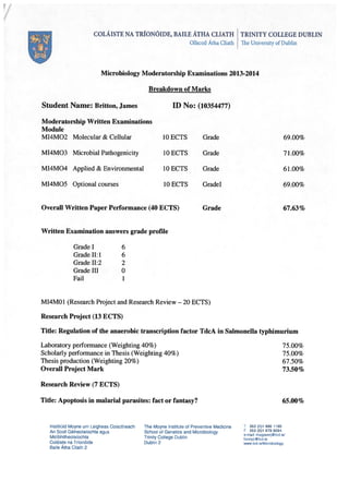
  • 18. Page | 15 FIG. 7. (A) Positive and negative fold change in tdcA expression between SL1344 wild type in aerobic conditions and the WT and selected mutants post anaerobic shock. (B) The same results as shown in (A) are shown here excluding the crp mutant for scale. Fold change is calculated relative to expression rates of control gene gmk. Binding affinities of CRP, FNR and H-NS to the tdcA promoter and CDS Using ChIP and qPCR analysis as described earlier the binding affinities of CRP, FNR and H- NS to the tdcA promoter and CDS was investigated under aerobic and anaerobic shock conditions. SL1344 strains which had the desired proteins tagged with a flag epitope were used for this experiment. In E. coli CRP binds to the promoter region of tdcA at -43.5 under anaerobic conditions (36; 46), however no similar investigations have taken place in Salmonella, although due to the high conservation of the CRP binding site in the Salmonella tdcA promoter it has been assumed that CRP likely plays a similar role in Salmonella. Similarly in FNR studies have shown the indirect effect it has on tdcA transcription in E. coli (6) however, no studies have confirmed this in the Salmonella homologue. As mentioned H-NS has been found to have an effect on tdcA expression in Salmonella (21) however its binding affinities to the region under anaerobic conditions remain unknown. The results of the ChIP analysis are shown in Fig. 8. As was expected CRP and FNR showed little binding under aerobic conditions. However under anaerobic shock conditions CRP binding increased both at the tdcA promoter and CDS over 4 and 3 fold respectively. FNR binding increased at the tdcA promoter region tested, however no binding activity was detected in the coding sequence. H-NS showed high relative levels of binding to both the tdcA promoter and CDS in the aerobically conditioned samples with fold
  • 19. Page | 16 changes of over 1,000 and 850 relative to the control respectively. Under anaerobic shock conditions the levels of H-NS binding were greatly reduced at both of the regions tested. (A) crp tdcA Prom oterA erobic tdcA Prom oterA naerobic tdcA C D S A erobic tdcA C D S A naerobic -2 0 2 4 6 FoldChange (B) fnr tdcA Prom oterA erobic tdcA Prom oterA naerobic tdcA C D S A erobic tdcA C D S A naerobic -2 0 2 4 6 FIG. 8. Fold change in binding affinity of (A) CRP, (B) FNR and (C) H-NS to the tdcA promoter and CDS under aerobic and anaerobic shock conditions. Fold change was calculated relative to the no antibody control using the 2-∆∆Ct method (25).
  • 20. Page | 17 DISCUSSION All attempts to make both SL1344 Flag-tdcA and ∆tdcA strains were unsuccessful. During the course of experimentation the experimental procedure for creation of these strains was altered in the hope of successful strain creation. As well as those changes described in the results section the method of preparing the cells for electroporation was altered by reducing all centrifugation steps by half in the hope of making the cells more permissible. Fresh stocks of all chemicals used in the Phenol – Chloroform extraction were also used to no avail. It was also found during experimentation that the temperature sensitive carbenicillin resistance of the pKD46 plasmid perhaps was not as temperature sensitive as one would like (Data not shown). This may suggest a fault in the pKD46 plasmid used as the temperature sensitivity may not be entirely reliable. The only component used in the creation of these strains which was not replaced during the course of the investigation was the primers ordered from Integrated DNA Technologies. These primers did amplify the desired regions of both the pSUB11 and pKD3 plasmids; however as is evident there was a failure for the PCR product to recombine into the chromosomal DNA of SL1344 pKD46. As other colleagues attempting similar projects had similar obstructions and that all primers were ordered at the same time it could be that there was a problem with the segment of the primers ordered homologous to the Salmonella chromosome. More interesting however are the results of the DNA binding and transcriptional analysis of tdcA. Expression of Salmonella tdcA increased 2-2.5 fold in anaerobically shocked cells in comparison with the wild type (Fig. 6). Other investigations have shown that maximal tdcA expression takes place under anaerobic shock conditions (20; 23), however there is a discrepancy between past work and that done here, past work has demonstrated tdcA expression under anaerobic shock rises over 1,000 fold (23). It may be that the conditions used during the course of this investigation were not truly anaerobic, as such all further conclusions must be viewed with scepticism. There is currently no evidence for the binding of CRP to the tdcA promoter in Salmonella. However CRP binding to the tdcA promoter at the position centred at -43.5 relative to the transcription start site has been shown to be essential to E. coli tdcA expression (36; 46). The tdcA promoter regions of both E. Coli and Salmonella are highly conserved (72% identity) and as such past studies have assumed similar mechanisms are at play in Salmonella (21). Using the virtual footprint promoter analysis and regulon prediction analysis programs on www.prodoric.de as described by Munch in 2005 (30) it was found that the tdcA promoter sequence contains one possible binding site for CRP. This binding site is quite similar in both position and sequence to that of the E. coli tdcA promoter. This suggests that the role of CRP in
  • 21. Page | 18 tdcA transcription in Salmonella may be not too dissimilar to E. coli as suggested in the past. If the ChIP results are to be believed then a similar mechanism may be at play in Salmonella, however, due to the fact that the ChIP procedure was carried out singly without replicates all results must be taken with a pinch of salt. CRP was found to bind at both the tdcA promoter and CDS under anaerobic shock (Fig. 8A). Binding at the promoter region of tdcA was expected (21) but the binding seen in the tdcA CDS was not. To give reason to this unusual result the limitations of the experiments carried out must be taken into account. During preparation of ChIP DNA a sonication step took place, during this all sample cells are subjected to ultrasonic frequencies which fragment the DNA. The average size of this fragmented DNA is ~500bp (9). Assuming fragments of 500bp were the norm one must also assume that immunoprecipitated DNA fragments would have been this length also. Continuing with this train of thought it can be speculated that if any immunoprecipitated DNA fragments contain the sequence to be amplified by any primers used then any positive result of the qPCR may mean binding of the DNA of the target protein up to 500bp upstream or downstream of the selected region. In this study the primer pairs used for amplification of the tdcA promoter and coding sequence were located at -212 to -298 and +79 to 190 respective to the transcription start site. Therefore for all positive ChIP results the range for the tdcA promoter and CDS primers must be extended. Using this logic and the fact that the CRP binding site in the tdcA promoter is located at -43.5 one can conclude that any positive result in the tdcA CDS could be the result of binding in the tdcA promoter. With this in mind and the fact that the CRP binding site is located less than 50bp upstream of the tdcA transcription start site it could be argued that the positive result shown by the tdcA CDS primers in the CRP ChIP experiment come as a result of CRP binding to the proximal binding site in the tdcA promoter. The results of the transcriptional analysis of tdcA in the crp mutant showed a large decrease in tdcA expression in comparison with the wild type. Taken together these results show an increased binding of CRP under anaerobic shock conditions and a necessity for CRP in tdcA expression. One may assume that similarly to the regulation of E. coli tdcA CRP binds to the tdcA promoter and promotes transcription. From the results produced it seems FNR may play a slightly different role in tdcA regulation than in E. coli. Under anaerobic shock conditions FNR showed high relative levels of binding to the tdcA promoter (Fig. 8B) which is in direct conflict of the findings of Chattophady et al (6) who found that in E. coli even though a possible FNR binding site is present in the tdcA promoter FNRs influence on tdcA expression is indirect, possibly acting through the accumulation of metabolites of anaerobic metabolism. A promoter analysis on tdcA using the
  • 22. Page | 19 virtual footprint program on www.prodoric.de (30) found possible binding sites of FNR to the tdcA promoter, a binding site at -150bp relative to the transcription start site was discovered. This could possibly be the region in which FNR binds to the promoter, however the limitations of ChIP discussed in relation to CRP may be at play here also. The region found has significant homology to the possible FNR binding site revealed by Chattophady (6) in E. coli. It is possible that since the divergence of the two species FNR has come to play a different role in tdcA expression, although this may not be the case. The RT-PCR analysis of tdcA transcription in the fnr mutant gave back a surprising result. In both replicates the levels of tdcA transcription rose (Fig. 7B) in the fnr mutant under anaerobic shock. Collectively these results show an alternative form of regulation by FNR on tdcA in Salmonella in which FNR binds directly to the tdcA promoter and inhibits the transcription of tdcA. In E. coli the reasoning for the anaerobic induction of tdcA is due to the rise in cAMP levels upon anaerobic shock (33) and partially to the build-up of anaerobic metabolites as a result of FNR activation (6). Previous studies have shown how expression of tdcA is maximized after 30 minutes of anaerobic shock but decreases thereafter. If FNR is indeed a negative regulator of tdcA transcription than it may be that FNR exhibits delayed binding. As shown in the past H-NS can influence FNR occupancy at promoter regions (31). The DNA binding analysis of H-NS to the tdcA region showed decreased binding post anaerobic shock (Fig. 8C). It is possible that FNR is only able to bind once H-NS has sufficiently dissociated. Assuming H-NS dissociation is not instantaneous one could speculate that this system provides a window of time when enough H- NS has dispersed but prior to FNR binding during which tdcA may be expressed. In past studies the fact that tdcA expression is limited, reaching maximum expression by 30 minutes and decreasing thereafter (20). This possible “window” of expression theory provides a mechanism for this. However investigations into both the exact timing of FNR binding and H-NS dissociation must take place before this can have any credibility. H-NS was found to have high levels of binding both at the tdcA promoter and CDS in cultures which did not undergo anaerobic shock (Fig. 8C). The levels of promoter and CDS binding were lowered considerably on anaerobic shock. A study on global H-NS binding conducted by Dillon et al (9) showed that the region of the chromosome in which the tdc operon resides is highly bound by H-NS. As a global transcription repressor H-NS often binds DNA in such a way as to exclude RNA polymerase and upon gene activation is removed (11). However removal of the hns gene resulted in reduced expression of tdcA under anaerobic shock. This finding is in contradiction to those of Kim et al (21) who found slightly increased expression of tdcA in an hns mutant. All transcriptional analyses were taken using cultures which had grown to an OD600 of
  • 23. Page | 20 0.3, this was considered mid log phase. The hns mutant grew considerably slower than all other strains used (Fig. 3B). The deletion of hns obviously had serious consequences on the fitness of the strain, possibly resulting in reduced tdcA expression; in hindsight the tdcA expression levels in the hns mutant under aerobic conditions should also have been tested. Also the fact that the tdcA gene resides in a region highly bound by H-NS (9) might suggest that the local structure of the DNA there may have been altered by its removal. Similarly to this in E. coli DNA supercoiling has been shown to alter the expression of tdcA (45). The tdc operon of Salmonella is under highly complex regulation by a number of different factors. Although the operon has changed much between Salmonella and E. coli since their divergence it still holds many similarities in regards to its regulation. CRP which holds principle control of the operon seems to play a very similar role in Salmonella as in E. coli however the regulation other less influential factors may vary as shown with FNR. There are still many unanswered questions on Salmonella tdcA expression. The roles of TdcA, IHF and other factors such as those involved in altering of DNA supercoiling have yet to be explored in Salmonella as they have in E. coli.
  • 24. Page | 21 Acknowledgements I would like to thank my supervisor Professor Shane Dillon for his guidance during this project. I would also like to thanks Professor Charles Dorman for the continued insight over the course of the investigation and Dr. Heather Quinn for all of the help and advice given throughout the project.
  • 25. Page | 22 REFERENCES 1. Altschul SF, Gish W, Miller W, Myers EW, Lipman DJ. 1990. Basic local alignment search tool. J Mol Biol 215:403-10 2. Bott M. 1997. Anaerobic citrate metabolism and its regulation in enterobacteria. Archives of microbiology 167:78-88 3. Bruckner R, Titgemeyer F. 2002. Carbon catabolite repression in bacteria: choice of the carbon source and autoregulatory limitation of sugar utilization. FEMS microbiology letters 209:141-8 4. Burman JD, Stevenson CE, Sawers RG, Lawson DM. 2007. The crystal structure of Escherichia coli TdcF, a member of the highly conserved YjgF/YER057c/UK114 family. BMC structural biology 7:30 5. Busby S, Ebright RH. 1997. Transcription activation at class II CAP-dependent promoters. Mol Microbiol 23:853-9 6. Chattopadhyay S, Wu Y, Datta P. 1997. Involvement of Fnr and ArcA in anaerobic expression of the tdc operon of Escherichia coli. Journal of bacteriology 179:4868-73 7. Corcoran CP, Dorman CJ. 2009. DNA relaxation-dependent phase biasing of the fim genetic switch in Escherichia coli depends on the interplay of H-NS, IHF and LRP. Mol Microbiol 74:1071-82 8. Datsenko KA, Wanner BL. 2000. One-step inactivation of chromosomal genes in Escherichia coli K-12 using PCR products. Proceedings of the National Academy of Sciences of the United States of America 97:6640-5 9. Dillon SC, Cameron ADS, Hokamp K, Lucchini S, Hinton JCD, Dorman CJ. 2010. Genome-wide analysis of the H-NS and Sfh regulatory networks in Salmonella Typhimurium identifies a plasmid-encoded transcription silencing mechanism. Molecular Microbiology 76:1250-65 10. Dillon SC, Espinosa E, Hokamp K, Ussery DW, Casadesus J, Dorman CJ. 2012. LeuO is a global regulator of gene expression in Salmonella enterica serovar Typhimurium. Mol Microbiol 85:1072-89 11. Dorman CJ. 2004. H-NS: a universal regulator for a dynamic genome. Nature reviews. Microbiology 2:391-400 12. Egan RM, Phillips AT. 1977. Requirements for induction of the biodegradative threonine dehydratase in Escherichia coli. Journal of bacteriology 132:370-6 13. Feldman DA, Datta P. 1975. Catabolite inactivation of biodegradative threonine dehydratase of Escherichia coli. Biochemistry 14:1760-7 14. Fink RC, Evans MR, Porwollik S, Vazquez-Torres A, Jones-Carson J, et al. 2007. FNR is a global regulator of virulence and anaerobic metabolism in Salmonella enterica serovar Typhimurium (ATCC 14028s). Journal of bacteriology 189:2262-73 15. Goss TJ, Schweizer HP, Datta P. 1988. Molecular characterization of the tdc operon of Escherichia coli K-12. Journal of bacteriology 170:5352-9 16. Hagewood BT, Ganduri YL, Datta P. 1994. Functional analysis of the tdcABC promoter of Escherichia coli: roles of TdcA and TdcR. Journal of bacteriology 176:6214-20 17. Hesslinger C, Fairhurst SA, Sawers G. 1998. Novel keto acid formate-lyase and propionate kinase enzymes are components of an anaerobic pathway in Escherichia coli that degrades L-threonine to propionate. Mol Microbiol 27:477-92 18. Inokuchi H, Ito R, Sekiguchi T, Sekiguchi M. 2013. Search for proteins required for accurate gene expression under oxidative stress: roles of guanylate kinase and RNA polymerase. J Biol Chem 288:32952-62 19. Kim M, Lim S, Kim D, Choy HE, Ryu S. 2009. A tdcA mutation reduces the invasive ability of Salmonella enterica serovar typhimurium. Molecules and cells 28:389-95
  • 26. Page | 23 20. Kim M, Lim S, Ryu S. 2011. Comparison of tdcA expression between Escherichia coli and Salmonella enterica serovar Typhimurium. Journal of microbiology and biotechnology 21:252-5 21. Kim MJ, Lim S, Ryu S. 2008. Molecular analysis of the Salmonella typhimurium tdc operon regulation. Journal of microbiology and biotechnology 18:1024-32 22. Kim W, Surette MG. 2004. Metabolic differentiation in actively swarming Salmonella. Mol Microbiol 54:702-14 23. Kroger C, Colgan A, Srikumar S, Handler K, Sivasankaran SK, et al. 2013. An infection- relevant transcriptomic compendium for Salmonella enterica Serovar Typhimurium. Cell host & microbe 14:683-95 24. Lim S, Kim M, Choi J, Ryu S. 2010. A mutation in tdcA attenuates the virulence of Salmonella enterica serovar Typhimurium. Molecules and cells 29:509-17 25. Livak KJ, Schmittgen TD. 2001. Analysis of relative gene expression data using real-time quantitative PCR and the 2(-Delta Delta C(T)) Method. Methods 25:402-8 26. Lostroh CP, Lee CA. 2001. The Salmonella pathogenicity island-1 type III secretion system. Microbes Infect 3:1281-91 27. Lu Z, Takeuchi M, Sato T. 2007. The LysR-type transcriptional regulator YofA controls cell division through the regulation of expression of ftsW in Bacillus subtilis. Journal of bacteriology 189:5642-51 28. Maddocks SE, Oyston PC. 2008. Structure and function of the LysR-type transcriptional regulator (LTTR) family proteins. Microbiology 154:3609-23 29. Meysman P, Sanchez-Rodriguez A, Fu Q, Marchal K, Engelen K. 2013. Expression divergence between Escherichia coli and Salmonella enterica serovar Typhimurium reflects their lifestyles. Mol Biol Evol 30:1302-14 30. Munch R, Hiller K, Grote A, Scheer M, Klein J, et al. 2005. Virtual Footprint and PRODORIC: an integrative framework for regulon prediction in prokaryotes. Bioinformatics 21:4187-9 31. Myers KS, Yan H, Ong IM, Chung D, Liang K, et al. 2013. Genome-scale analysis of escherichia coli FNR reveals complex features of transcription factor binding. PLoS genetics 9:20 32. Oh MK, Liao JC. 2000. Gene expression profiling by DNA microarrays and metabolic fluxes in Escherichia coli. Biotechnol Prog 16:278-86 33. Phillips AT, Egan RM, Lewis B. 1978. Control of biodegradative threonine dehydratase inducibility by cyclic AMP in energy-restricted Escherichia coli. Journal of bacteriology 135:828-40 34. Sambrook J, Russell DW. 2006. Purification of nucleic acids by extraction with phenol:chloroform. CSH Protoc 1 35. Sawers G. 1998. The anaerobic degradation of L-serine and L-threonine in enterobacteria: networks of pathways and regulatory signals. Archives of microbiology 171:1-5 36. Sawers G. 2001. A novel mechanism controls anaerobic and catabolite regulation of the Escherichia coli tdc operon. Mol Microbiol 39:1285-98 37. Schweizer HP, Datta P. 1989. Identification and DNA sequence of tdcR, a positive regulatory gene of the tdc operon of Escherichia coli. Mol Gen Genet 218:516-22 38. Shizuta Y, Hayaishi O. 1970. Regulation of biodegradative threonine deaminase synthesis in Escherichia coli by cyclic adenosine 3',5'-monophosphate. J Biol Chem 245:5416-23 39. Sperandio V, Li CC, Kaper JB. 2002. Quorum-sensing Escherichia coli regulator A: a regulator of the LysR family involved in the regulation of the locus of enterocyte effacement pathogenicity island in enterohemorrhagic E. coli. Infect Immun 70:3085-93 40. Spiro S, Guest JR. 1990. FNR and its role in oxygen-regulated gene expression in Escherichia coli. FEMS microbiology reviews 6:399-428
  • 27. Page | 24 41. Sumantran VN, Schweizer HP, Datta P. 1990. A novel membrane-associated threonine permease encoded by the tdcC gene of Escherichia coli. Journal of bacteriology 172:4288-94 42. Umbarger HE, Brown B. 1957. Threonine deamination in Escherichia coli. II. Evidence for two L-threonine deaminases. Journal of bacteriology 73:105-12 43. Untergasser A, Cutcutache I, Koressaar T, Ye J, Faircloth BC, et al. 2012. Primer3--new capabilities and interfaces. Nucleic acids research 40:e115 44. Uzzau S, Figueroa-Bossi N, Rubino S, Bossi L. 2001. Epitope tagging of chromosomal genes in Salmonella. Proceedings of the National Academy of Sciences of the United States of America 98:15264-9 45. Wu Y, Datta P. 1995. Influence of DNA topology on expression of the tdc operon in Escherichia coli K-12. Mol Gen Genet 247:764-7 46. Wu Y, Patil RV, Datta P. 1992. Catabolite gene activator protein and integration host factor act in concert to regulate tdc operon expression in Escherichia coli. Journal of bacteriology 174:6918-27