
Recommended
Recommended
More Related Content
Similar to Role of pro-brain-derived neurotrophic factor (proBDNF)to ma.docx
Similar to Role of pro-brain-derived neurotrophic factor (proBDNF)to ma.docx (20)
More from joellemurphey
More from joellemurphey (20)
Recently uploaded
Recently uploaded (20)
Role of pro-brain-derived neurotrophic factor (proBDNF)to ma.docx
- 1. Role of pro-brain-derived neurotrophic factor (proBDNF) to mature BDNF conversion in activity-dependent competition at developing neuromuscular synapses H. Shawn Jea,b,c,1, Feng Yanga,b,d,1, Yuanyuan Jia,e, Guhan Nagappana,e, Barbara L. Hempsteadf, and Bai Lua,b,e,2 aSection on Neural Development and Plasticity, National Institute of Child Health and Human Development, Bethesda, MD 20892-3714; bGenes, Cognition, and Psychosis Program (GCAP), National Institute of Mental Health, Bethesda, MD 20892-3714; cProgram in Neuroscience and Behavioral Disorders, Duke– National University of Singapore (Duke-NUS) Graduate Medical School, 169857, Singapore; dLieber Institute for Brain Development, The Johns Hopkins University Medical Campus, Baltimore, MD 21205; eR&D China, GlaxoSmithKline, Pudong, Shanghai 201203, China; and fDivision of Hematology, Department of Medicine, Weill Medical College, Cornell University, New York, NY 10021 Edited* by Richard L. Huganir, The Johns Hopkins University School of Medicine, Baltimore, MD, and approved August 20, 2012 (received for review May 10, 2012) Formation of specific neuronal connections often involves compe- tition between adjacent axons, leading to stabilization of the active terminal, while retraction of the less active ones. The underlying
- 2. molecular mechanisms remain unknown. We show that activity- dependent conversion of pro–brain-derived neurotrophic factor (proBDNF) to mature (m)BDNF mediates synaptic competition. Stim- ulation of motoneurons triggers proteolytic conversion of proBDNF to mBDNF at nerve terminals. In Xenopus nerve–muscle cocultures, in which two motoneurons innervate one myocyte, proBDNF- p75NTR signaling promotes retraction of the less active terminal, whereas mBDNF–tyrosine-related kinase B (TrkB) p75NTR (p75 neu- rotrophin receptor) facilitates stabilization of the active one. Thus, proBDNF and mBDNF may serve as potential “punishment” and “re- ward” signals for inactive and active terminals, respectively, and activity-dependent conversion of proBDNF to mBDNF may regulate synapse elimination. neuromuscular junction | pro-neurotrophin | synapse competition The nervous system responds to experience by altering thenumber and strength of synaptic connections (1). Activity- dependent synaptic competition, a general process seen in many parts of the developing nervous system, plays a critical role in shaping patterns of neuronal connections (2–7). At the neuro- muscular junction (NMJ), for example, multiple axons compete for the same postsynaptic muscle cell during early postnatal life until all but one is eliminated (8–10). Extensive experimental data support the view that the more active terminal or “cartel” gets stabilized, whereas less active ones withdraw, resulting in
- 3. canonical elimination of polyneuronal innervation (8, 11). It is generally believed that this synaptic competition is mediated by a “punishment” or “elimination” signal, produced by the post- synaptic cell, that causes the retraction of the inactive terminals, as well as a “protective” or “reward” signal that stabilizes the active terminal (10–12). Despite significant efforts over decades, the identity of the punishment or reward signals remains un- known (4, 13). This is due at least in part, to the experimental difficulties in manipulating gene expression selectively in one of the competing axons. Brain-derived neurotrophic factor (BDNF) has been recog- nized as a key regulator of synapse development and plasticity (14, 15). This is because BDNF is the only neurotrophin in- disputably secreted in an activity-dependent manner (15). In- deed, activity-dependent secretion of BDNF has been shown to be critical for hippocampus-dependent memory in human (16, 17). Like all neurotrophins, BDNF is initially synthesized as a precursor (proBDNF), which is subsequently cleaved to generate mature (m)BDNF. proBDNF interacts preferentially with the pan-neurotrophin receptor p75 (p75NTR), whereas mBDNF selectively binds and activates the receptor tyrosine kinase TrkB (18, 19). Cumulative evidence supports a “yin-yang hypothesis,” in which pro- and mBDNF elicit opposite biological effects by activating two distinct receptor systems (20). For ex- ample, proBDNF, if not processed, promotes long-term de- pression (LTD) through the activation of p75NTR in the hippocampus (21, 22). In contrast, mBDNF-TrkB signaling is essential for the early phase of long-term potentiation (E-LTP) (23–25). Moreover, recent studies indicate that a significant proportion of BDNF in the brain is secreted in the proform (26–
- 4. 28), and extracellular conversion of proBDNF to mBDNF by the tissue plasminogen activator (tPA)/plasmin protease system is critical for late-phase LTP (29). The expression of proBDNF and p75NTR in rodents is developmentally regulated, with the highest levels in the first and second postnatal week, correlating well with the timings of synapse formation (28). Therefore, proteolytic cleavage of proBDNF represents an important mechanism by which the opposing cellular actions of proBDNF and mBDNF may be regulated (20). The opposing nature of proBDNF and mBDNF prompted us to hypothesize that proBDNF and mBDNF might serve as punish- ment and reward signals, respectively, during synaptic competition at the developing NMJs. In this study, we developed a triplet sys- tem that allows alteration of gene function in one of two distinctly labeled axons that innervate a single, unlabeled myocyte. Our study suggests that proBDNF serves as a general “punishment signal” that causes p75NTR-expressing motor terminals to retract, whereas at the active terminal, secretion/activation of extracellular protease(s) converts proBDNF to mBDNF, which serves as a re- ward signal to stabilize the terminal. Results Activity-Dependent Synaptic Competition in Xenopus Nerve– Muscle Cocultures. Xenopus nerve–muscle coculture system was used to study the activity-dependent synaptic competition. We
- 5. developed a cell-culture system, in which an unlabeled myocyte was inner- vated by two spinal neurons (one labeled in green and the other in red, respectively; Fig. 1A). This was accomplished by injecting either FITC-dextran (green) or rhodamine-dextran (red) into a single dorsal animal blastomere at the 8- or 16-cell stage and mixing the neural tubes from embryos that were injected with two different fluorophores to prepare dissociated nerve–muscle Author contributions: H.S.J., F.Y., and B.L. designed research; H.S.J., F.Y., Y.J., and G.N. performed research; B.L.H. contributed new reagents/analytic tools; H.S.J., F.Y., and Y.J. analyzed data; and H.S.J., F.Y., and B.L. wrote the paper. The authors declare no conflict of interest. *This Direct Submission article had a prearranged editor. 1H.S.J. and F.Y. contributed equally to this work. 2To whom correspondence should be addressed. E-mail: [email protected] This article contains supporting information online at www.pnas.org/lookup/suppl/doi:10. 1073/pnas.1207767109/-/DCSupplemental. 15924–15929 | PNAS | September 25, 2012 | vol. 109 | no. 39 www.pnas.org/cgi/doi/10.1073/pnas.1207767109 mailto:[email protected] http://www.pnas.org/lookup/suppl/doi:10.1073/pnas.120776710 9/-/DCSupplemental http://www.pnas.org/lookup/suppl/doi:10.1073/pnas.120776710 9/-/DCSupplemental www.pnas.org/cgi/doi/10.1073/pnas.1207767109
- 6. cocultures (30). Instead of employing electrical stimulation using a glass electrode, which might cause mechanical damages on neurons, we stimulated one of the spinal neurons by local pho- tolysis of caged glutamate (MNI-glutamate; 50 μM) using a multiphoton microscope (31). Photo-uncaging of caged gluta- mate at a neuronal soma induced a marked potentiation of synaptic transmission, which lasted for more than 60 min (Fig. S1A). This synaptic potentiation was completely blocked by the sodium channel blocker tetrodotoxin (TTX) (1 μM), demon- strating the requirement of action potentials (Fig. S1A). Fur- thermore, synaptic potentiation was only observed when a laser beam was applied within 25 μm from the neuronal cell body, suggesting that the photo-uncaging occurred locally (Fig. S1B). A brief episode (250 ms) of photolysis of MNI-glutamate was applied to one of the two neurons, and the resulting morpho- logical changes in synaptic terminals from both neurons, which were innervating a single myocyte, were monitored by dual- color, time-lapse confocal imaging. Upon stimulation of a red neuron, the axon terminal of the unstimulated neuron (green terminal) gradually withdrew from the previously innervated muscle, whereas the terminal of the stimulated neuron (red terminal) remained stable and occasionally extended (Fig. 1B and Movie S1). Conversely, stimulation of a green neuron triggered the retraction of the red terminal (Fig. S2). Because stimulation of one neuron always resulted in the retraction of the unstimulated neuron, regardless of whether it was red or green, we randomly stimulated neurons in subsequent experiments based on the convenience of photo-uncaging. We performed 11 experiments involving preferential photolysis of one neuron. In all 11 cases, the axon terminals of the unstimulated neurons retracted to varying degrees. In six cases, axon terminals of stimulated neu-
- 7. rons showed slight expansion or elongation (Fig. 1 B and C). These results suggest that a postsynaptically derived local pun- ishment signal may trigger synaptic retraction. Activity-Dependent Cleavage of Secreted proBDNF at NMJ. Given that proBDNF and mBDNF could elicit opposite effects, we hypothesized that proBDNF and mBDNF might serve as pun- ishment and reward signals, respectively. Furthermore, we hypoth- esized that the proteolytic conversion of proBDNF to mBDNF might be essential for synaptic competition and elimination at the developing NMJs. First, we tested whether neuronal activity could activate proteases to process proBDNF to mBDNF using a fluorogenic proteolytic beacon assay. The fluorogenic beacon consisted of synthetic peptides, harboring the propeptide cleav- age sequences (MSMRVRR↓HSD) within proBDNF, and these synthetic peptides were flanked by a fluorophore at the N terminus and a quencher at its C terminus (Fig. 2A). Small size peptides allowed both a fluorophore and a quencher within proximity, thereby quenching the fluorescence via fluorescence resonance energy transfer (FRET). Upon cleavage of peptides by a pro- tease(s), which specifically recognizes the proBDNF cleavage sequence, the fluorogenic beacon would emit the fluorescence signal because of the separation of a fluorophore from a quencher (Fig. 2A). In addition, we immobilized the fluorogenic beacon to polystyrene beads to minimize diffusion in culture medium. Next, we investigated whether stimulation of neurons could activate proteases at the nerve terminals. The fluorogenic beacons were placed on the axonal process or termini of spinal neurons, and neurons were subsequently stimulated either by an
- 8. electrical stimulation or by photo-uncaging of MNI-glutamate (Fig. 2B, bright field). After stimulation, the fluorescence intensity of the beads on the axon terminals or axonal processes (e.g., yellow arrows in Fig. 2B) greatly increased (Fig. 2 B and C), suggesting cleavage of the peptide by proteases secreted from the axon. In contrast, no fluorescence change was observed from beads, which were not in contact with the axonal process or termini (Fig. 2B, “free beads,” white arrow). Time-lapse imaging indicated that the increase in fluorescence occurred relatively quickly: the first surge came within minutes, and the florescence increase reached its peak within 20 min (Fig. 2C). Pretreatment with a mixture of protease inhibitors prevented the increase in fluorescence from beads on axon terminals (Fig. 2D). Remarkably, inhibitors for tPA and furin, the extracellular and intracellular proteases known to cleave proBDNF in the brain (29, 32), did not inhibit the fluorescence increase (Fig. 2D). In contrast, matrix metalloprotease (MMP) inhibitors successfully blocked the stimulus-induced increase in fluorescence. Further analyses indicated that MMP3 and MMP9, but not MMP2, MMP8, or MMP13, could increase fluorescence signal on the fluorogenic beacons (Fig. 2D). Finally, when the beacon-containing beads were placed on muscle cells, stimulation of muscle cells did not cause an increase in fluorescence (Fig. 2D), N N
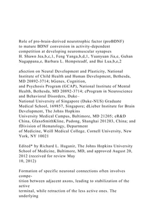
- 9. M stim. unstim.A N M N B C -5 0 5 10 15 20 unstimulated neuron (11) stimulated neuron (11) Distance(µm) * st im . un st im .
- 10. Elongation Retraction M er ge d 30 60 90 1200 Fig. 1. Activity-dependent synaptic competition in culture. (A) Schematic diagram (Left) and confocal image (Right) showing a triplet in which a spherical myocyte (M) (indicated by white dotted lines) is innervated by two spinal neurons (N), one labeled with FITC (green) and one labeled in rhodamine (red), in nerve–muscle coculture. (B) Time course of synaptic competition. A higher magnification of A is shown. Stimulation of the red neuron, by photo-uncaging of MNI-glutamate in the soma area using a two- photon laser, caused the unstimulated (green) axon terminal to retract from the synaptic target (indicated by a yellow arrow, upper row). In contrast, the axon terminal (red) from the stimulated neuron did not retract, but elongated a little (white arrows, middle row). The phase and two-color fluorescence images of the triplet at multiple time points (lower row). (Scale bar: 10 μm.) The phase (Bottom) and two-color fluorescence
- 11. images (Top and Middle) of the triplet at “0” and “120” min are shown. (C) Quantifi- cation of axonal retraction and elongation measured 120 min after stimu- lation of one of the neurons in the triplets. *P < 0.01. Je et al. PNAS | September 25, 2012 | vol. 109 | no. 39 | 15925 N EU R O SC IE N C E http://www.pnas.org/lookup/suppl/doi:10.1073/pnas.120776710 9/- /DCSupplemental/pnas.201207767SI.pdf?targetid=nameddest=S F1 http://www.pnas.org/lookup/suppl/doi:10.1073/pnas.120776710 9/- /DCSupplemental/pnas.201207767SI.pdf?targetid=nameddest=S F1 http://www.pnas.org/lookup/suppl/doi:10.1073/pnas.120776710 9/- /DCSupplemental/pnas.201207767SI.pdf?targetid=nameddest=S F1 http://www.pnas.org/lookup/suppl/doi:10.1073/pnas.120776710 9/- /DCSupplemental/pnas.201207767SI.pdf?targetid=nameddest=S
- 12. F1 http://www.pnas.org/lookup/suppl/doi:10.1073/pnas.120776710 9/-/DCSupplemental/sm01.avi http://www.pnas.org/lookup/suppl/doi:10.1073/pnas.120776710 9/-/DCSupplemental/sm01.avi http://www.pnas.org/lookup/suppl/doi:10.1073/pnas.120776710 9/- /DCSupplemental/pnas.201207767SI.pdf?targetid=nameddest=S F2 indicating that muscle cells do not secrete proteases that could process proBDNF. We further examined whether postsynaptic muscle cells se- crete proBDNF upon stimulation. Because of unavailability of a proBDNF-specific ELISA and limited number of muscle cells in the Xenopus cocultures (<1,000 myocytes), it was not feasible to measure proBDNF secretion from muscle cells using existing biochemical techniques. Thus, we used cell surface immuno- staining to measure proBDNF secretion, given that proBDNF is positively charged at physiological pH and could be associated with a negatively charged cell membrane upon secretion (27, 33). The muscle cell cultures were depolarized by high-K+ treatment (50 mM) for 5 min, fixed, and processed for cell surface immu- nofluorescence staining under membrane impermeable condi- tions. A proBDNF-specific monoclonal antibody was used for cell- surface staining of secreted proBDNF (27). Although proBDNF was barely detectable on the muscle cell surface at rest (control), the proBDNF immunoreactivity increased dramatically (88%) upon depolarization (Fig. S3A). This increase was even more pronounced when extracellular cleavage of proBDNF was
- 13. blocked by general MMPs inhibitors (indicated as MMP-In. in Fig. S3A). To measure the protease-mediated conversion of endogenous proBDNF to mBDNF at the neuromuscular synapses, we per- formed cell-surface immunostaining using a specific antibody against mBDNF. We applied glutamate, instead of high K+, to selectively depolarize neurons, because muscle cells do not ex- press glutamate receptors (34). After neuronal depolarization, cultures were subsequently fixed and processed for surface im- munostaining using an mBDNF-specific antibody (27). In control conditions, there was no mBDNF staining (Fig. S3B, Left). Upon glutamate application (10 mM; 5 min), we observed a dramatic, threefold increase in surface mBDNF staining at the synaptic junction (Fig. S3B, Center). This increase in mBDNF immuno- reactivity was observed only in myocytes that were innervated by a spinal neuron but not in the neighboring noninnervated myocytes (Fig. S3B, Center). This suggested that the release and/ or activation of proteases at the nerve terminal converted proBDNF to mBDNF at the active synaptic terminals. Further- more, when the cultures were pretreated with the MMP inhib- itors, the glutamate-induced increase in mBDNF staining was completely abolished (Fig. S3B, Right). Taken together, these results supported the notion that activity-dependent conversion of proBDNF to mBDNF occurred at the developing Xenopus NMJ in situ. Synaptic Stabilization During Competition Mediated by mBDNF/TrkB. Previously, we have shown that exogenous proBDNF triggers synaptic depression and subsequent retraction of axon terminal
- 14. through p75NTR in Xenopus neuromuscular synapses (35). Based on robust secretion of proBDNF upon synaptic depolarization, we reasoned that active terminals might cleave proBDNF to mBDNF, which protects these active terminals from proBDNF- mediated synaptic depression and retraction. To test this, we knocked down endogenous TrkB in one of neurons in our triplet system using a morpholino (Fig. S4A; also see ref. 36). Control experiments indicated that the expression of the TrkB morpho- lino in presynaptic neurons blocked the mBDNF-mediated syn- aptic potentiation (Fig. S4 B and C). When the TrkB morpholino was selectively expressed in one of the spinal neurons in our triplet system, stimulation of the TrkB morpholino-expressing neuron (green) resulted in retraction of both green and red terminals (Fig. 3A, yellow arrows). Quantitative analysis of 10 triplets indicated that both stimulated and unstimulated termi- nals retracted ∼12 μm within 120 min (Fig. 3C). Next, we reasoned that if the activation of TrkB by mBDNF was critical to prevent synaptic retraction, supply of exogenous BDNF would prevent synaptic competition. Indeed, in a nerve–muscle culture treated with mBDNF (25 ng/mL) for 2 h, stimulation of the red neuron did not cause retraction of the green terminal (Fig. 3B and Movie S2). Moreover, application of exogenous mBDNF even elicited axonal elongation from the stimulated neurons (Fig. 3 B and C, white arrowheads). These results suggest that mBDNF plays an active role in preventing synaptic retraction of the active terminals through TrkB signaling during synaptic competition. Synaptic Retraction During Competition Mediated by proBDNF/p75NTR.
- 15. To determine whether this activity-based synaptic retraction was mediated through proBDNF/p75NTR signaling, we knocked down endogenous p75NTR by using FITC-conjugated p75NTR siRNA that were targeted to all Xenopus p75NTR isoforms (p75NTRa and p75NTRb) (37). The p75NTR siRNA was introduced into a single neuron in our triplet system using embryo-injection techniques. A B C D ROX-MSM RVRR /HSD-QXL610 Fluorophore Quencher Proteolytic cleavage Emit fluorescence proBDNF cleavage sequences 0 min 6 min Bright field Before stim. After stim. 0.8 1 1.2 1.4
- 16. 1.6 1.8 0 5 10 15 20 25 30 min N or m al iz ed fl uo re sc en ce in te ns ity (F /F o)
- 17. Control (8) Protease In.(7) Free beads (9) 0.8 1 1.2 1.4 1.6 1.8 9 7 7 6 5 7 8 6 98 8 6 * * * * * * N or m al
- 21. P 3 In . M M P 8 In . M M P 9 In . Fig. 2. Activity-dependent cleavage of proBDNF detected by fluorogenic probes. (A) Schematic dia- gram depicting the design of a fluorogenic in- dicator of proBDNF cleavage. A peptide containing the proBDNF cleavage site was placed between a fluorophore and a quencher. Upon proteolytic cleavage, the quencher was dissociated from the fluorophore, leading to emission of fluorescence. (B) Sample images of fluorogenic probes in phase (Left) and fluorescence (Right) before and after neuronal stimulation. Polystyrene beads soaked with fluorogenic probes were positioned on (yellow
- 22. arrows) or near (white arrows) an axonal process (indicated by white dotted lines), respectively, by micromanipulation. A neuronal cell body was stimulated by photolysis of caged-glutamate (MNI- glutamate) with a UV laser. Note that after stimu- lation, only beads on, but not near, the axonal processes showed an increase in fluorescence. (C) Time course of cleavage-dependent increase in fluorescence intensity upon stimulation of spinal neurons. Fluorescence intensities measured on contacted beads or free beads were normalized to that at “0” time point and presented as ratios (F/F0) over time. (D) Quantification of relative fluorescence intensities in beads 30 min after stimulation of neurons under various conditions. Concentrations of protease inhibitors (In.): general protease inhibitor mixture, 50 μM; pan MMP inhibitor, 60 μM; tPA inhibitor, 10 μM; furin inhibitor, 40 μM; MMP2 inhibitor, 60 μM; MMP3 inhibitor, 50 nM; MMP8 inhibitor, 20 nM; MMP9 inhibitor, 50 μM; MMP13 inhibitor, 80 nM. B, beads; M, muscle cell; N, neuron. 15926 | www.pnas.org/cgi/doi/10.1073/pnas.1207767109 Je et al. http://www.pnas.org/lookup/suppl/doi:10.1073/pnas.120776710 9/- /DCSupplemental/pnas.201207767SI.pdf?targetid=nameddest=S F3 http://www.pnas.org/lookup/suppl/doi:10.1073/pnas.120776710 9/- /DCSupplemental/pnas.201207767SI.pdf?targetid=nameddest=S F3 http://www.pnas.org/lookup/suppl/doi:10.1073/pnas.120776710 9/- /DCSupplemental/pnas.201207767SI.pdf?targetid=nameddest=S
- 23. F3 http://www.pnas.org/lookup/suppl/doi:10.1073/pnas.120776710 9/- /DCSupplemental/pnas.201207767SI.pdf?targetid=nameddest=S F3 http://www.pnas.org/lookup/suppl/doi:10.1073/pnas.120776710 9/- /DCSupplemental/pnas.201207767SI.pdf?targetid=nameddest=S F3 http://www.pnas.org/lookup/suppl/doi:10.1073/pnas.120776710 9/- /DCSupplemental/pnas.201207767SI.pdf?targetid=nameddest=S F3 http://www.pnas.org/lookup/suppl/doi:10.1073/pnas.120776710 9/- /DCSupplemental/pnas.201207767SI.pdf?targetid=nameddest=S F4 http://www.pnas.org/lookup/suppl/doi:10.1073/pnas.120776710 9/- /DCSupplemental/pnas.201207767SI.pdf?targetid=nameddest=S F4 http://www.pnas.org/lookup/suppl/doi:10.1073/pnas.120776710 9/-/DCSupplemental/sm02.avi www.pnas.org/cgi/doi/10.1073/pnas.1207767109 Western blot analysis revealed significant reduction of endogenous p75NTR protein in neural tubes derived from embryos injected with p75NTR siRNA, but not in those injected with scrambled siRNA for p75NTR (35). We reasoned that the blockade of p75NTR would block synapse elimination of less active neurons. Indeed, when photolysis was applied to the red neuron, the axon terminal of a p75NTR siRNA-expressing neuron did not retract over a long
- 24. period (Fig. 4A). In seven triplets, four showed no retraction, whereas the other three showed reduced retraction of the unstimulated termi- nals after 2hrs (Fig. 4 A and C). Furthermore, an axon expressing scrambled siRNA still retracted when the competing axon was stimulated (Fig. S5). Because p75NTR siRNA and p75NTR mor- pholino target different regions of p75NTR, this experiment con- firmed that blockade of p75NTR signaling abolished synaptic re- traction. Taken together, these data support the hypothesis that synaptic retraction was mediated by presynaptic p75NTR signaling during synaptic competition. The results above, together with the finding that neuronal stimulation triggered protease secretion/activation at nerve ter- minals (Fig. 2), prompted us to speculate that active terminals were spared from synaptic retraction because of proteolytic cleavage of proBDNF. If this were true, then blockade of proBDNF cleavage would trigger retraction of all axon terminals during synaptic competition. To test this hypothesis, we treated nerve–muscle cocultures with a mixture of protease inhibitors for 10 min before neuronal stimulation. These inhibitors were able to block proteases that could cleave proBDNF (Fig. 2D). Remark- ably, axon terminals from red and green spinal neurons retracted simultaneously from the synaptic site (Movie S3). Quantitative analysis indicated that both stimulated and unstimulated terminals
- 25. retracted ∼10 μm (Fig. 4C). Because MMP inhibitors, particularly MMP3/9 inhibitors, blocked activity-induced proBDNF cleavage at nerve terminals (Fig. 2D), we further tested whether inhibition of MMPs could perturb synapse elimination. Indeed, pretreat- ment with a combination of both MMP3 and MMP9 inhibitors triggered synaptic retraction upon stimulation of the competing axon, indicating that MMP3/9 are the candidate proteases that convert proBDNF to mBDNF at the active synaptic terminal (Fig. S6 and Movie S4). Taken together, these data supported the hy- pothesis that the activation of presynaptic p75NTR by muscle- de- rived proBDNF triggered the retraction of less active axonal terminals during synaptic competition. Discussion For over a half century, activity-dependent synaptic competition/ elimination has been one of the central issues in developmental neurobiology. However, the underlying mechanisms remain un- known. Because the competition depends on activity and occurs locally, the experimental challenge has been to selectively acti- vate one of the competing terminals and to perform molecular manipulation selectively in competing axons (38). In the present study, we modified the Xenopus embryo-injection protocol, so that we could reliably obtain triplets, in which an unlabeled myocyte is innervated by two distinctly labeled axons. Confocal live-cell imaging and local glutamate uncaging using a multi- photon laser enabled us to visualize and trigger synaptic re- traction of individual axons in triplets. To demonstrate activity- dependent secretion of proBDNF and its conversion to mBDNF, we performed cell surface immunofluorescence staining on cultured cells, using specific antibodies against proBDNF and
- 26. mBDNF. FRET-based fluorogenic probes were used to monitor the activation of protease activities locally at axonal terminals in real time. Moreover, embryo injection techniques were used to selectively inhibit TrkB or p75NTR in one neuron, but not in others, in triplets. By combining these techniques, we provided the evi- dence for a local and activity-dependent activation of specific proteases that could convert proBDNF to mBDNF in neuromus- cular synapses. Given that proBDNF and mBDNF often elicit opposite biological effects (20, 28), this finding may help us un- derstand how proBDNF-to-mBDNF conversion is regulated via neuronal activity. Moreover, we found that the blockade of p75NTR signaling attenuated synapse elimination, whereas the blockade of TrkB signaling, or inhibition of proBDNF cleavage by metalloproteases, promoted synaptic retraction of both in- nervated axon terminals in triplets. Taken together, these findings suggest a model for synapse elimination in which the activity- dependent conversion of proBDNF to mBDNF selectively sta- bilized active terminals, whereas inactive terminals were elimi- nated in response to proBDNF and subsequent activation of p75NTR signaling (Fig. S7). Activity-Dependent proBDNF Cleavage Converts a “Synaptotoxin” to a “Synaptotrophin.” Two theories have been proposed to explain activity-dependent synaptic competition and elimination. The “synaptotoxin” theory suggests that the postsynaptic cell produces destabilizing or “toxic” signals such as proteases that retrogradely punish and remove presynaptic terminals. This theory (11) was based largely on the observation that inhibition of protease ac- tivity (39, 40), particularly thrombin, by the naturally occurring
- 27. protease inhibitor nexin I (41, 42), attenuated synapse elimina- tion. However, specific target(s) of the proteases have not been identified yet. More importantly, selective protection of the active terminal by local secretion of protease inhibitors has not been demonstrated. It is also unclear what axons are competing for, if the fate of an axon is dependent only on its intrinsic activity (11). An alternative is the “synaptotrophin” hypothesis, which suggests that axons compete with each other for a limited supply of trophic A B C -10 -5 0 5 10 15 20 Elongation Retraction unstimulated neuron stimulated neuron BDNF (4) TrkB morpholino (10) Distance (µm) unstimulated neuron stimulated neuron B
- 28. D N F (2 5n g/ m l) S tim . U n st im . 0 30 60 90 120 0 9030 60 120 U ns tim . S
- 29. tim . Tr K B -m or p. Fig. 3. Synaptic stabilization mediated by mBDNF/TrkB. (A) Down-regula- tion of TrkB by TrkB-morpholino (TrkB-morp.) led to retraction of both stimulated and unstimulated axon terminals. In a triplet system, stimulation of a neuron expressing TrkB morpholino (green) resulted in retraction of the red axon as well as the green axon. (Scale bar: 10 μm.) (B) Treatment with mBDNF prevented activity-dependent synaptic retraction. The culture was treated with mBDNF before stimulating the soma of a red neuron. The terminals from both red and green neurons remained in the synaptic site without any retraction. (Scale bar: 20 μm.) (C) Quantification of axonal re- traction and elongation measured 120 min after stimulation of one of the neurons in the triplets. Je et al. PNAS | September 25, 2012 | vol. 109 | no. 39 | 15927
- 31. fails to explain: (i) how the trophic factor is secreted locally from the postsynaptic cell only near the active terminal, but not at the inactive terminal; and (ii) how the active terminal preferentially binds, uptakes, or signals the trophic factor, if any. Neither local secretion nor preferential signaling has been reported at NMJs to support the synaptotrophin hypothesis. Moreover, synaptic elim- ination is an active process that requires a punishment signal, ultimately resulting in the loss of all, but one protected terminal (13). Thus, the synaptotrophin theory fails to explain how inactive terminals initiate withdrawal and why the absence of trophic support leads to synaptic depression and retraction. Our data support a model that both the punishment and re- ward signals are originated from the same molecule, namely BDNF (Fig. S7). In this model, presynaptic activity drives the secretion of proBDNF from postsynaptic muscle cells, which serves as a default “punishment signal” to actively retract af- ferent terminals through p75NTR. mBDNF, on the other hand, serves as a reward signal for which all terminals compete. A major conceptual breakthrough to the model is the activity-de- pendent conversion of proBDNF to mBDNF by active axon terminals. Locally secreted proteases at the active terminal not only block the punishment signal (proBDNF), but also generate a reward signal (mBDNF), which stabilizes the terminal by activating TrkB. Further work is necessary to substantiate this model in vivo. proBDNF-mBDNF As Punishment–Reward Signals in Synapse Elimination. Using cultured neurons, computational modeling, and knockout animals, two recent studies reported mechanisms for synaptic
- 32. competition and axonal pruning in the sympathetic nervous system (43, 44). In this system, target-derived NGF, through TrkA, provides protection and prosurvival of active axons, which are further strengthened and stabilized by increasing TrkA transcription. On the other hand, NGF-induced synthesis and secretion of BDNF and neurotrophin-4 (NT-4), through p75NTR, facilitate axonal degeneration and pruning of the competing axons (43–45). Although these experiments were largely carried out in cell culture, the basic premise is quite similar: TrkA mediates reward, whereas p75NTR mediates punishment. There are several important differences between the present study and those published works. First, activity-dependent secretion of the reward (NGF) or punishment signal (BDNF) in the superior cervical ganglia (SCG) neurons was not demonstrated. In the Xenopus neuromuscular system, we showed that BDNF is se- creted from postsynaptic muscle cells. Thus, the target cell (myocyte) is an important player in synaptic (as opposed to ax- onal) competition. Second, it is widely accepted that motoneurons express only TrkB, but TrkC and TrkA expression is controversial. In addition, negligible levels of NGF are detectable in the neu- romuscular system (46) and TrkB expression in sympathetic neurons is extremely low (47). Therefore, application of exces- sive BDNF, even with low affinity, can only bind to p75NTR, trig- gering axonal pruning. Third, and most importantly, in SCG, two separate ligands (NGF and BDNF) are needed for reward and punishment signals, respectively. At the NMJ, the reward and punishment signals are derived from the same molecule, depending on proteolytic cleavage. Our model suggests that the punish- ment signal (proBDNF) acts on all competing axons to promote
- 33. elimination, and the active axon is spared because it activates ex- tracellular protease(s) that convert the punishment (proBDNF) signal to a reward (mBDNF) signal. Several issues require further investigation. First, what is the identity and source of the protease(s)? Our imaging experiments (Figs. 2 and 3) point to metalloproteases, particularly MMP3 and MMP9, as candidates that cleave proBDNF at the active terminal during synaptic competition in Xenopus NMJ. Fluorogenic bea- con experiments suggest that the MMPs are derived from axonal terminal, but not from muscle cells (Fig. 2D). Indeed, these pro- teases are expressed in motor neurons and are highly enriched at the NMJs (48–50). Further work is necessary to identify the spe- cific proteases required at mouse NMJs in vivo. Second, contrary to our findings, several previous studies have shown that inhibition of protease activity attenuates synaptic elimination (39, 40) and even increases numbers of acetylcholine receptor (AChR) in the postsynaptic muscle (51, 52). One way to reconcile these two sets of data is that inhibition of a protease “X,” the function of which is to degrade MMPs, may result in an enhanced conversion of proBDNF to mBDNF by MMPs, leading to attenuation of syn- aptic elimination. Moreover, our results suggest that proBDNF is derived from postsynaptic muscle cells but not presynaptic motor neurons. It remains to be established whether muscle cells are the only source of proBDNF or whether other cell types, such as Schwann cells, also produce and secrete proBDNF at the NMJ
- 34. in vivo. With the currently available technologies, it is not possible to demonstrate the secretion of BDNF (more specifically proBDNF) at the NMJs in vivo. An important future experiment is to dem- onstrate that proBDNF is both secreted and cleaved locally at the active terminal but not at the inactive terminal in vivo. Finally, activation of p75NTR may be a general mechanism for activity- dependent synapse retraction. Indeed, in sympathetic neurons, BDNF (possibly proBDNF) acts through p75NTR to suppress A B C Distance (µm)-5 0 5 10 15 20 p75NTR siRNA(7) Protease inhibitors (7) Elongation Retraction unstimulated neuron stimulated neuron unstimulated neuron stimulated neuron p7 5N
- 35. TR -s iR N A P ro te as e in hi bi to rs 0 120 60 30 un st im . st im . 90
- 36. un st im . st im . 0 30 60 90 120 Fig. 4. Synaptic retraction mediated by proBDNF/p75NTR. (A) Down-regu- lation of p75NTR by siRNA prevents activity-dependent synaptic retraction. Fluorescence images show a double-innervated myocyte (white dotted lines) by a neuron expressing p75NTR siRNAs (green) and a rhodamine-labeled control neuron (red). The soma of the red neuron (outside the field) was stimulated by photo-uncaging and the axon terminals from both neurons were monitored by time-lapse microscopy. (Scale bar: 20 μm.) Even after 120 min, the axon terminals (white arrow) of both red and green neurons remained unchanged at the synaptic site. (B) Time-lapse images showing retraction of both stimulated (red) and unstimulated (green) axons in the presence of a mixture of protease inhibitors (50 μM). (Scale bar: 20 μm.) (C)
- 37. Quantification of axonal retraction and elongation measured 120 min after stimulation of one of the neurons in the triplets. 15928 | www.pnas.org/cgi/doi/10.1073/pnas.1207767109 Je et al. http://www.pnas.org/lookup/suppl/doi:10.1073/pnas.120776710 9/- /DCSupplemental/pnas.201207767SI.pdf?targetid=nameddest=S F7 www.pnas.org/cgi/doi/10.1073/pnas.1207767109 inactive axons, whereas active axons (depolarized by high K+) of the same neurons are spared (45). It will be extremely important to test whether protease-mediated conversion of proBDNF to mBDNF also contributes to activity-dependent synaptic compe- tition in the central nervous system (CNS), an example being ocular dominance formation within the visual cortex. Experimental Procedures Full details are contained in SI Experimental Procedures. Use and care of animals in this study abided by the guidelines of the institutional Animal Care and Use Committee at the National Institutes of Health NIH. Embryo Injection and Xenopus Nerve–Muscle Coculture. Xenopus nerve–mus- cle cocultures were prepared as described (53). Morpholinos, siRNAs, or cDNAs were injected into one of the blastomeres at the 2- to 4-cell or 8- to 16-cell
- 38. stage as described (53). Immunocytochemistry, Confocal Microscopy Image Analysis, and Statistics. Xenopus nerve–muscle cocultures were immunostained, maintained, and analyzed as described previously (35). See SI Experimental Procedures for details. Image analysis was performed by investigators who were blinded to experimental conditions. ACKNOWLEDGMENTS. We thank Drs. Phillip Nelson, Eugene Zaitsev, Keri Martinowitch, Jay Chang, and Newton Woo for thoughtful comments and suggestions and Regeneron Pharmaceuticals for providing recombi- nant BDNF. We also thank Drs. Bruce Carter, Mark Bothwell, Moses Chao, and Phil Barker for antibodies to p75NTR and Louis Reichardt and Moses Chao for antibodies to TrkB. Microscopy imaging was performed at the Porter Neuroscience Center Light Imaging Facility with the assistance of Dr. Carolyn Smith (NIH). This work was supported by the National In- stitute of Mental Health (NIMH) and National Institute of Child Health and Human Development (NICHD) intramural research programs (B.L.) and grants from the NIH and Muscular Dystrophy Association (MDA) (to B.H.).
- 39. 1. Katz LC, Shatz CJ (1996) Synaptic activity and the construction of cortical circuits. Science 274:1133–1138. 2. Constantine-Paton M, Cline HT, Debski E (1990) Patterned activity, synaptic conver- gence, and the NMDA receptor in developing visual pathways. Annu Rev Neurosci 13: 129–154. 3. Goda Y, Davis GW (2003) Mechanisms of synapse assembly and disassembly. Neuron 40:243–264. 4. Lichtman JW, Colman H (2000) Synapse elimination and indelible memory. Neuron 25: 269–278. 5. Taha SA, Stryker MP (2005) Molecular substrates of plasticity in the developing visual cortex. Prog Brain Res 147:103–114. 6. Hensch TK (2005) Critical period plasticity in local cortical circuits. Nat Rev Neurosci 6: 877–888. 7. Debski EA, Cline HT (2002) Activity-dependent mapping in the retinotectal projection. Curr Opin Neurobiol 12:93–99. 8. Lichtman JW, Balice-Gordon RJ (1990) Understanding synaptic competition in theory and in practice. J Neurobiol 21:99–106. 9. Sanes JR, Lichtman JW (1999) Development of the vertebrate neuromuscular junction.
- 40. Annu Rev Neurosci 22:389–442. 10. Wyatt RM, Balice-Gordon RJ (2003) Activity-dependent elimination of neuromuscular synapses. J Neurocytol 32:777–794. 11. Nguyen QT, Lichtman JW (1996) Mechanism of synapse disassembly at the developing neuromuscular junction. Curr Opin Neurobiol 6:104–112. 12. Jennings C (1994) Developmental neurobiology. Death of a synapse. Nature 372: 498–499. 13. Snider WD, Lichtman JW (1996) Are neurotrophins synaptotrophins? Mol Cell Neu- rosci 7:433–442. 14. Poo MM (2001) Neurotrophins as synaptic modulators. Nat Rev Neurosci 2:24–32. 15. Lu B (2003) BDNF and activity-dependent synaptic modulation. Learn Mem 10:86–98. 16. Egan MF, et al. (2003) The BDNF val66met polymorphism affects activity-dependent secretion of BDNF and human memory and hippocampal function. Cell 112:257–269. 17. Lu B (2003) Pro-region of neurotrophins: Role in synaptic modulation. Neuron 39: 735–738. 18. Chao MV, Bothwell M (2002) Neurotrophins: To cleave or not to cleave. Neuron 33: 9–12. 19. Ibáñez CF (2002) Jekyll-Hyde neurotrophins: The story of
- 41. proNGF. Trends Neurosci 25: 284–286. 20. Lu B, Pang PT, Woo NH (2005) The yin and yang of neurotrophin action. Nat Rev Neurosci 6:603–614. 21. Woo NH, et al. (2005) Activation of p75NTR by proBDNF facilitates hippocampal long- term depression. Nat Neurosci 8:1069–1077. 22. Rösch H, Schweigreiter R, Bonhoeffer T, Barde YA, Korte M (2005) The neurotrophin receptor p75NTR modulates long-term depression and regulates the expression of AMPA receptor subunits in the hippocampus. Proc Natl Acad Sci USA 102:7362–7367. 23. Korte M, et al. (1995) Hippocampal long-term potentiation is impaired in mice lacking brain-derived neurotrophic factor. Proc Natl Acad Sci USA 92:8856–8860. 24. Figurov A, Pozzo-Miller LD, Olafsson P, Wang T, Lu B (1996) Regulation of synaptic responses to high-frequency stimulation and LTP by neurotrophins in the hippo- campus. Nature 381:706–709. 25. Patterson SL, et al. (1996) Recombinant BDNF rescues deficits in basal synaptic transmission and hippocampal LTP in BDNF knockout mice. Neuron 16:1137–1145. 26. Teng HK, et al. (2005) ProBDNF induces neuronal apoptosis
- 42. via activation of a re- ceptor complex of p75NTR and sortilin. J Neurosci 25:5455– 5463. 27. Nagappan G, et al. (2009) Control of extracellular cleavage of ProBDNF by high fre- quency neuronal activity. Proc Natl Acad Sci USA 106:1267– 1272. 28. Yang J, et al. (2009) Neuronal release of proBDNF. Nat Neurosci 12:113–115. 29. Pang PT, et al. (2004) Cleavage of proBDNF by tPA/plasmin is essential for long-term hippocampal plasticity. Science 306:487–491. 30. Moody SA (1989) Quantitative lineage analysis of the origin of frog primary motor and sensory neurons from cleavage stage blastomeres. J Neurosci 9:2919–2930. 31. Matsuzaki M, et al. (2001) Dendritic spine geometry is critical for AMPA receptor expression in hippocampal CA1 pyramidal neurons. Nat Neurosci 4:1086–1092. 32. Seidah NG, Benjannet S, Pareek S, Chrétien M, Murphy RA (1996) Cellular processing of the neurotrophin precursors of NT3 and BDNF by the mammalian proprotein convertases. FEBS Lett 379:247–250. 33. Blöchl A, Thoenen H (1996) Localization of cellular storage compartments and sites of constitutive and activity-dependent release of nerve growth factor (NGF) in primary
- 43. cultures of hippocampal neurons. Mol Cell Neurosci 7:173–190. 34. Fu WM, Liou JC, Lee YH, Liou HC (1995) Potentiation of neurotransmitter release by activation of presynaptic glutamate receptors at developing neuromuscular synapses of Xenopus. J Physiol 489:813–823. 35. Yang F, et al. (2009) Pro-BDNF-induced synaptic depression and retraction at de- veloping neuromuscular synapses. J Cell Biol 185:727–741. 36. Du JL, Poo MM (2004) Rapid BDNF-induced retrograde synaptic modification in a developing retinotectal system. Nature 429:878–883. 37. Hutson LD, Bothwell M (2001) Expression and function of Xenopus laevis p75(NTR) suggest evolution of developmental regulatory mechanisms. J Neurobiol 49:79–98. 38. Buffelli M, et al. (2003) Genetic evidence that relative synaptic efficacy biases the outcome of synaptic competition. Nature 424:430–434. 39. Connold AL, Evers JV, Vrbová G (1986) Effect of low calcium and protease inhibitors on synapse elimination during postnatal development in the rat soleus muscle. Brain Res 393:99–107. 40. Vrbová G, Fisher TJ (1989) The effect of inhibiting the calcium activated neutral protease, on motor unit size after partial denervation of the rat soleus muscle. Eur J Neurosci 1:616–625.
- 44. 41. Liu Y, Fields RD, Festoff BW, Nelson PG (1994) Proteolytic action of thrombin is re- quired for electrical activity-dependent synapse reduction. Proc Natl Acad Sci USA 91: 10300–10304. 42. Nelson PG, Lanuza MA, Jia M, Li MX, Tomas J (2003) Phosphorylation reactions in activity-dependent synapse modification at the neuromuscular junction during de- velopment. J Neurocytol 32:803–816. 43. Deppmann CD, et al. (2008) A model for neuronal competition during development. Science 320(5874):369–373. 44. Singh KK, et al. (2008) Developmental axon pruning mediated by BDNF-p75NTR- mediated axon degeneration. Nat Neurosci 11(6):649–658. 45. Singh KK, Miller FD (2005) Activity regulates positive and negative neurotrophin- derived signals to determine axon competition. Neuron 45:837– 845. 46. Pitts EV, Potluri S, Hess DM, Balice-Gordon RJ (2006) Neurotrophin and Trk-mediated signaling in the neuromuscular system. Int Anesthesiol Clin 44:21–76. 47. Fagan AM, et al. (1996) TrkA, but not TrkC, receptors are essential for survival of sympathetic neurons in vivo. J Neurosci 16:6208–6218. 48. Kherif S, Dehaupas M, Lafuma C, Fardeau M, Alameddine
- 45. HS (1998) Matrix metal- loproteinases MMP-2 and MMP-9 in denervated muscle and injured nerve. Neuro- pathol Appl Neurobiol 24:309–319. 49. VanSaun M, Werle MJ (2000) Matrix metalloproteinase-3 removes agrin from synaptic basal lamina. J Neurobiol 43:140–149. 50. Schoser BG, Blottner D (1999) Matrix metalloproteinases MMP-2, MMP-7 and MMP-9 in denervated human muscle. Neuroreport 10:2795–2797. 51. Werle MJ, VanSaun M (2003) Activity dependent removal of agrin from synaptic basal lamina by matrix metalloproteinase 3. J Neurocytol 32:905–913. 52. VanSaun M, Herrera AA, Werle MJ (2003) Structural alterations at the neuromuscular junctions of matrix metalloproteinase 3 null mutant mice. J Neurocytol 32:1129–1142. 53. Je HS, et al. (2011) Presynaptic protein synthesis required for NT-3-induced long-term synaptic modulation. Mol Brain 4:1. Je et al. PNAS | September 25, 2012 | vol. 109 | no. 39 | 15929 N EU R O SC IE
- 46. N C E http://www.pnas.org/lookup/suppl/doi:10.1073/pnas.120776710 9/- /DCSupplemental/pnas.201207767SI.pdf?targetid=nameddest=S TXT http://www.pnas.org/lookup/suppl/doi:10.1073/pnas.120776710 9/- /DCSupplemental/pnas.201207767SI.pdf?targetid=nameddest=S TXT Supporting Information Je et al. 10.1073/pnas.1207767109 SI Experimental Procedures DNA Constructs, Embryo Injection, and Xenopus Nerve–Muscle Coculture. Xenopus egg laying was induced by injecting female Xenopus with human chronic gonadotrophin (hCG) (Sigma). Re- sulting eggs were fertilized with sperm derived from male testis. Morpholinos, siRNA, or cDNAs (1 μg/μL) were mixed with fluorescence dye (FITC or rhodamine) or GFP mRNA (1 μg/mL) in a 1:1 ratio, injected into one of the blastomeres at the 2- to 4- cell or 8- to 16-cell stage using the Picospritzer pressure ejector (Parker Hannifin). One day after injection, the neural tube and associated myotomal tissues were dissected, dissociated in Ca2+- Mg2+-free medium [58.2 mM NaCl, 0.7 mM KCl, 0.3 mM EDTA (pH 7.4)] for 15–20 min, and plated on glass coverslips. Cells were grown for 1 d in culture medium consisting (vol/vol) of 50% (vol/vol) L-15 medium, 1% (vol/vol) FCS, and 49% (vol/vol) Ringer’s solution [117.6 mM NaCl, 2 mM CaCl2, 2.5 mM KCl,
- 47. 10 mM Hepes (pH 7.6)]. Western Blot Analysis. Xenopus embryos at stage 22 were quickly homogenized in extraction buffer I of “native membrane protein extraction kit” according to manufacturer’s instructions (pro- teoExtact; Calbiochem) in the presence of protease inhibitor mixture (set III; Calbiochem). After high-speed centrifugation (14,000 × g), the supernatants were transferred to a fresh tube containing 300 μL of Freon (1,1,2-trichlorotrifluoroethane; Sigma) vortexed for 1 min, incubated on ice for 5 min, and centrifuged again to remove yolk protein. To measure proBDNF expression, immunoprecipitation was performed. The super- natants (cytoplasmic fraction) were measured for protein con- centration, and an equal amount of protein was then precleared and immunoprecipitated by chicken anti-human BDNF (de- scribed in ref. 1; 2 μg for 500 μg of total protein) and chicken IgY agarose (Chemicon). The precipitated materials were subject to Western blotting using polyclonal anti-BDNF (N-20; Santa Cruz; 1:1,500). For membrane proteins (p75NTR and TrkB), the in- soluble pellets after cytoplasmic extraction were solubilized in modified RIPA buffer containing 1% SDS. Following centrifu- gation at 10,000 × g to remove insoluble material, the superna- tant was measured for protein concentration and 30 μg of protein was separated on SDS/PAGE and blotted onto Im- mobilon-P membrane (Millipore). The blots were probed with the following primary antibodies: polyclonal antibody against mouse intracellular domain of p75NTR (kindly provided by Bruce Carter, Vanderbilt University, Nashville, TN; 1:3,000); chicken polyclonal anti-human TrkB (kindly provided by Louis Reich- ardt, University of California, San Francisco; 1:1,000); and polyclonal rabbit anti-tubulin (Abcam; 1:5,000). After thorough washes, the blots were reacted with a secondary antibody con-
- 48. jugated with HRP. Signals were detected by ECL-plus kit (GE Healthcare) exposed on HyperFilm for band intensities in the linear range (Amersham Biosciences), scanned, and quantified using ImageJ version 1.37. To determine the expression of proBDNF in muscle cells, 10- to 50-mg muscle tissues from the hindlimbs of neonatal mice or Xenopus were homogenized in modified RIPA buffer with pro- tease inhibitor mixture, sonicated, and extracted for 15 min by increasing SDS and Triton X-100 to 1% and then centrifuged to remove the insoluble material. The supernatant was estimated for protein concentration and 30 μg of protein was separated on 4–12% Bis-Tris (NuPAGE), transferred to Immobilon-P mem- brane, blocked using 5% BSA in TBST, probed overnight with chicken polyclonal proBDNF antibody (Chemicon) in 3% BSA/ Tris-buffered saline (TBS) with 1% Triton X-100 (TBS-T), washed, and developed using anti-chicken secondary antibody conjugated to HRP (IgY-HRP; 1:5,000; Promega) and an ECL- plus kit (GE Healthcare). Morpholino and siRNA. Position 45–67 relative to the start codon of the ORF, which is shared between the two Xenopus p75NTR genes [p75NTRa and p75NTRb (2)], was selected (5′-AGAGC- CAUGUUGGUCAGGGCA-3′) to generate p75NTR siRNA. The 23-nt sense and 23-nt antisense strands with two base overhangs (AA) were chemically synthesized by Dharmacon in deprotected and desalted form. Scrambled p75NTR siRNA (5′- GCUGGCCGAGGCUUAAGAGAU-3′) was used as a control. Antisense morpholino oligonucleotides, which achieve their ef- fects by inhibiting translation initiation through its binding to the 5′ UTR of the target mRNA, were used to down-regulate the expression of Xenopus genes. The Xenopus p75NTR morpholino
- 49. sequence was 5′-CCATGCTGATCCTAGAAAGCTGATG-3′. Its scrambled control was 5′-CTACTGCAAACATGGTACT- GCTAGG-3′ (invert antisense). Anti-Xenopus TrkB morpholino was generated as described previously (5′-CCACTGGAT- CCCCCCTAGAATGGAG-3′) (3). Its scrambled control was 5′-CACCTAACATGGGGGGTACCTGAGG-3′ (invert anti- sense). Anti-Xenopus BDNF morpholino (5′-CTCACCTGAT- GGAACTTATTTTAGC-3′), which is specific to Xenopus BDNF, and the control morpholino oligonucleotides with a scrambled sequence (5′-CCTCTTACCTCAGTTACAATGTATA-3′) were synthesized by Gene Tools. To visualize their distributions, all morpholino oligonucleotides were tagged with a fluorophore (FITC). The effectiveness of p75NTR siRNA, TrkB morpholino, and BDNF morpholino was confirmed by Western blotting (Fig. S4 and see ref. 4). All of the electrophysiological and imaging studies on synaptic depression and retraction were done in a blind manner such that the experimenter did not know whether or not the fluorescent cells expressed siRNAs/morpholinos for scrambled or target genes. Dual Color Labeling of Spinal Neurons for Synaptic Competition. To selectively label spinal neurons, a single dorsal, animal blastomere at the 8- or 16-cell stage was injected with fluorescent dyes (FITC- or rhodamine-conjugated dextran, 10,000 kDa). This protocol has been shown to selectively label spinal neurons but not other nonneuronal cells (5). Down-regulation of gene expression in a subset of Xenopus spinal neurons or muscle cells was achieved by embryo-injection techniques (6). FITC-conjugated TrkB mor- pholino (3), FITC-conjugated BDNF morpholino, or p75NTR
- 50. siRNA was mixed with GFP mRNA (1 μg/mL) in a 1:1 ratio and injected into the Xenopus embryos at the two-cell stage. One day after injection, the neural tube and associated myotomal tissues were dissected from differently injected embryos and mixed, and nerve–muscle cocultures were prepared as described (7). Time-Lapse Microscopy. Confocal imaging was performed using an inverted Zeiss LSM 510META laser scanning microscope with a ×25 (1.0 NA) or ×40 (1.3 NA) oil immersion objective (Zeiss). For dual- or triple-color imaging, excitation lines of an argon laser of 488 nm and two helium lasers of 543 nm and 640 nm were used. Fluorescence was detected using a 458-/514-nm di- chroic beam splitter and a 530- to 560-nm bandpass filter for FITC, a 580- to 620-nm bandpass filter for rhodamine, and a 650-nm long-pass filter for Cy5. With the narrow band-pass filters, any crossover or bleed-through of fluorescence was eliminated. Time- Je et al. www.pnas.org/cgi/content/short/1207767109 1 of 8 www.pnas.org/cgi/content/short/1207767109 lapse scanning was performed using Zeiss LSM 510 imaging system software. Postacquisition images were processed with the LSM 5 Image Browser (Zeiss) and Adobe Photoshop 7.0 software. One phase-contrast image and subsequent fluorescent images were recorded every 5 or 10 min with Z-series stack at 1.0-μm intervals. Expression of fluorescent dyes or siRNA did not affect axon morphology compared with uninjected controls. Axons were reconstructed 3D. For time-lapse confocal imaging,
- 51. identical settings (acquisition speed, pinhole size, laser in- tensities, dichroics, filters, size of uncaging region of interest (ROI), uncaging iterations, etc.) were used except for the gain, because of the heterogeneity of fluorescence signals. We also tested the photobleaching effects. Even after 6 h, the level of photobleaching was less than 10% of fluorescence signal. Fur- thermore, to circumvent focal point changes during long-term imaging, several Z-stacks (7–10 stacks) were acquired and later projected to 2D with maximum intensity. After image acquisi- tion, threshold was adjusted for better visualization. Finally, we used high-powered differential interference contrast (DIC) optics to trace the remnant of the axon terminals to ensure the entire axon arbor was efficiently visualized. Two-Photon Uncaging. Two-photon laser-scanning microscope was directly coupled to a Mira Ti:sapphire laser (100-fs pulses; 76 MHz, pumped by a 5-W Verdi laser; Coherent). For maximum uncaging of MNI-glutamate (50 μM; Tocris), the Ti:sapphire laser (725 nm) was beamed at the soma of a single neuron, with a laser power of 5 mW and duration of 250 ms. The x-y scanning of a ROI was comprised of 25 by 25 pixels (1 pixel, 0.45 μm) and was performed at single z-axis planes 25 times using Zeiss LSM510 imaging system software. Scanning speed of each pixel was 2.5 μs. FRET-Based Molecular Beacons. The secretion/activation of proBDNF- cleaving proteases was detected by a FRET-based fluorescence beacon, which is a peptide containing the cleavage sequences (MSMRVRRHSD) of proBDNF, flanked by a fluorophore [rho- damine (ROX)] at the N terminus and a quencher at its C terminus. In the intact FRET peptide, the fluorescence of ROX is
- 52. quenched by QXL610. Upon cleavage, the fluorophore is dissociated from the quencher, generating fluorescence, which can be monitored at excitation/emission wavelengths of 567/591 nm. To facilitate the detection of proteolytic cleavage locally on axonal terminals, polystyrene beads containing the fluorogenic peptide beacons were prepared. Polystyrene beads (10 μm in diameter; Polysciences) were rinsed in 50 volumes of PBS four to six times, incubated in 5mg/mL heparin for 1 h at room temperature, and soaked in peptide beacons (final concentration: 250 μg/mL, from a 1 mg/mL stock in DMSO, 0.5% BSA in PBS) for 2 h at room temperature with gentle agitation. The beads were then rinsed in PBS three times and applied gently near neurons in the culture dishes. The beads were manipulated into contact with neuronal axons by a glass pipette controlled by a micromanipulator. Control beads were soaked in 0.5% BSA in PBS in the same manner. Various protease inhibitors (Sigma) were applied 10 min before stimulation of spinal neurons by photo-uncaging. Fluorescence changes in beads were monitored using Olympus IX70 inverted microscope equip- ped with a Hamamatsu ORCA-ER cooled CCD camera, and data were processed off-line with the IPLab software (Scanalytics). Quantification of Axon Elongation and Retraction. Using an ROI tool from the IPLab software, the optical center of mass (COM) of a given axon terminal was identified. For single muscle-in- nervating axons, the distances between the COM of the AChR
- 53. fluorescence signal and the COM of axon terminal were deter- mined at different time points after proBDNF application. For dually innervated axons, the distances between the COM of a single muscle cell and the COM of the two competing terminals were measured. The extent of retraction (or elongation) was calculated by subtracting the distance at a given time point after neuronal stimulation with that at before stimulation. The values for mul- tiple axons in the same condition were pooled and averaged. Immunofluorescence Staining of Xenopus Cultures. Xenopus neurons and muscle cells grown on coverslips were washed with PBS (PBS), fixed with 4% paraformaldehyde in PBS for 30 min at room temperature, incubated in 0.1% NaBH4 (sodium borohy- drate) in PBS to reduce auto-fluorescence, and blocked using 5% nonfat milk in PBS for 60 min. For proBDNF surface staining, the cultures were incubated with proBDNF antibody (Chemicon; 1:200) in 5% nonfat milk overnight at 4 °C. For mBDNF surface staining, cells were reacted with a newly gen- erated antibody specific for mBDNF (1:250) in 5% BSA. The cultures were then reacted with secondary antibody (Alexa Fluor antibodies from Molecular Probes) for 1 h. Following PBS washes, the coverslips were rinsed with water and then mounted on medium containing Mowiol 4-88 and the antifade medium DABCO (1,4-diazabicyclo[2.2.2]octane). Images were acquired using a 63× oil plan Apochromat lens (1.4 NA) and multitrack option in the Zeiss confocal LSM 510 Meta for all samples on the same day under identical conditions (laser power, pinhole, gain, and offset for two different colors). For quantitative anal- ysis of proBDNF or mBDNF fluorescence signals, we first sub- tracted background and adjusted the threshold to 50% over the background fluorescence intensity by averaging the numbers obtained from three nonfluorescent areas. The dynamic range of
- 54. fluorescence intensity values were confined to arbitral fluores- cence unit in 8 bits [in pixels; 0 (minimum) – 255 (maximum)] to normalize fluorescent signals from differently treated groups. Average intensities from all positively stained spots along syn- aptic area in a myocyte were obtained using the ROI tool in IPLab software. Then, the average intensity values of fluores- cence signal in a given synapse were averaged and presented as mean ± SEM. For immunofluorescence staining of TrkB, p75NTR, and in- tracellular proBDNF, similar procedures were used except that cells were permeabilized with 0.05% Triton X-100 in PBS for 5 min. After blocking, cells were incubated with TrkB antibody (Santa Cruz; 1:50) or p75 NTR ICD antibody (kindly provided by Bruce Carter; 1:100) overnight at 4 °C. 1. Lee R, Kermani P, Teng KK, Hempstead BL (2001) Regulation of cell survival by secreted proneurotrophins. Science 294:1945–1948. 2. Hutson LD, Bothwell M (2001) Expression and function of Xenopus laevis p75(NTR) suggest evolution of developmental regulatory mechanisms. J Neurobiol 49:79–98. 3. Du JL, Poo MM (2004) Rapid BDNF-induced retrograde synaptic modification in a developing retinotectal system. Nature 429:878–883. 4. Yang F, et al. (2009) Pro-BDNF-induced synaptic depression and retraction at developing neuromuscular synapses. J Cell Biol 185:727–741. 5. Moody SA (1989) Quantitative lineage analysis of the origin
- 55. of frog primary motor and sensory neurons from cleavage stage blastomeres. J Neurosci 9:2919–2930. 6. Wang T, Xie KW, Lu B (1995) Neurotrophins promote maturation of developing neuromuscular synapses. J Neurosci 15:4796–4805. 7. Je HS, Zhou J, Yang F, Lu B (2005) Distinct mechanisms for neurotrophin-3-induced acute and long-term synaptic potentiation. J Neurosci 25:11719– 11729. Je et al. www.pnas.org/cgi/content/short/1207767109 2 of 8 www.pnas.org/cgi/content/short/1207767109 A 1minMNI-glutamate uncaging 60 min Control TTX B 0 5 0 0 1 0 0 0
- 56. 1 5 0 0 0 5 0 1 0 0 1 5 0 Distance between neuronal soma and uncaging spot ( m) S S C fr eq ue nc y no rm al iz ed to un tr ea te d
- 57. co nt ro l ( % ) 25 m MNI-glutamate uncaging Fig. S1. Stimulation of spinal neurons by photo-uncaging of MNI-glutamate. Caged-glutamate compound (MNI-glutamate, 50 μM) was added to the culture medium. Synaptic currents were recorded from a myocyte innervated by spinal neurons. A laser spot of 10 by 10 μm was applied to the cell body region of the spinal neurons for 250 ms (indicated by the upward line). (A) Glutamate uncaging on spinal neuron resulted in a dramatic increase in spontaneous synaptic current (SSC) frequency, which lasted for more than 1 h (Upper). The effect of photo-uncaging is completely blocked by inclusion of TTX (1 μM) in the medium (Lower). (B) Relationship between percentage increase in SSC frequency and the distance of uncaging spot from neuronal cell body. Note that uncaging further than 25 μm from cell body did not evoke increase in SSC frequency (n = 15). S tim .
- 58. U ns tim . 0 30 60 90 120 M er ge d Fig. S2. Reverse case of Fig. 1, in which the soma of a green neuron (outside the field) is stimulated. The axon terminal of a red neuron (yellow arrow) (Lower) retracted, whereas the terminal of a green neuron (Upper) did not. A myocyte (also contained a small amount of red dye) was indicated with white dotted lines. The phase (Bottom) and two-color fluorescence images (Top and Middle) of the triplet at “0” and “80” min are shown. Je et al. www.pnas.org/cgi/content/short/1207767109 3 of 8 www.pnas.org/cgi/content/short/1207767109 Fl uo re sc en
- 59. ce in te ns ity (A U ) Control + high K+/MMP-In. + high K+ 0 0 .2 0 .4 0 .6 0 .8 1 1 .2 1 .4 * * 30 57 53 +Glu. + Glu./MMP-In.Control B
- 61. ni ng Fl uo re sc en ce in te ns ity (A U ) Control (9) + Glu./MMP-In. (12) + Glu. (24) C M M
- 62. M M M M M N N N A Fig. S3. Quantification of cell surface staining of secreted proBDNF and mBDNF. (A) Cultures were treated with drugs as indicated, lightly fixed under nonpermeable conditions, and processed for surface immunocytochemistry using a polyclonal antibody specific for proBDNF. Number associated with legends represent number of myocytes. *P < 0.01. (B) Cell surface staining of secreted mBDNF. Cultures were treated with drugs as indicated, lightly fixed under nonpermeable conditions, and processed for surface immunocytochemistry using a polyclonal antibody specific for mBDNF. mBDNF immunoreactivities are shown as inset images on top of DIC images. Two bigger circles (inside of a middle, lower image) indicate the enlargement of the areas on muscle cells. Please note more mBDNF immunoreactivity on a muscle cell, innervated by a spinal neuron. (Scale bar: 20 μm.) (C) Quantification of the immunofluorescence is shown
- 63. as bar graph on the right. Number associated with legends represents number of myocytes. “+Glu./MMP-In” means cells were treated with glutamate (10 mM) in the presence of MMP inhibitors (60 μM). M, muscle cell; N, neuron. *P < 0.01. Je et al. www.pnas.org/cgi/content/short/1207767109 4 of 8 www.pnas.org/cgi/content/short/1207767109 B mBDNF mBDNF control TrkB morpholino N+ 5min 5min 50 0p A 1min 0 1 2
- 64. 3 4 - mBDNF + mBDNF N or m al iz ed S S C fr eq ue nc y N-N+ M+ TrkB morpholino * 8 9 5 * C
- 67. ph . TrkB tubulin Fig. S4. Reduction of endogenous TrkB levels by embryo injection of morpholino. TrkB morpholinos, or its scrambled analogs, were introduced into the developing Xenopus by embryo injection. Neural tubes were dissected and proteins were extracted. Western blots were performed using TrkB specific an- tibody. The blot was also probed with an anti-tubulin antibody for loading controls. (A) Down-regulation of TrkB expression by TrkB morpholino. Note a reduction of TrkB expression in embryos injected with TrkB morpholino but not in those with scrambled morpholino. (B and C) Blockade of BDNF-induced synaptic potentiation by presynaptic expression of TrkB morpholino. BDNF (25 ng/mL) was applied directly to 1-d-old nerve–muscle cultures expressing TrkB morpholino either presynaptically (N+) or postsynaptically (M+). An example (B) and the summary (C) of the effect of TrkB morpholino on acute synaptic potentiation induced by BDNF are shown. Each data point represents normalized SSCs (averaged from 10 min of recording) from a single synapse before and after BDNF application. Note that presynaptic, but not postsynaptic expression of TrkB morpholino prevents BDNF- induced acute synaptic potentiation of SSC frequency. Je et al. www.pnas.org/cgi/content/short/1207767109 5 of 8
- 68. www.pnas.org/cgi/content/short/1207767109 0 0 20 40 60 120 20 40 60 120 S cr am bl ed p7 5N TR si R N A U ns tim . S tim
- 69. . Fig. S5. Time course showing retraction of an axon from a neuron expressing scrambled p75NTR siRNA. Stimulation of the red neuron resulted in the retraction of green terminal (yellow arrow) expressing scrambled p75NTR siRNA but not red terminal (white arrow) expressing true p75NTR siRNA. Phase and two-color fluorescence images at “0” min are shown on the left. S tim . U ns tim . MMP 3 & 9 In. Fig. S6. Synaptic retraction mediated by inhibition of MMP3 and -9 metalloproteases. Inhibition of MMP3 and -9 cleavage leads to retraction of both axon terminals coinnervating the same myocyte. The culture was pretreated with both MMP3 inhibitor (50 nM) and MMP9 inhibitor (50 μM) for 15 min. After stimulating the soma of the red neuron, both red and green terminals (yellow arrows) retracted from their prior synaptic sites. Win Lose Synaptic
- 70. retraction p75NTR Less active More active proBDNF protease Trk mBDNF proBDNF Fig. S7. Activity-dependent conversion of proBDNF to mBDNF: a model for activity-dependent synaptic competition and elimination at the NMJ. During neuromuscular development, presynaptic innervation drives the production and secretion of proBDNF from the postsynaptic muscle cell. At the active axonal terminal, proteases secreted into the synaptic cleft convert proBDNF to mBDNF, which binds TrkB and triggers a series of downstream signaling, leading to the stabilization of the synaptic connection. By contrast, relatively fewer active terminals are unable to convert proBDNF, which binds p75NTR, to facilitate synaptic depression and eventually retraction of these terminals. Je et al. www.pnas.org/cgi/content/short/1207767109 6 of 8
- 71. www.pnas.org/cgi/content/short/1207767109 Movie S1. Activity-dependent synaptic competition in culture. Neurons were separately labeled with FITC-conjugated (green) and rhodamine-conjugated (red) dextran, respectively, by embryo injection. Stimulation of the red neuron, by photo-uncaging of MNI-glutamate in the soma area using a two photon laser, caused the unstimulated (green) axon terminal to retract from the synaptic target (indicated by a yellow arrow) (Upper). In contrast, the axon terminal (red) from the stimulated neuron sustained (indicated by a white arrow) (Lower). Movie S1 Movie S2. Treatment with mBDNF prevents activity-dependent synaptic retraction. The culture was treated with mBDNF for 15 min, before stimulating the soma of a red neuron. The terminals from both red and green neurons remained in the synaptic site without any retraction. Movie S2 Movie S3. Inhibition of proteolytic cleavage leads to retraction of both axon terminals coinnervating the same myocyte. The culture was pretreated with protease inhibitors for 15 min. After stimulating the soma of the red neuron, both red and green terminals (yellow arrows) retracted from their prior synaptic sites. Movie S3 Je et al. www.pnas.org/cgi/content/short/1207767109 7 of 8
- 72. http://www.pnas.org/lookup/suppl/doi:10.1073/pnas.120776710 9/-/DCSupplemental/sm01.avi http://www.pnas.org/lookup/suppl/doi:10.1073/pnas.120776710 9/-/DCSupplemental/sm02.avi http://www.pnas.org/lookup/suppl/doi:10.1073/pnas.120776710 9/-/DCSupplemental/sm03.avi www.pnas.org/cgi/content/short/1207767109 Movie S4. Inhibition of proteolytic cleavage by MMP3/MMP9 leads to retraction of both axon terminals coinnervating the same myocyte. The culture was pretreated with combined MMP3 inhibitor and MMP9 inhibitor for 15 min. After stimulating the soma of the red neuron, both red and green terminals (yellow arrows) retracted from their prior synaptic site. Movie S4 Je et al. www.pnas.org/cgi/content/short/1207767109 8 of 8 http://www.pnas.org/lookup/suppl/doi:10.1073/pnas.120776710 9/-/DCSupplemental/sm04.avi www.pnas.org/cgi/content/short/1207767109