SlideShare a Scribd company logo
1 of 157
Neurons, Sense Organs, and
Nervous Systems
34
Chapter 34 Neurons, Sense Organs, and Nervous Systems
Key Concepts
34.1 Nervous Systems Are Composed of Neurons and Glial
Cells
34.2 Neurons Generate Electric Signals by Controlling Ion
Distributions
34.3 Neurons Communicate with Other Cells
at Synapses
34.4 Sensory Processes Provide Information on an
Animal’s External Environment and Internal Status
34.5 Neurons Are Organized into Nervous Systems
Chapter 34 Opening Question
How might the star-nosed mole’s brain be
specialized to process information from its
nose?
34.1
Nervous Systems are Composed of
Neurons and Glial Cells
Concept 34.1 Nervous Systems Are Composed of Neurons and
Glial Cells
Animals need a way to transmit signals at high speeds
from place to place within their bodies (e.g., to avoid
danger).
Mammalian neurons transmit signals at 20–100 meters
per second.
Concept 34.1 Nervous Systems Are Composed of Neurons and
Glial Cells
Neurons are excitable cells—they can generate and
transmit electrical signals, called action potentials.
Cell membranes ordinarily have electrical polarity:
the outside is more positive than the inside.
An impulse, or action potential, is a state of
reversed polarity.
Concept 34.1 Nervous Systems Are Composed of Neurons and
Glial Cells
In an excitable cell, an action potential generated at
one point propagates over the whole membrane.
The region of depolarization moves along the cell
membrane, and the membrane is said to “conduct”
the impulse.
Concept 34.1 Nervous Systems Are Composed of Neurons and
Glial Cells
Concept 34.1 Nervous Systems Are Composed of Neurons and
Glial Cells
Neurons (nerve cells) are specially adapted to
generate electric signals, usually in the form of action
potentials.
Neurons must make contact with target cells.
Synapse: cell-to-cell contact point specialized for
signal transmission
A signal arrives at the synapse by way of the
presynaptic cell and leaves by way of the
postsynaptic cell.
Concept 34.1 Nervous Systems Are Composed of Neurons and
Glial Cells
Most neurons have four regions:
• Dendrites—carry signals to
the cell body
• Cell body—contains nucleus
and most organelles
• Axon—conducts action
potentials away from the cell
body; can be very long
• Presynaptic axon terminals
—make contact with other
cells
Figure 34.2 A Generalized Neuron
Concept 34.1 Nervous Systems Are Composed of Neurons and
Glial Cells
A neuron is said to innervate (provide neural imput)
the cells that the axon terminals contact.
Axons of many neurons often travel together in
bundles called nerves.
Nerve refers only to axon bundles outside the brain
and spinal cord.
In the brain or spinal cord they are called tracts.
Concept 34.1 Nervous Systems Are Composed of Neurons and
Glial Cells
Glial cells, or glia, are not excitable. They have
several functions:
• Help orient neurons toward their target cells
during embryonic development
• Provide metabolic support for neurons
• Help regulate composition of extracellular
fluids and perform immune functions
• Assist signal transmission across synapses
Concept 34.1 Nervous Systems Are Composed of Neurons and
Glial Cells
Oligodendrocytes are glia that
insulate axons in the brain and
spinal cord.
Schwann cells insulate axons in
nerves outside of these areas.
The glial membranes form a
nonconductive sheath called
myelin.
Myelin-coated axons are white
matter and areas of cell bodies
are gray matter.
Figure 34.3 Electrically Insulating an Axon
34.2
Neurons Generate Electric Signals by
Controlling Ion Distributions
Click-and-Learn (HHMI)
View this Click-and-Learn about Neurons
Be sure to watch the embedded videos.
Concept 34.2 Neurons Generate Electric Signals by Controlling
Ion Distributions
Current: flow of electric charges from place to
place; in cells, current is based on flow of ions
such as Na+
Voltage, or electrical potential difference exists
if positive charges are concentrated in one place
and negative charges are concentrated in a
different place.
Voltages produce currents because opposite
charges attract and will move toward one another
if given a chance.
Concept 34.2 Neurons Generate Electric Signals by Controlling
Ion Distributions
No voltage
differences exist
within open
solutions such
as the intracellular
fluids.
Voltage
differences exist
only across
membranes such
as the cell
membrane.
Concept 34.2 Neurons Generate Electric Signals by Controlling
Ion Distributions
Concept 34.2 Neurons Generate Electric Signals by Controlling
Ion Distributions
The voltage across a membrane is called membrane
potential and is easily measured.
Resting neuron: membrane potential is the resting
potential, typically –60 to –70 millivolts (mV)
• Negative sign means the inside of the cell is
electrically negative relative to the outside.
Figure 34.4 Measuring the Membrane Potential
Concept 34.2 Neurons Generate Electric Signals by Controlling
Ion Distributions
Membrane potential can change rapidly, and only
relatively small numbers of positive charges need to
move through the membrane for this change of
membrane potential to occur.
Composition of the bulk solutions (the intra- and
extracellular fluids) does not change.
Concept 34.2 Neurons Generate Electric Signals by Controlling
Ion Distributions
Ion redistribution occurs through membrane channel
proteins and ion transporters in the membrane.
Sodium–potassium pump—uses energy from ATP
to move 3 Na+
ions to the outside and 2 K+
to the
inside; establishes concentration gradients of these
ions
Concept 34.2 Neurons Generate Electric Signals by Controlling
Ion Distributions
Potassium channels (K+
) are open in the resting
membrane.
K+
ions diffuse out of the cell through leak channels
and leave behind negative charges within the cell.
K+
ions diffuse back into the cell because of the
negative electrical potential.
At this equilibrium point, there is no net movement of
K+
; called the equilibrium potential of K+
.
Concept 34.2 Neurons Generate Electric Signals by Controlling
Ion Distributions
Diffusion of ions is controlled by concentration effect and
electrical effect. When they are equal,
electrochemical equilibrium is reached.
Concept 34.2 Neurons Generate Electric Signals by Controlling
Ion Distributions
Electrochemical equilibrium is called the
equilibrium potential of the ion, calculated by the
Nernst equation:
Concept 34.2 Neurons Generate Electric Signals by Controlling
Ion Distributions
Most ion channels are “gated”—they open and close
under certain conditions. Most are closed in a
resting neuron, which is why K+
leak channels
determine resting membrane potential.
• Voltage-gated channels open or close in
response to changes in membrane potential
• Stretch-gated channels respond to tension
applied to cell membrane
• Ligand-gated channels open or close when a
specific chemical (ligand) binds to the channel
protein.
Figure 34.5 Three Types of Gated Ion Channels (Part 1)
• Voltage-gated channels open or close in response
to changes in membrane potential
Figure 34.5 Three Types of Gated Ion Channels (Part 2)
• Stretch-gated channels respond to tension applied
to cell membrane
Figure 34.5 Three Types of Gated Ion Channels (Part 3)
• Ligand-gated channels open or close when a
specific chemical (ligand) binds to the channel
protein.
Concept 34.2 Neurons Generate Electric Signals by Controlling Ion
Distributions
Opening and closing gated channels can alter
membrane potential.
If Na+
channels open, Na+
diffuses into the neuron
because it is more concentrated outside the cell,
and the cell membrane is more negative on the
inside.
When membrane becomes less negative on the
inside, the membrane is depolarized.
The membrane is hyperpolarized if the charge on
the inside becomes more negative.
Concept 34.2 Neurons Generate Electric Signals by Controlling Ion
Distributions
Membrane potential can be graded or all-or-none.
Graded membrane potentials are changes from the
resting potential that are less than the threshold of –50
mV.
• Graded means any value of the membrane potential
is possible
• Caused by various ion channels opening or closing
• Only spread a short distance
Concept 34.2 Neurons Generate Electric Signals by Controlling Ion
Distributions
Graded membrane potentials spread only a short
distance.
Concept 34.2 Neurons Generate Electric Signals by Controlling
Ion Distributions
If neuron depolarizes to
the –50 mV threshold,
an all-or-none event
occurs: an action
potential is generated.
Action potentials are not
graded (always the
same size) and do not
become smaller, they
stay the same in size as
they propagate along the
cell membrane.
Figure 34.6 Graded and All-or-None Changes in Membrane Potential
A membrane
potential of
-50 mV or greater
isneeded to start
and action
potential.
Action potentials
stay thesamesize
asthey move
along the
membrane.
Concept 34.2 Neurons Generate Electric Signals by Controlling
Ion Distributions
Graded changes can give rise to all-or-
none changes by being summed
together; provides a mechanism for
integrating signals.
A key area for this integration is the
axon hillock, where action potentials
are most often generated.
Graded changes resulting from
multiple signals reaching the
dendrites, spread to the axon hillock,
where all the depolarizations and
hyperpolarizations sum (add
together).
Concept 34.2 Neurons Generate Electric Signals by Controlling
Ion Distributions
An action potential (nerve impulse) is a rapid, large
change in membrane potential that reverses
membrane polarity.
The membrane depolarizes from –65 mV at rest to about
+40 mV (depolarization).
It is localized and brief but is propagated with no loss of
size—an action potential at one location causes
currents to flow that depolarize neighboring regions.
Concept 34.2 Neurons Generate Electric Signals by Controlling
Ion Distributions
When membrane potential reaches threshold, many
voltage-gated Na+
channels open quickly, and Na+
rushes into the axon.
The influx of positive ions (Na+
) causes more
depolarization, and an action potential occurs.
Figure 34.7 Production of an Action Potential (Part 3)
Concept 34.2 Neurons Generate Electric Signals by Controlling
Ion Distributions
The axon quickly returns to resting potential:
• Voltage-gated Na+
channels close
• Voltage-gated K+
channels open slowly and stay
open longer—K+
moves out
Concept 34.2 Neurons Generate Electric Signals by Controlling
Ion Distributions
Positive feedback during depolarization:
• When the membrane is partially depolarized, some
Na+
channels open; as Na+
starts to diffuse into the
cell, more depolarization occurs, opening more
channels.
• This continues until all voltage-gated Na+
channels
open and maximum depolarization occurs.
Figure 34.8 Positive Feedback Plays a Key Role in the Generation of an Action Potential
Positive Feedback!
Concept 34.2 Neurons Generate Electric Signals by Controlling Ion
Distributions
Action potential travels in only one direction:
• After the action potential, Na+
channels cannot
open again for a brief period (refractory period)
and cannot depolarize.
• Thus the action potential can only propagate in the
direction of the axon terminals.
Why can action potentials only travel in one direction?
Concept 34.2 Neurons Generate Electric Signals by Controlling Ion
Distributions
Action potentials travel faster in larger diameter axons.
Myelination by glial cells also increases speed of action
potentials.
The nodes of Ranvier are gaps where the axon is not
covered by myelin.
Action potentials are generated only at the nodes and
jump from node to node (saltatory conduction).
Concept 34.2 Neurons Generate Electric Signals by Controlling
Ion Distributions
Concept 34.3 Neurons Communicate with Other Cells at
Synapses
Neurons communicate with other neurons or target cells
at synapses.
Chemical synapse: a very narrow space between cells
(synaptic cleft) that an action potential cannot cross
• When an action potential arrives at the end of the
presynaptic cell, a neurotransmitter is released that
diffuses across the space.
Concept 34.3 Neurons Communicate with Other Cells at
Synapses
Concept 34.3 Neurons Communicate with Other Cells at
Synapses
Neurotransmitters diffuse across the synaptic cleft very
rapidly (short distance).
They bind to receptors on the postsynaptic cell
membrane, which generates another action
potential or other change.
Neurotransmitters are quickly removed from the cleft—
to end signal transmission—by enzymatic breakdown,
uptake by other neurons or glial cells, or reuptake by
the presynaptic cell.
Concept 34.3 Neurons Communicate with Other Cells at
Synapses
Electrical synapse: cells are joined by gap junctions
where the cytoplasm is continuous; signals cross
with essentially no delay
• They occur where very fast, invariant signal
transmission is needed, such as neurons that
control escape swimming in some fish.
• Also occur where many cells must be stimulated
to act together, such as fish electric organs.
Concept 34.3 Neurons Communicate with Other Cells at
Synapses
Neuromuscular
junctions: chemical
synapses between
motor neurons and
skeletal muscle cells.
The axon of the
presynaptic cell
branches close to the
muscle cell, creating
several axon
terminals (boutons)
that synapse with the
muscle cell.
Figure 34.9 Synaptic Transmission at a Vertebrate Neuromuscular Junction (Part 1)
Figure 34.9 Synaptic Transmission at a Vertebrate Neuromuscular Junction (Part 2)
Concept 34.3 Neurons Communicate with Other Cells at
Synapses
An action potential causes voltage-gated
Ca+
channels to open in the
presynaptic membrane, allowing Ca+
to
flow in.
This induces release of the
neurotransmitter acetylcholine (ACh)
(one of the most common
neurotransmitters in vertebrates and
invertebrates – can be inhibitory or
excitatory):
• ACh is stored in vesicles that fuse
with the cell membrane to release
ACh into the cleft by exocytosis.
Figure 34.9 Synaptic Transmission at a Vertebrate Neuromuscular Junction (Part 3)
Concept 34.3 Neurons Communicate with Other Cells at
Synapses
ACh diffuses across the cleft and binds to receptors on
the postsynaptic cell.
These receptors allow Na+
and K+
to flow through, and
the increase in Na+
depolarizes the membrane.
If it reaches threshold, more Na+
voltage-gated channels
are activated and an action potential is generated.
Concept 34.3 Neurons Communicate with Other Cells at
Synapses
Three categories of neurotransmitters:
• Amino acids—glutamate, glycine, and
γ-aminobutyric acid (GABA)
• Biogenic amines include acetylcholine, dopamine,
epinephrine, norepinephrine, and serotonin
 Active in the CNS and PNS
• A variety of peptides (strings of amino acids)
 Active in the brain
There are also gases such as nitric oxide and carbon
monoxide
 Are local regulators in the PNS
Concept 34.3 Neurons Communicate with Other Cells at
Synapses
In the brain, a postsynaptic neuron
may have chemical synapses with
hundreds or thousands of
presynaptic neurons, which may use
different neurotransmitters.
Receptors for a given neurotransmitter
on the postsynaptic cell may be of
different types with different actions.
This complexity in synapse function
helps explain the complexity of brain
function.
Figure 34.10 Synapses on a Single Postsynaptic Neuron in the Brain of a Mouse
Concept 34.3 Neurons Communicate with Other Cells at
Synapses
Neurotransmitter receptors:
• Ionotropic receptors are ligand-gated ion channels
—cause changes in ion movement; response is fast
and short-lived.
• Metabotropic receptors are G protein-linked
receptors that produce second messengers that
induce signaling cascades; responses are slower
and longer-lived.
Concept 34.3 Neurons Communicate with Other Cells at
Synapses
Synapses
Excitatory synapses produce graded membrane
depolarizations called excitatory postsynaptic
potentials (EPSPs); shift membrane potential
towards threshold.
Inhibitory synapses shift membrane potential
away from threshold; produce graded membrane
hyperpolarizations called inhibitory
postsynaptic potentials (IPSPs).
Concept 34.3 Neurons Communicate with Other Cells at
Synapses
Each EPSP or IPSP is usually less than 1 mV, and
disappears in 10–20 milliseconds.
They are graded potentials, typically produced at
synapses on dendrites and the cell body.
They affect membrane potential at the axon hillock,
where action potentials are generated.
Summation of the graded potentials is both temporal
(must be present at the same time), and spatial.
Figure 34.11 A Postsynaptic Neuron Carries Out Spatial Summation
Concept 34.3 Neurons Communicate with Other Cells at
Synapses
The postsynaptic cell sums the excitatory and
inhibitory input.
Summation determines whether the postsynaptic cell
produces action potentials.
If the sum of EPSPs and IPSPs at the axon hillock
is great enough to reach threshold, an action
potential is produced.
Concept 34.3 Neurons Communicate with Other Cells at
Synapses
Synaptic plasticity: synapses in an individual can
undergo long-term changes in functional properties
and physical shape during the individual’s lifetime.
This may be one of the major mechanisms of
learning.
• Experiences at one time in life produce long-term
changes in synapses, so that future experiences
are processed by the nervous system in altered
ways.
Concept 34.3 Neurons Communicate with Other Cells at
Synapses
Sea hares (mollusks) pull their gills inside
when certain parts of the body are touched:
Concept 34.3 Neurons Communicate with Other Cells at
Synapses
They withdraw their gills more vigorously if they
have previously been exposed to a noxious
agent (sensitization).
The synapses between the sensory neurons
and the motor neurons for gill withdrawal are
functionally strengthened—more
neurotransmitter is released per impulse.
The postsynaptic cell is thus excited to a
greater degree.
Concept 34.3 Neurons Communicate with Other Cells at
Synapses
In mammals, the hippocampus is associated
with spatial learning and memory formation.
Concept 34.3 Neurons Communicate with Other Cells at
Synapses
In studies of mice brains, when a circuit is
repeatedly stimulated, the postsynaptic
structures physically grow and the synapses
strengthen functionally.
The postsynaptic receptor molecules increase,
increasing response.
Synaptic plasticity has been shown to depend
on second messengers, altered protein
synthesis, and altered gene transcription.
Concept 34.4 Sensory Processes Provide Information on an
Animal’s External Environment and Internal Status
Animals need information about their external
environments to move, locate food, find mates,
and avoid danger.
They also need information regarding internal
conditions, such as partial pressure of O2 and CO2
in the blood, and tension in contracting muscles.
Concept 34.4 Sensory Processes Provide Information on an
Animal’s External Environment and Internal Status
Sensory receptor cells: neurons specialized for
sensory transduction—transforming the energy of a
stimulus into an electric signal.
The electric signal generates action potentials. Action
potentials convey the sensory information to the
brain or other areas of the nervous system.
Sensory receptor cells are highly specific in the
stimuli to which they respond.
Concept 34.4 Sensory Processes Provide Information on an
Animal’s External Environment and Internal Status
Sensory receptor proteins: membrane proteins in
sensory receptor cells that initially detect a stimulus
They produce graded membrane potentials (receptor
potentials).
Receptor cell membranes are often modified to have
a large surface area, such as microvilli, cilia, or
folding. This allows more receptor molecules and
greater sensitivity.
Concept 34.4 Sensory Processes Provide Information on an
Animal’s External Environment and Internal Status
Cone cells in vertebrate eyes have highly folded
membranes with great numbers of photoreceptor
molecules.
Concept 34.4 Sensory Processes Provide Information on an
Animal’s External Environment and Internal Status
Ionotropic receptor cells:
receptor proteins are typically
stimulus-gated Na+
channels.
Stimulus opens the channel, Na+
moves in, and receptor potential
is generated.
Metabotropic receptor cells are
typically G protein-linked
receptors; activation leads to
change in a second messenger.
This can directly or indirectly
produce a receptor potential.
Figure 34.12 Sensory Receptor Cells Are Ionotropic or Metabotropic
Concept 34.4 Sensory Processes Provide Information on an
Animal’s External Environment and Internal Status
In the brain, different regions receive and process
information from different sensory systems.
Examples:
• Axons from the eyes travel to the visual cortex
• Axons from the inner ear travel to the auditory
cortex
Intensity of sensations is coded by the frequency of
the action potentials.
Concept 34.4 Sensory Processes Provide Information on an
Animal’s External Environment and Internal Status
Mechanoreceptors respond to mechanical distortion
of the cell membrane; most are ionotropic.
Stretch receptor cells in muscles respond when the
muscle contracts.
Concept 34.4 Sensory Processes Provide Information on an
Animal’s External Environment and Internal Status
Stretch receptors are
specialized neurons.
When cell membranes of
the dendrites are
stretched, Na+
channels
open and produce a
graded receptor potential
that spreads to the axon
hillock.
Action potentials are
generated if
depolarization is above
threshold.
Figure 34.13 Stimulating a Crayfish Stretch Receptor Cell Produces Electric Signals
Concept 34.4 Sensory Processes Provide Information on an
Animal’s External Environment and Internal Status
Stretch receptors in the biceps help adjust the muscle’s
strength of contraction to match the load the muscle
must sustain.
Receptors in the muscle detect how much the muscle is
being lengthened by a load; this information is used to
keep the arm stable as the load increases.
Figure 34.14 Negative Feedback: Sensory Signals from Stretch Receptors Enable the
Nervous System to Control Muscle Contraction
Sense of Smell
Concept 34.4 Sensory Processes Provide Information on an
Animal’s External Environment and Internal Status
Smell
The sense of smell (olfaction) involves metabotropic
receptors.
Chemicals detected by smell are odorants. The sensory
cells are chemoreceptors.
In mammals, the receptors are in the lining of the nasal
cavity. Axons extend to the olfactory bulb in the brain.
Odorants must be dissolved in the liquid mucus to be
detected.
Concept 34.4 Sensory Processes Provide Information on an
Animal’s External Environment and Internal Status
When an odorant binds to a receptor protein, it
activates a G protein, which activates a second
messenger.
The second messenger opens ion channels and an
influx of Na+
depolarizes the olfactory neuron.
Concept 34.4 Sensory Processes Provide Information on an
Animal’s External Environment and Internal Status
Different receptor cells have different G proteins that
bind different odorants.
With tens of thousands of olfactory receptor cells—each
with one type of receptor
protein—a wide range of odorants can
be detected.
The combination of olfactory cells stimulated
by a particular odorant is unique to that odorant. The
brain can interpret the pattern
of signals as pointing to a particular smell.
Concept 34.4 Sensory Processes Provide Information on an
Animal’s External Environment and Internal Status
Some animals have extremely
specific olfactory receptor cells that
are extremely sensitive.
Some male moths have thousands
of receptors on their antennae that
respond to pheromones emitted by
the females.
They can detect a female when the
pheromone level is as low as
0.000000000000001% of the air
molecules.
Concept 34.4 Sensory Processes Provide Information on an
Animal’s External Environment and Internal Status
Sense of Hearing
Concept 34.4 Sensory Processes Provide Information on an
Animal’s External Environment and Internal Status
Hearing
Auditory systems use mechanoreceptors to sense
sound pressure waves.
Many hearing organs have a membrane that
moves in and out when sound pressure waves hit
it.
In mammals, this is the tympanic membrane (ear
drum).
Concept 34.4 Sensory Processes Provide Information on an
Animal’s External Environment and Internal Status
The middle ear is an air-filled
cavity with three bones
(ossicles)—the malleus, incus,
stapes.
They transmit vibrations of the
tympanic membrane to another
membrane, the oval window.
The oval window connects to the
cochlea, a coiled, fluid-filled
tube where sound energy is
transduced into electric signals.
Figure 34.15 The Human Ear (Part 1)
Figure 34.15 The Human Ear (Part 2)
Concept 34.4 Sensory Processes Provide Information on an
Animal’s External Environment and Internal Status
The cochlea has three parallel canals
separated by membranes.
The basilar membrane changes in width and
stiffness over its length.
Different pitches, or frequency of vibration,
cause different regions of the basilar
membrane to oscillate.
Figure 34.15 The Human Ear (Part 3)
Figure 34.16 Frequencies in Sound Pressure Waves Are Transduced into Signals from
Distinctive Sets of Hair Cells (Part 1)
Figure 34.16 Frequencies in Sound Pressure Waves Are Transduced into Signals from
Distinctive Sets of Hair Cells (Part 2)
Concept 34.4 Sensory Processes Provide Information on an
Animal’s External Environment and Internal Status
The organ of Corti, on the basilar membrane, has
mechanoreceptor cells called hair cells.
Hair cells have projections called stereocilia that
project into the semi-rigid tectorial membrane. When
the basilar membrane moves, the stereocilia are
bent.
Hair cells transduce the bending motions of the
stereocilia into electric signals and synapse with
neurons that produce action potentials.
Figure 34.15 The Human Ear (Part 4)
Sense of Sight
Concept 34.4 Sensory Processes Provide Information on an
Animal’s External Environment and Internal Status
Photoreceptors—receptor cells sensitive to
light
Photoreceptors are metabotropic; the receptor
proteins are all in the family of pigments
called rhodopsins, which act as G protein–
linked receptors.
Rhodopsin consists of the protein opsin and a
light-absorbing group, 11-cis-retinal.
Concept 34.4 Sensory Processes Provide Information on an
Animal’s External Environment and Internal Status
When 11-cis-retinal absorbs light, it changes to
the isomer all-trans-retinal, which changes the
conformation of opsin.
Concept 34.4 Sensory Processes Provide Information on an
Animal’s External Environment and Internal Status
An advantage of metabotropic control is that signals
can be amplified.
With large numbers of rhodopsin molecules and
strong amplification, some photoreceptor cells
undergo a measurable change in membrane
potential in response to just a single photon of light.
Concept 34.4 Sensory Processes Provide Information on an
Animal’s External Environment and Internal Status
Vertebrates have image-forming eyes, like a camera.
Concept 34.4 Sensory Processes Provide Information on an
Animal’s External Environment and Internal Status
Connective tissue forms the transparent cornea
on front of eye.
Iris (pigmented)—controls amount of light
reaching photoreceptors; opening is the pupil
Lens—crystalline protein, focuses image, can
change shape for focusing
Retina—photosensitive layer at the back of the
eye
Concept 34.4 Sensory Processes Provide Information on an
Animal’s External Environment and Internal Status
The retina has several types of neurons including the
photoreceptors (rods and cones).
Rods and cones have large membrane surface area
and many rhodopsin molecules. In humans, rods
outnumber cones.
Rods are more sensitive to light; important in dim light
situations. Cones provide color vision.
Figure 34.17 Rods and Cones
Concept 34.4 Sensory Processes Provide Information on an
Animal’s External Environment and Internal Status
Humans have three types of cone cells with slightly
different opsin molecules—they absorb different
wavelengths of light.
This allows the brain to interpret input from the
different cones as a full range of color.
Concept 34.4 Sensory Processes Provide Information on an
Animal’s External Environment and Internal Status
Rod and cone cells only produce graded
membrane potentials (not action potentials).
When stimulated, they hyperpolarize—the
opposite of other sensory cells responding to
stimuli.
In darkness, Na+
channels are open and Na+
continually enters the cells. When stimulated,
the 2nd
messenger closes the channels, and
inside of membrane becomes more negative.
Figure 34.18 A Rod Cell Responds to Light (Part 1)
Figure 34.18 A Rod Cell Responds to Light (Part 2)
Concept 34.4 Sensory Processes Provide Information on an
Animal’s External Environment and Internal Status
The effect of the hyperpolarization is graded,
depending on light intensity.
Rod and cone cells synapse with other neurons
in the retina and relay signals to them by
graded neurotransmitter release.
Concept 34.4 Sensory Processes Provide Information on an
Animal’s External Environment and Internal Status
The retina has four types of integrating neurons
arranged in layers.
Rods and cones synapse with bipolar cells,
which synapse with ganglion cells.
Horizontal cells and amacrine cells
communicate laterally within the retina.
Ganglion cells are the only ones that produce
action potentials. Their axons converge to
form the optic nerves.
Figure 34.19 The Human Retina
Concept 34.4 Sensory Processes Provide Information on an
Animal’s External Environment and Internal Status
Each ganglion cell has a receptive field—a
defined, circular field formed by the cells from
which it receives signals.
Different ganglion cells are excited by light
falling on the center versus the periphery of
the receptive field.
This enables ganglion cells to communicate
information to the brain about visual patterns
such as spots, edges, and areas of contrast.
Concept 34.4 Sensory Processes Provide Information on an
Animal’s External Environment and Internal Status
About 1–2% of ganglion cells are photosensitive. The
receptor protein is believed to be melanopsin.
They provide information on the presence of light and
its intensity.
This information is relayed to the suprachiasmatic
nuclei, where it is used to entrain the master circadian
biological clock.
It also regulates pupil size so they are large in dim light
and small in bright light.
Concept 34.4 Sensory Processes Provide Information on an
Animal’s External Environment and Internal Status
Arthropods have compound eyes consisting of
units called ommatidia.
Each ommatidium has a lens to focus light onto
photoreceptor cells containing rhodopsin.
Each ommatidium points in a slightly different
direction. The more ommatidia, the higher the
image resolution. Fast-flying predators such
as dragonflies have up to 30,000.
Figure 34.20 Ommatidia: The Functional Units of Arthropod Eyes
Concept 34.4 Sensory Processes Provide Information on an
Animal’s External Environment and Internal Status
Some animals have evolved unusual sensory
abilities to sense information such as electric
fields or Earth’s magnetic field.
Some animals perceive their environment very
differently than humans do, for example,
hearing sound frequencies that we cannot
hear, or seeing electromagnetic wavelengths
that we cannot see.
Concept 34.4 Sensory Processes Provide Information on an
Animal’s External Environment and Internal Status
Our eyes can see wavelengths between 0.4
and 0.7 μm.
Infrared wavelengths—longer than 0.7 μm
Ultraviolet wavelengths—shorter than 0.4 μm
Some snakes can see infrared wavelengths,
detecting warmth in darkness, which helps
them find prey.
Concept 34.4 Sensory Processes Provide Information on an
Animal’s External Environment and Internal Status
Pit vipers have infrared sensing organs in pits
near the eyes.
Concept 34.4 Sensory Processes Provide Information on an
Animal’s External Environment and Internal Status
Many animals can see ultraviolet wave-
lengths—bees, some birds.
Male and female cedar waxwings look alike to us, but
researchers have discovered that the birds
themselves can tell the difference, with UV
wavelengths.
Concept 34.4 Sensory Processes Provide Information on an
Animal’s External Environment and Internal Status
Some animals can sense electric fields.
Some fish produce weak electric fields and detect
distortions in these fields caused by objects in their
environment.
The fish can perceive objects even in complete darkness.
Concept 34.5 Neurons Are Organized into Nervous Systems
The cnidarians have nerve nets, the most
simple type of nervous system.
Concept 34.5 Neurons Are Organized into Nervous Systems
Evolution of nervous systems followed two major
trends:
• Centralization—integrating neurons became
clustered together in centralized organs (e.g.,
brain and spinal cord).
• Cephalization—major integrating areas
became concentrated toward the anterior end of
the animal (head).
The anterior end meets the environment first;
sensory organs are also concentrated there.
Figure 34.21 The Organization of Vertebrate and Arthropod Nervous Systems
Organization of the
Nervous System
Concept 34.5 Neurons Are Organized into Nervous Systems
Central nervous system
(CNS)—brain and spinal cord
Composed mostly of
integrating neurons and glial
cells; it must interact with
sensors and effectors.
Effectors are cells or tissues
that perform actions, that
“carry out orders,” such as
muscle cells.
Concept 34.5 Neurons Are Organized into Nervous Systems
Peripheral nervous
system (PNS)—
neurons outside the
CNS
Sensors bring sensory
information from sense
organs to the CNS and
carry orders from the
CNS to effectors.
Nerves are bundles of
axons in the PNS.
Concept 34.5 Neurons Are Organized into Nervous Systems
Kinds of Neurons
Interneurons—neurons confined to the CNS
Sensory neurons—sensory receptor cells or neurons
that carry signals from sensory cells to the CNS
(afferent neurons)
Efferent neurons—convey signals from the CNS to
muscles or other effectors
Motor neurons—carry signals to skeletal muscles
Concept 34.5 Neurons Are Organized into Nervous Systems
Autonomic nervous system (ANS)—controls
effectors other than skeletal muscles (autonomic
effectors)
Controls smooth muscle in organs, exocrine glands,
some endocrine glands, acid secreting cells in the
stomach, and other effectors.
ANS has 3 divisions:
• Enteric
• Sympathetic
• Parasympathetic
Concept 34.5 Neurons Are Organized into Nervous Systems
Vertebrate ANS (Autonomic
nervous system) has three
divisions:
• Enteric division—nerve
cells internal to the gut
wall
• Sympathetic division
prepares the body for
emergencies—“fight or
flight”
• Parasympathetic
division slows the heart,
lowers blood pressure and
increases digestion
Figure 34.22 The Autonomic Nervous System
Concept 34.5 Neurons Are Organized into Nervous Systems
Autonomic efferent pathways begin with preganglionic
neurons with cell bodies in the CNS.
Axons of preganglionic neurons synapse on a second
neuron outside the CNS in a collection of nerve cell
bodies called a ganglion.
The second neuron is postganglionic—its axon leaves
the ganglion and synapses in the target organs.
Concept 34.5 Neurons Are Organized into Nervous Systems
Parasympathetic preganglionic neurons exit the
CNS from the brain and sacral region of the spinal
cord.
• The ganglia are near the target organs.
Sympathetic preganglionic neurons exit the CNS at
the thoracic and lumbar regions of the spinal cord.
• Most of the ganglia lie next to the spinal cord.
Concept 34.5 Neurons Are Organized into Nervous Systems
Sympathetic postganglionic neurons use
norepinephrine as the neurotransmitter.
Parasympathetic postganglionic neurons use
acetylcholine.
In organs that receive both inputs, the target cells
usually respond in opposite ways.
Concept 34.5 Neurons Are Organized into Nervous Systems
The sympathetic and parasympathetic divisions often
work in opposition; acting together, they can adjust
effector functions up or down as needed.
Example: Sympathetic stimulation of the pace maker
causes the heart rate to increase, and
parasympathetic stimulation causes it to decrease.
Concept 34.5 Neurons Are Organized into Nervous Systems
The fight-or-flight response is an effect of the
sympathetic division.
When activated, it increases the heart rate, force of
contraction, and cardiac output; it dilates lung
passageways and increases release of glucose from
the liver.
At the same time, it reduces less urgent activities such
as digestion.
Concept 34.5 Neurons Are Organized into Nervous Systems
Many neurons that control skeletal muscles enter or
leave the CNS in spinal nerves.
Spinal nerves have both sensory and motor neurons.
Figure 34.23 Spinal Nerves Connect to the Spinal Cord at Regular Intervals
Concept 34.5 Neurons Are Organized into Nervous Systems
Spinal reflex—afferent
information converts to efferent
activity without going through
the brain
The knee-jerk reflex:
• Stretch receptors in the
patellar tendon send action
potentials to the spinal cord.
• The sensory neuron
synapses with a motor
neuron, sending an action
potential to the leg muscle.
Figure 34.24 The Spinal Cord Coordinates the Knee-Jerk Reflex
The Brain
Concept 34.5 Neurons Are Organized into Nervous Systems
Vertebrate brains have three
main regions: forebrain,
midbrain, and hindbrain.
The brain and spinal cord
must pass through the
medulla oblongata, the
most posterior part of the
hindbrain.
• This area has changed
little over the course of
vertebrate evolution.
Figure 34.25 Evolution of the Vertebrate Brain
Concept 34.5 Neurons Are Organized into Nervous Systems
In contrast, the cerebral hemispheres have
undergone dramatic changes.
• They are important in carrying out high-
order sensory, motor, and integrative
functions.
The evolution of enhanced functionality in
mammals and birds has gone hand in hand
with large increases in the numbers of
neurons and larger brain size.
Concept 34.5 Neurons Are Organized into Nervous Systems
However, it is important to note that some animals
with small brains exhibit stunning behavioral
capacities.
In humans, all available evidence indicates that
individual intelligence is not correlated with individual
brain size.
Concept 34.5 Neurons Are Organized into Nervous Systems
Cerebral cortex—outer-most layer of the
cerebral hemispheres, with many cell bodies
• It is folded into convolutions, which
increases its size.
The left side of the body is served mostly by
the right side of the brain, and vice versa.
In each cerebral hemisphere, specific regions
are specialized to carry out specific sensory
and motor functions.
Figure 34.26 The Human Left Cerebral Hemisphere (Part 1)
Figure 34.26 The Human Left Cerebral Hemisphere (Part 2)
Concept 34.5 Neurons Are Organized into Nervous Systems
Combined sensory and
motor functions often
occur in localized brain
areas.
Imaging methods such
as PET allow us to
visualize areas where
neurons exhibit
increased electrical
activity correlated with
specific activities, such
as language functions.
Figure 34.27 Imaging Techniques Reveal Active Parts of the Brain
Concept 34.5 Neurons Are Organized into Nervous Systems
Functional magnetic
resonance imaging (fMRI)
is another technique to
pinpoint brain activity.
Example: In a person
experiencing fear,
increased activity is seen
in the amygdala in the
forebrain.
Even memories of
frightening situations can
activate the amygdala.
Figure 34.28 Source of the Fear Response
Concept 34.5 Neurons Are Organized into Nervous Systems
Parts of the brain that serve various anatomical
regions of the body are physically related to
each other in ways that mirror the rest of the
body.
Example: Map of the somatosensory (“body
sensing”) part of the cerebral cortex
• The size of each body part in the drawing
reflects the amount of cortical area devoted
to the part.
Figure 34.29 The Body Is Represented in Maplike Ways in the Primary Somatosensory and
Primary Motor Parts of the Cerebral Cortex
Answer to Opening Question
Large areas of a star-nosed mole’s
cerebral cortex are devoted to
processing sensory information
from the tentacles of its nose.
A map of the cortical region
receiving sensory input from all 11
tentacles shows that it exactly
mirrors the arrangement of the
tentacles.
How might the star-nosed mole’s brain be
specialized to process information from its
nose?
Figure 34.30 Representation of the Nose of the Star-nosed Mole in the Cerebral Cortex

More Related Content

Similar to Po l2e ch34 lecture neurons, sense organs, and nervous systems edited sphs

Bio Potential and Bio Electrodes
Bio Potential and Bio ElectrodesBio Potential and Bio Electrodes
Bio Potential and Bio ElectrodesBurdwan University
 
HAP 1 anatomy physiology and pathophysio
HAP 1 anatomy physiology and pathophysioHAP 1 anatomy physiology and pathophysio
HAP 1 anatomy physiology and pathophysiochristinajohn24
 
Nerve cells, Nervous communication & its link to the celllular signalling
Nerve cells, Nervous communication & its link to the celllular signallingNerve cells, Nervous communication & its link to the celllular signalling
Nerve cells, Nervous communication & its link to the celllular signallingSabahat Ali
 
Nerve-actionpotential-.pptx
Nerve-actionpotential-.pptxNerve-actionpotential-.pptx
Nerve-actionpotential-.pptxsanjeev saini
 
Neuron & its structural & functional type by Murtaza Syed
Neuron & its structural & functional type by Murtaza SyedNeuron & its structural & functional type by Murtaza Syed
Neuron & its structural & functional type by Murtaza SyedMurtaza Syed
 
Neurotransmission in Central nervous system.
Neurotransmission in Central nervous system.Neurotransmission in Central nervous system.
Neurotransmission in Central nervous system.KISHANKUMAR651836
 
Introduction to neurons
Introduction to neuronsIntroduction to neurons
Introduction to neuronsmahnoorIjaz6
 
Presentation on second topic.pptx
Presentation on second topic.pptxPresentation on second topic.pptx
Presentation on second topic.pptxafsanaali7
 
NeuronsJFJKJFJKDFJKJKFKJGJKERGJEFGJEGJKEDJKFKDFDKF.pptx
NeuronsJFJKJFJKDFJKJKFKJGJKERGJEFGJEGJKEDJKFKDFDKF.pptxNeuronsJFJKJFJKDFJKJKFKJGJKERGJEFGJEGJKEDJKFKDFDKF.pptx
NeuronsJFJKJFJKDFJKJKFKJGJKERGJEFGJEGJKEDJKFKDFDKF.pptxssakher
 
6-Membrane Potential.pptx
6-Membrane Potential.pptx6-Membrane Potential.pptx
6-Membrane Potential.pptxTemam1
 
Nerve muscle physiology
Nerve muscle physiologyNerve muscle physiology
Nerve muscle physiologyAnshika Singh
 
Eagleman Ch 3. Neurons and Synapses2.pptx
Eagleman Ch 3. Neurons and Synapses2.pptxEagleman Ch 3. Neurons and Synapses2.pptx
Eagleman Ch 3. Neurons and Synapses2.pptxnowsheranss185151
 
Nerve muscle physiology1 /certified fixed orthodontic courses by Indian denta...
Nerve muscle physiology1 /certified fixed orthodontic courses by Indian denta...Nerve muscle physiology1 /certified fixed orthodontic courses by Indian denta...
Nerve muscle physiology1 /certified fixed orthodontic courses by Indian denta...Indian dental academy
 
PY3.2 Describe the types, functions & properties of nerve fiber.pptx
PY3.2 Describe the types, functions & properties of nerve fiber.pptxPY3.2 Describe the types, functions & properties of nerve fiber.pptx
PY3.2 Describe the types, functions & properties of nerve fiber.pptxShikha Saxena
 
Muscloskeletal system.pdf
Muscloskeletal system.pdfMuscloskeletal system.pdf
Muscloskeletal system.pdfssuser7d3c1b
 

Similar to Po l2e ch34 lecture neurons, sense organs, and nervous systems edited sphs (20)

Bio Potential and Bio Electrodes
Bio Potential and Bio ElectrodesBio Potential and Bio Electrodes
Bio Potential and Bio Electrodes
 
HAP 1 anatomy physiology and pathophysio
HAP 1 anatomy physiology and pathophysioHAP 1 anatomy physiology and pathophysio
HAP 1 anatomy physiology and pathophysio
 
Nerve cells, Nervous communication & its link to the celllular signalling
Nerve cells, Nervous communication & its link to the celllular signallingNerve cells, Nervous communication & its link to the celllular signalling
Nerve cells, Nervous communication & its link to the celllular signalling
 
Nerve-actionpotential-.pptx
Nerve-actionpotential-.pptxNerve-actionpotential-.pptx
Nerve-actionpotential-.pptx
 
Neuron & its structural & functional type by Murtaza Syed
Neuron & its structural & functional type by Murtaza SyedNeuron & its structural & functional type by Murtaza Syed
Neuron & its structural & functional type by Murtaza Syed
 
Neurotransmission in Central nervous system.
Neurotransmission in Central nervous system.Neurotransmission in Central nervous system.
Neurotransmission in Central nervous system.
 
Introduction to neurons
Introduction to neuronsIntroduction to neurons
Introduction to neurons
 
Nerve physiology
Nerve physiologyNerve physiology
Nerve physiology
 
Presentation on second topic.pptx
Presentation on second topic.pptxPresentation on second topic.pptx
Presentation on second topic.pptx
 
Nervous System.pptx
Nervous System.pptxNervous System.pptx
Nervous System.pptx
 
NeuronsJFJKJFJKDFJKJKFKJGJKERGJEFGJEGJKEDJKFKDFDKF.pptx
NeuronsJFJKJFJKDFJKJKFKJGJKERGJEFGJEGJKEDJKFKDFDKF.pptxNeuronsJFJKJFJKDFJKJKFKJGJKERGJEFGJEGJKEDJKFKDFDKF.pptx
NeuronsJFJKJFJKDFJKJKFKJGJKERGJEFGJEGJKEDJKFKDFDKF.pptx
 
Electrophysiology
ElectrophysiologyElectrophysiology
Electrophysiology
 
6-Membrane Potential.pptx
6-Membrane Potential.pptx6-Membrane Potential.pptx
6-Membrane Potential.pptx
 
Nervous Transduction
Nervous TransductionNervous Transduction
Nervous Transduction
 
Nerve muscle physiology
Nerve muscle physiologyNerve muscle physiology
Nerve muscle physiology
 
Neuron
Neuron Neuron
Neuron
 
Eagleman Ch 3. Neurons and Synapses2.pptx
Eagleman Ch 3. Neurons and Synapses2.pptxEagleman Ch 3. Neurons and Synapses2.pptx
Eagleman Ch 3. Neurons and Synapses2.pptx
 
Nerve muscle physiology1 /certified fixed orthodontic courses by Indian denta...
Nerve muscle physiology1 /certified fixed orthodontic courses by Indian denta...Nerve muscle physiology1 /certified fixed orthodontic courses by Indian denta...
Nerve muscle physiology1 /certified fixed orthodontic courses by Indian denta...
 
PY3.2 Describe the types, functions & properties of nerve fiber.pptx
PY3.2 Describe the types, functions & properties of nerve fiber.pptxPY3.2 Describe the types, functions & properties of nerve fiber.pptx
PY3.2 Describe the types, functions & properties of nerve fiber.pptx
 
Muscloskeletal system.pdf
Muscloskeletal system.pdfMuscloskeletal system.pdf
Muscloskeletal system.pdf
 

More from James Franks

Po l2e ch44 lecture ecological communites edited sphs
Po l2e ch44 lecture ecological communites edited sphsPo l2e ch44 lecture ecological communites edited sphs
Po l2e ch44 lecture ecological communites edited sphsJames Franks
 
Po l2e ch43 lecture ecological and evolutionary consequences of interactions ...
Po l2e ch43 lecture ecological and evolutionary consequences of interactions ...Po l2e ch43 lecture ecological and evolutionary consequences of interactions ...
Po l2e ch43 lecture ecological and evolutionary consequences of interactions ...James Franks
 
Ch. 42 PowerPoint POL2e
Ch. 42 PowerPoint POL2eCh. 42 PowerPoint POL2e
Ch. 42 PowerPoint POL2eJames Franks
 
Po l2e ch41 lecture the distribution of earths ecological systems edited sphs
Po l2e ch41 lecture the distribution of earths ecological systems edited sphsPo l2e ch41 lecture the distribution of earths ecological systems edited sphs
Po l2e ch41 lecture the distribution of earths ecological systems edited sphsJames Franks
 
Po l2e ch02.1 2.4 lecture-the chemistry and energy of life edited sphs2
Po l2e ch02.1 2.4 lecture-the chemistry and energy of life edited sphs2Po l2e ch02.1 2.4 lecture-the chemistry and energy of life edited sphs2
Po l2e ch02.1 2.4 lecture-the chemistry and energy of life edited sphs2James Franks
 
Po l2e ch01 lecture principles of life edited sphs
Po l2e ch01 lecture principles of life edited sphsPo l2e ch01 lecture principles of life edited sphs
Po l2e ch01 lecture principles of life edited sphsJames Franks
 
Po l2e ch29.6 lecture fundamentals of animal function homeostasis and feedbac...
Po l2e ch29.6 lecture fundamentals of animal function homeostasis and feedbac...Po l2e ch29.6 lecture fundamentals of animal function homeostasis and feedbac...
Po l2e ch29.6 lecture fundamentals of animal function homeostasis and feedbac...James Franks
 
Po l2e ch35.1 35.2 lecture-control by the endocrine and nervous systems edite...
Po l2e ch35.1 35.2 lecture-control by the endocrine and nervous systems edite...Po l2e ch35.1 35.2 lecture-control by the endocrine and nervous systems edite...
Po l2e ch35.1 35.2 lecture-control by the endocrine and nervous systems edite...James Franks
 
Po l2e ch05.1 5.4 lecture-cell membranes and signaling edited sphs
Po l2e ch05.1 5.4 lecture-cell membranes and signaling edited sphsPo l2e ch05.1 5.4 lecture-cell membranes and signaling edited sphs
Po l2e ch05.1 5.4 lecture-cell membranes and signaling edited sphsJames Franks
 
Po l2e ch05.5 5.6 lecture-cell membranes and signaling edited sphs
Po l2e ch05.5 5.6 lecture-cell membranes and signaling edited sphsPo l2e ch05.5 5.6 lecture-cell membranes and signaling edited sphs
Po l2e ch05.5 5.6 lecture-cell membranes and signaling edited sphsJames Franks
 

More from James Franks (10)

Po l2e ch44 lecture ecological communites edited sphs
Po l2e ch44 lecture ecological communites edited sphsPo l2e ch44 lecture ecological communites edited sphs
Po l2e ch44 lecture ecological communites edited sphs
 
Po l2e ch43 lecture ecological and evolutionary consequences of interactions ...
Po l2e ch43 lecture ecological and evolutionary consequences of interactions ...Po l2e ch43 lecture ecological and evolutionary consequences of interactions ...
Po l2e ch43 lecture ecological and evolutionary consequences of interactions ...
 
Ch. 42 PowerPoint POL2e
Ch. 42 PowerPoint POL2eCh. 42 PowerPoint POL2e
Ch. 42 PowerPoint POL2e
 
Po l2e ch41 lecture the distribution of earths ecological systems edited sphs
Po l2e ch41 lecture the distribution of earths ecological systems edited sphsPo l2e ch41 lecture the distribution of earths ecological systems edited sphs
Po l2e ch41 lecture the distribution of earths ecological systems edited sphs
 
Po l2e ch02.1 2.4 lecture-the chemistry and energy of life edited sphs2
Po l2e ch02.1 2.4 lecture-the chemistry and energy of life edited sphs2Po l2e ch02.1 2.4 lecture-the chemistry and energy of life edited sphs2
Po l2e ch02.1 2.4 lecture-the chemistry and energy of life edited sphs2
 
Po l2e ch01 lecture principles of life edited sphs
Po l2e ch01 lecture principles of life edited sphsPo l2e ch01 lecture principles of life edited sphs
Po l2e ch01 lecture principles of life edited sphs
 
Po l2e ch29.6 lecture fundamentals of animal function homeostasis and feedbac...
Po l2e ch29.6 lecture fundamentals of animal function homeostasis and feedbac...Po l2e ch29.6 lecture fundamentals of animal function homeostasis and feedbac...
Po l2e ch29.6 lecture fundamentals of animal function homeostasis and feedbac...
 
Po l2e ch35.1 35.2 lecture-control by the endocrine and nervous systems edite...
Po l2e ch35.1 35.2 lecture-control by the endocrine and nervous systems edite...Po l2e ch35.1 35.2 lecture-control by the endocrine and nervous systems edite...
Po l2e ch35.1 35.2 lecture-control by the endocrine and nervous systems edite...
 
Po l2e ch05.1 5.4 lecture-cell membranes and signaling edited sphs
Po l2e ch05.1 5.4 lecture-cell membranes and signaling edited sphsPo l2e ch05.1 5.4 lecture-cell membranes and signaling edited sphs
Po l2e ch05.1 5.4 lecture-cell membranes and signaling edited sphs
 
Po l2e ch05.5 5.6 lecture-cell membranes and signaling edited sphs
Po l2e ch05.5 5.6 lecture-cell membranes and signaling edited sphsPo l2e ch05.5 5.6 lecture-cell membranes and signaling edited sphs
Po l2e ch05.5 5.6 lecture-cell membranes and signaling edited sphs
 

Recently uploaded

“Oh GOSH! Reflecting on Hackteria's Collaborative Practices in a Global Do-It...
“Oh GOSH! Reflecting on Hackteria's Collaborative Practices in a Global Do-It...“Oh GOSH! Reflecting on Hackteria's Collaborative Practices in a Global Do-It...
“Oh GOSH! Reflecting on Hackteria's Collaborative Practices in a Global Do-It...Marc Dusseiller Dusjagr
 
Presentation by Andreas Schleicher Tackling the School Absenteeism Crisis 30 ...
Presentation by Andreas Schleicher Tackling the School Absenteeism Crisis 30 ...Presentation by Andreas Schleicher Tackling the School Absenteeism Crisis 30 ...
Presentation by Andreas Schleicher Tackling the School Absenteeism Crisis 30 ...EduSkills OECD
 
Student login on Anyboli platform.helpin
Student login on Anyboli platform.helpinStudent login on Anyboli platform.helpin
Student login on Anyboli platform.helpinRaunakKeshri1
 
POINT- BIOCHEMISTRY SEM 2 ENZYMES UNIT 5.pptx
POINT- BIOCHEMISTRY SEM 2 ENZYMES UNIT 5.pptxPOINT- BIOCHEMISTRY SEM 2 ENZYMES UNIT 5.pptx
POINT- BIOCHEMISTRY SEM 2 ENZYMES UNIT 5.pptxSayali Powar
 
Sanyam Choudhary Chemistry practical.pdf
Sanyam Choudhary Chemistry practical.pdfSanyam Choudhary Chemistry practical.pdf
Sanyam Choudhary Chemistry practical.pdfsanyamsingh5019
 
The basics of sentences session 2pptx copy.pptx
The basics of sentences session 2pptx copy.pptxThe basics of sentences session 2pptx copy.pptx
The basics of sentences session 2pptx copy.pptxheathfieldcps1
 
Advanced Views - Calendar View in Odoo 17
Advanced Views - Calendar View in Odoo 17Advanced Views - Calendar View in Odoo 17
Advanced Views - Calendar View in Odoo 17Celine George
 
Arihant handbook biology for class 11 .pdf
Arihant handbook biology for class 11 .pdfArihant handbook biology for class 11 .pdf
Arihant handbook biology for class 11 .pdfchloefrazer622
 
Activity 01 - Artificial Culture (1).pdf
Activity 01 - Artificial Culture (1).pdfActivity 01 - Artificial Culture (1).pdf
Activity 01 - Artificial Culture (1).pdfciinovamais
 
How to Make a Pirate ship Primary Education.pptx
How to Make a Pirate ship Primary Education.pptxHow to Make a Pirate ship Primary Education.pptx
How to Make a Pirate ship Primary Education.pptxmanuelaromero2013
 
Employee wellbeing at the workplace.pptx
Employee wellbeing at the workplace.pptxEmployee wellbeing at the workplace.pptx
Employee wellbeing at the workplace.pptxNirmalaLoungPoorunde1
 
Kisan Call Centre - To harness potential of ICT in Agriculture by answer farm...
Kisan Call Centre - To harness potential of ICT in Agriculture by answer farm...Kisan Call Centre - To harness potential of ICT in Agriculture by answer farm...
Kisan Call Centre - To harness potential of ICT in Agriculture by answer farm...Krashi Coaching
 
Organic Name Reactions for the students and aspirants of Chemistry12th.pptx
Organic Name Reactions  for the students and aspirants of Chemistry12th.pptxOrganic Name Reactions  for the students and aspirants of Chemistry12th.pptx
Organic Name Reactions for the students and aspirants of Chemistry12th.pptxVS Mahajan Coaching Centre
 
Beyond the EU: DORA and NIS 2 Directive's Global Impact
Beyond the EU: DORA and NIS 2 Directive's Global ImpactBeyond the EU: DORA and NIS 2 Directive's Global Impact
Beyond the EU: DORA and NIS 2 Directive's Global ImpactPECB
 
Software Engineering Methodologies (overview)
Software Engineering Methodologies (overview)Software Engineering Methodologies (overview)
Software Engineering Methodologies (overview)eniolaolutunde
 
Nutritional Needs Presentation - HLTH 104
Nutritional Needs Presentation - HLTH 104Nutritional Needs Presentation - HLTH 104
Nutritional Needs Presentation - HLTH 104misteraugie
 
Mastering the Unannounced Regulatory Inspection
Mastering the Unannounced Regulatory InspectionMastering the Unannounced Regulatory Inspection
Mastering the Unannounced Regulatory InspectionSafetyChain Software
 
A Critique of the Proposed National Education Policy Reform
A Critique of the Proposed National Education Policy ReformA Critique of the Proposed National Education Policy Reform
A Critique of the Proposed National Education Policy ReformChameera Dedduwage
 
Web & Social Media Analytics Previous Year Question Paper.pdf
Web & Social Media Analytics Previous Year Question Paper.pdfWeb & Social Media Analytics Previous Year Question Paper.pdf
Web & Social Media Analytics Previous Year Question Paper.pdfJayanti Pande
 

Recently uploaded (20)

“Oh GOSH! Reflecting on Hackteria's Collaborative Practices in a Global Do-It...
“Oh GOSH! Reflecting on Hackteria's Collaborative Practices in a Global Do-It...“Oh GOSH! Reflecting on Hackteria's Collaborative Practices in a Global Do-It...
“Oh GOSH! Reflecting on Hackteria's Collaborative Practices in a Global Do-It...
 
Presentation by Andreas Schleicher Tackling the School Absenteeism Crisis 30 ...
Presentation by Andreas Schleicher Tackling the School Absenteeism Crisis 30 ...Presentation by Andreas Schleicher Tackling the School Absenteeism Crisis 30 ...
Presentation by Andreas Schleicher Tackling the School Absenteeism Crisis 30 ...
 
Student login on Anyboli platform.helpin
Student login on Anyboli platform.helpinStudent login on Anyboli platform.helpin
Student login on Anyboli platform.helpin
 
POINT- BIOCHEMISTRY SEM 2 ENZYMES UNIT 5.pptx
POINT- BIOCHEMISTRY SEM 2 ENZYMES UNIT 5.pptxPOINT- BIOCHEMISTRY SEM 2 ENZYMES UNIT 5.pptx
POINT- BIOCHEMISTRY SEM 2 ENZYMES UNIT 5.pptx
 
Código Creativo y Arte de Software | Unidad 1
Código Creativo y Arte de Software | Unidad 1Código Creativo y Arte de Software | Unidad 1
Código Creativo y Arte de Software | Unidad 1
 
Sanyam Choudhary Chemistry practical.pdf
Sanyam Choudhary Chemistry practical.pdfSanyam Choudhary Chemistry practical.pdf
Sanyam Choudhary Chemistry practical.pdf
 
The basics of sentences session 2pptx copy.pptx
The basics of sentences session 2pptx copy.pptxThe basics of sentences session 2pptx copy.pptx
The basics of sentences session 2pptx copy.pptx
 
Advanced Views - Calendar View in Odoo 17
Advanced Views - Calendar View in Odoo 17Advanced Views - Calendar View in Odoo 17
Advanced Views - Calendar View in Odoo 17
 
Arihant handbook biology for class 11 .pdf
Arihant handbook biology for class 11 .pdfArihant handbook biology for class 11 .pdf
Arihant handbook biology for class 11 .pdf
 
Activity 01 - Artificial Culture (1).pdf
Activity 01 - Artificial Culture (1).pdfActivity 01 - Artificial Culture (1).pdf
Activity 01 - Artificial Culture (1).pdf
 
How to Make a Pirate ship Primary Education.pptx
How to Make a Pirate ship Primary Education.pptxHow to Make a Pirate ship Primary Education.pptx
How to Make a Pirate ship Primary Education.pptx
 
Employee wellbeing at the workplace.pptx
Employee wellbeing at the workplace.pptxEmployee wellbeing at the workplace.pptx
Employee wellbeing at the workplace.pptx
 
Kisan Call Centre - To harness potential of ICT in Agriculture by answer farm...
Kisan Call Centre - To harness potential of ICT in Agriculture by answer farm...Kisan Call Centre - To harness potential of ICT in Agriculture by answer farm...
Kisan Call Centre - To harness potential of ICT in Agriculture by answer farm...
 
Organic Name Reactions for the students and aspirants of Chemistry12th.pptx
Organic Name Reactions  for the students and aspirants of Chemistry12th.pptxOrganic Name Reactions  for the students and aspirants of Chemistry12th.pptx
Organic Name Reactions for the students and aspirants of Chemistry12th.pptx
 
Beyond the EU: DORA and NIS 2 Directive's Global Impact
Beyond the EU: DORA and NIS 2 Directive's Global ImpactBeyond the EU: DORA and NIS 2 Directive's Global Impact
Beyond the EU: DORA and NIS 2 Directive's Global Impact
 
Software Engineering Methodologies (overview)
Software Engineering Methodologies (overview)Software Engineering Methodologies (overview)
Software Engineering Methodologies (overview)
 
Nutritional Needs Presentation - HLTH 104
Nutritional Needs Presentation - HLTH 104Nutritional Needs Presentation - HLTH 104
Nutritional Needs Presentation - HLTH 104
 
Mastering the Unannounced Regulatory Inspection
Mastering the Unannounced Regulatory InspectionMastering the Unannounced Regulatory Inspection
Mastering the Unannounced Regulatory Inspection
 
A Critique of the Proposed National Education Policy Reform
A Critique of the Proposed National Education Policy ReformA Critique of the Proposed National Education Policy Reform
A Critique of the Proposed National Education Policy Reform
 
Web & Social Media Analytics Previous Year Question Paper.pdf
Web & Social Media Analytics Previous Year Question Paper.pdfWeb & Social Media Analytics Previous Year Question Paper.pdf
Web & Social Media Analytics Previous Year Question Paper.pdf
 

Po l2e ch34 lecture neurons, sense organs, and nervous systems edited sphs

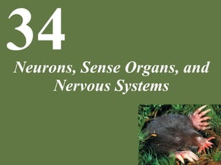
  • 1. Neurons, Sense Organs, and Nervous Systems 34
  • 2. Chapter 34 Neurons, Sense Organs, and Nervous Systems Key Concepts 34.1 Nervous Systems Are Composed of Neurons and Glial Cells 34.2 Neurons Generate Electric Signals by Controlling Ion Distributions 34.3 Neurons Communicate with Other Cells at Synapses 34.4 Sensory Processes Provide Information on an Animal’s External Environment and Internal Status 34.5 Neurons Are Organized into Nervous Systems
  • 3. Chapter 34 Opening Question How might the star-nosed mole’s brain be specialized to process information from its nose?
  • 4. 34.1 Nervous Systems are Composed of Neurons and Glial Cells
  • 5. Concept 34.1 Nervous Systems Are Composed of Neurons and Glial Cells Animals need a way to transmit signals at high speeds from place to place within their bodies (e.g., to avoid danger). Mammalian neurons transmit signals at 20–100 meters per second.
  • 6. Concept 34.1 Nervous Systems Are Composed of Neurons and Glial Cells Neurons are excitable cells—they can generate and transmit electrical signals, called action potentials. Cell membranes ordinarily have electrical polarity: the outside is more positive than the inside. An impulse, or action potential, is a state of reversed polarity.
  • 7. Concept 34.1 Nervous Systems Are Composed of Neurons and Glial Cells In an excitable cell, an action potential generated at one point propagates over the whole membrane. The region of depolarization moves along the cell membrane, and the membrane is said to “conduct” the impulse.
  • 8. Concept 34.1 Nervous Systems Are Composed of Neurons and Glial Cells
  • 9. Concept 34.1 Nervous Systems Are Composed of Neurons and Glial Cells Neurons (nerve cells) are specially adapted to generate electric signals, usually in the form of action potentials. Neurons must make contact with target cells. Synapse: cell-to-cell contact point specialized for signal transmission A signal arrives at the synapse by way of the presynaptic cell and leaves by way of the postsynaptic cell.
  • 10. Concept 34.1 Nervous Systems Are Composed of Neurons and Glial Cells Most neurons have four regions: • Dendrites—carry signals to the cell body • Cell body—contains nucleus and most organelles • Axon—conducts action potentials away from the cell body; can be very long • Presynaptic axon terminals —make contact with other cells
  • 11. Figure 34.2 A Generalized Neuron
  • 12. Concept 34.1 Nervous Systems Are Composed of Neurons and Glial Cells A neuron is said to innervate (provide neural imput) the cells that the axon terminals contact. Axons of many neurons often travel together in bundles called nerves. Nerve refers only to axon bundles outside the brain and spinal cord. In the brain or spinal cord they are called tracts.
  • 13. Concept 34.1 Nervous Systems Are Composed of Neurons and Glial Cells Glial cells, or glia, are not excitable. They have several functions: • Help orient neurons toward their target cells during embryonic development • Provide metabolic support for neurons • Help regulate composition of extracellular fluids and perform immune functions • Assist signal transmission across synapses
  • 14. Concept 34.1 Nervous Systems Are Composed of Neurons and Glial Cells Oligodendrocytes are glia that insulate axons in the brain and spinal cord. Schwann cells insulate axons in nerves outside of these areas. The glial membranes form a nonconductive sheath called myelin. Myelin-coated axons are white matter and areas of cell bodies are gray matter.
  • 15. Figure 34.3 Electrically Insulating an Axon
  • 16. 34.2 Neurons Generate Electric Signals by Controlling Ion Distributions
  • 17. Click-and-Learn (HHMI) View this Click-and-Learn about Neurons Be sure to watch the embedded videos.
  • 18. Concept 34.2 Neurons Generate Electric Signals by Controlling Ion Distributions Current: flow of electric charges from place to place; in cells, current is based on flow of ions such as Na+ Voltage, or electrical potential difference exists if positive charges are concentrated in one place and negative charges are concentrated in a different place. Voltages produce currents because opposite charges attract and will move toward one another if given a chance.
  • 19. Concept 34.2 Neurons Generate Electric Signals by Controlling Ion Distributions No voltage differences exist within open solutions such as the intracellular fluids. Voltage differences exist only across membranes such as the cell membrane.
  • 20. Concept 34.2 Neurons Generate Electric Signals by Controlling Ion Distributions
  • 21. Concept 34.2 Neurons Generate Electric Signals by Controlling Ion Distributions The voltage across a membrane is called membrane potential and is easily measured. Resting neuron: membrane potential is the resting potential, typically –60 to –70 millivolts (mV) • Negative sign means the inside of the cell is electrically negative relative to the outside.
  • 22. Figure 34.4 Measuring the Membrane Potential
  • 23. Concept 34.2 Neurons Generate Electric Signals by Controlling Ion Distributions Membrane potential can change rapidly, and only relatively small numbers of positive charges need to move through the membrane for this change of membrane potential to occur. Composition of the bulk solutions (the intra- and extracellular fluids) does not change.
  • 24. Concept 34.2 Neurons Generate Electric Signals by Controlling Ion Distributions Ion redistribution occurs through membrane channel proteins and ion transporters in the membrane. Sodium–potassium pump—uses energy from ATP to move 3 Na+ ions to the outside and 2 K+ to the inside; establishes concentration gradients of these ions
  • 25. Concept 34.2 Neurons Generate Electric Signals by Controlling Ion Distributions Potassium channels (K+ ) are open in the resting membrane. K+ ions diffuse out of the cell through leak channels and leave behind negative charges within the cell. K+ ions diffuse back into the cell because of the negative electrical potential. At this equilibrium point, there is no net movement of K+ ; called the equilibrium potential of K+ .
  • 26. Concept 34.2 Neurons Generate Electric Signals by Controlling Ion Distributions Diffusion of ions is controlled by concentration effect and electrical effect. When they are equal, electrochemical equilibrium is reached.
  • 27. Concept 34.2 Neurons Generate Electric Signals by Controlling Ion Distributions Electrochemical equilibrium is called the equilibrium potential of the ion, calculated by the Nernst equation:
  • 28. Concept 34.2 Neurons Generate Electric Signals by Controlling Ion Distributions Most ion channels are “gated”—they open and close under certain conditions. Most are closed in a resting neuron, which is why K+ leak channels determine resting membrane potential. • Voltage-gated channels open or close in response to changes in membrane potential • Stretch-gated channels respond to tension applied to cell membrane • Ligand-gated channels open or close when a specific chemical (ligand) binds to the channel protein.
  • 29. Figure 34.5 Three Types of Gated Ion Channels (Part 1) • Voltage-gated channels open or close in response to changes in membrane potential
  • 30. Figure 34.5 Three Types of Gated Ion Channels (Part 2) • Stretch-gated channels respond to tension applied to cell membrane
  • 31. Figure 34.5 Three Types of Gated Ion Channels (Part 3) • Ligand-gated channels open or close when a specific chemical (ligand) binds to the channel protein.
  • 32. Concept 34.2 Neurons Generate Electric Signals by Controlling Ion Distributions Opening and closing gated channels can alter membrane potential. If Na+ channels open, Na+ diffuses into the neuron because it is more concentrated outside the cell, and the cell membrane is more negative on the inside. When membrane becomes less negative on the inside, the membrane is depolarized. The membrane is hyperpolarized if the charge on the inside becomes more negative.
  • 33. Concept 34.2 Neurons Generate Electric Signals by Controlling Ion Distributions Membrane potential can be graded or all-or-none. Graded membrane potentials are changes from the resting potential that are less than the threshold of –50 mV. • Graded means any value of the membrane potential is possible • Caused by various ion channels opening or closing • Only spread a short distance
  • 34. Concept 34.2 Neurons Generate Electric Signals by Controlling Ion Distributions Graded membrane potentials spread only a short distance.
  • 35. Concept 34.2 Neurons Generate Electric Signals by Controlling Ion Distributions If neuron depolarizes to the –50 mV threshold, an all-or-none event occurs: an action potential is generated. Action potentials are not graded (always the same size) and do not become smaller, they stay the same in size as they propagate along the cell membrane.
  • 36. Figure 34.6 Graded and All-or-None Changes in Membrane Potential A membrane potential of -50 mV or greater isneeded to start and action potential. Action potentials stay thesamesize asthey move along the membrane.
  • 37. Concept 34.2 Neurons Generate Electric Signals by Controlling Ion Distributions Graded changes can give rise to all-or- none changes by being summed together; provides a mechanism for integrating signals. A key area for this integration is the axon hillock, where action potentials are most often generated. Graded changes resulting from multiple signals reaching the dendrites, spread to the axon hillock, where all the depolarizations and hyperpolarizations sum (add together).
  • 38. Concept 34.2 Neurons Generate Electric Signals by Controlling Ion Distributions An action potential (nerve impulse) is a rapid, large change in membrane potential that reverses membrane polarity. The membrane depolarizes from –65 mV at rest to about +40 mV (depolarization). It is localized and brief but is propagated with no loss of size—an action potential at one location causes currents to flow that depolarize neighboring regions.
  • 39. Concept 34.2 Neurons Generate Electric Signals by Controlling Ion Distributions When membrane potential reaches threshold, many voltage-gated Na+ channels open quickly, and Na+ rushes into the axon. The influx of positive ions (Na+ ) causes more depolarization, and an action potential occurs.
  • 40. Figure 34.7 Production of an Action Potential (Part 3)
  • 41. Concept 34.2 Neurons Generate Electric Signals by Controlling Ion Distributions The axon quickly returns to resting potential: • Voltage-gated Na+ channels close • Voltage-gated K+ channels open slowly and stay open longer—K+ moves out
  • 42. Concept 34.2 Neurons Generate Electric Signals by Controlling Ion Distributions Positive feedback during depolarization: • When the membrane is partially depolarized, some Na+ channels open; as Na+ starts to diffuse into the cell, more depolarization occurs, opening more channels. • This continues until all voltage-gated Na+ channels open and maximum depolarization occurs.
  • 43. Figure 34.8 Positive Feedback Plays a Key Role in the Generation of an Action Potential Positive Feedback!
  • 44. Concept 34.2 Neurons Generate Electric Signals by Controlling Ion Distributions Action potential travels in only one direction: • After the action potential, Na+ channels cannot open again for a brief period (refractory period) and cannot depolarize. • Thus the action potential can only propagate in the direction of the axon terminals. Why can action potentials only travel in one direction?
  • 45. Concept 34.2 Neurons Generate Electric Signals by Controlling Ion Distributions Action potentials travel faster in larger diameter axons. Myelination by glial cells also increases speed of action potentials. The nodes of Ranvier are gaps where the axon is not covered by myelin. Action potentials are generated only at the nodes and jump from node to node (saltatory conduction).
  • 46. Concept 34.2 Neurons Generate Electric Signals by Controlling Ion Distributions
  • 47. Concept 34.3 Neurons Communicate with Other Cells at Synapses Neurons communicate with other neurons or target cells at synapses. Chemical synapse: a very narrow space between cells (synaptic cleft) that an action potential cannot cross • When an action potential arrives at the end of the presynaptic cell, a neurotransmitter is released that diffuses across the space.
  • 48. Concept 34.3 Neurons Communicate with Other Cells at Synapses
  • 49. Concept 34.3 Neurons Communicate with Other Cells at Synapses Neurotransmitters diffuse across the synaptic cleft very rapidly (short distance). They bind to receptors on the postsynaptic cell membrane, which generates another action potential or other change. Neurotransmitters are quickly removed from the cleft— to end signal transmission—by enzymatic breakdown, uptake by other neurons or glial cells, or reuptake by the presynaptic cell.
  • 50. Concept 34.3 Neurons Communicate with Other Cells at Synapses Electrical synapse: cells are joined by gap junctions where the cytoplasm is continuous; signals cross with essentially no delay • They occur where very fast, invariant signal transmission is needed, such as neurons that control escape swimming in some fish. • Also occur where many cells must be stimulated to act together, such as fish electric organs.
  • 51. Concept 34.3 Neurons Communicate with Other Cells at Synapses Neuromuscular junctions: chemical synapses between motor neurons and skeletal muscle cells. The axon of the presynaptic cell branches close to the muscle cell, creating several axon terminals (boutons) that synapse with the muscle cell.
  • 52. Figure 34.9 Synaptic Transmission at a Vertebrate Neuromuscular Junction (Part 1)
  • 53. Figure 34.9 Synaptic Transmission at a Vertebrate Neuromuscular Junction (Part 2)
  • 54. Concept 34.3 Neurons Communicate with Other Cells at Synapses An action potential causes voltage-gated Ca+ channels to open in the presynaptic membrane, allowing Ca+ to flow in. This induces release of the neurotransmitter acetylcholine (ACh) (one of the most common neurotransmitters in vertebrates and invertebrates – can be inhibitory or excitatory): • ACh is stored in vesicles that fuse with the cell membrane to release ACh into the cleft by exocytosis.
  • 55. Figure 34.9 Synaptic Transmission at a Vertebrate Neuromuscular Junction (Part 3)
  • 56. Concept 34.3 Neurons Communicate with Other Cells at Synapses ACh diffuses across the cleft and binds to receptors on the postsynaptic cell. These receptors allow Na+ and K+ to flow through, and the increase in Na+ depolarizes the membrane. If it reaches threshold, more Na+ voltage-gated channels are activated and an action potential is generated.
  • 57. Concept 34.3 Neurons Communicate with Other Cells at Synapses Three categories of neurotransmitters: • Amino acids—glutamate, glycine, and γ-aminobutyric acid (GABA) • Biogenic amines include acetylcholine, dopamine, epinephrine, norepinephrine, and serotonin  Active in the CNS and PNS • A variety of peptides (strings of amino acids)  Active in the brain There are also gases such as nitric oxide and carbon monoxide  Are local regulators in the PNS
  • 58. Concept 34.3 Neurons Communicate with Other Cells at Synapses In the brain, a postsynaptic neuron may have chemical synapses with hundreds or thousands of presynaptic neurons, which may use different neurotransmitters. Receptors for a given neurotransmitter on the postsynaptic cell may be of different types with different actions. This complexity in synapse function helps explain the complexity of brain function.
  • 59. Figure 34.10 Synapses on a Single Postsynaptic Neuron in the Brain of a Mouse
  • 60. Concept 34.3 Neurons Communicate with Other Cells at Synapses Neurotransmitter receptors: • Ionotropic receptors are ligand-gated ion channels —cause changes in ion movement; response is fast and short-lived. • Metabotropic receptors are G protein-linked receptors that produce second messengers that induce signaling cascades; responses are slower and longer-lived.
  • 61. Concept 34.3 Neurons Communicate with Other Cells at Synapses Synapses Excitatory synapses produce graded membrane depolarizations called excitatory postsynaptic potentials (EPSPs); shift membrane potential towards threshold. Inhibitory synapses shift membrane potential away from threshold; produce graded membrane hyperpolarizations called inhibitory postsynaptic potentials (IPSPs).
  • 62. Concept 34.3 Neurons Communicate with Other Cells at Synapses Each EPSP or IPSP is usually less than 1 mV, and disappears in 10–20 milliseconds. They are graded potentials, typically produced at synapses on dendrites and the cell body. They affect membrane potential at the axon hillock, where action potentials are generated. Summation of the graded potentials is both temporal (must be present at the same time), and spatial.
  • 63. Figure 34.11 A Postsynaptic Neuron Carries Out Spatial Summation
  • 64. Concept 34.3 Neurons Communicate with Other Cells at Synapses The postsynaptic cell sums the excitatory and inhibitory input. Summation determines whether the postsynaptic cell produces action potentials. If the sum of EPSPs and IPSPs at the axon hillock is great enough to reach threshold, an action potential is produced.
  • 65. Concept 34.3 Neurons Communicate with Other Cells at Synapses Synaptic plasticity: synapses in an individual can undergo long-term changes in functional properties and physical shape during the individual’s lifetime. This may be one of the major mechanisms of learning. • Experiences at one time in life produce long-term changes in synapses, so that future experiences are processed by the nervous system in altered ways.
  • 66. Concept 34.3 Neurons Communicate with Other Cells at Synapses Sea hares (mollusks) pull their gills inside when certain parts of the body are touched:
  • 67. Concept 34.3 Neurons Communicate with Other Cells at Synapses They withdraw their gills more vigorously if they have previously been exposed to a noxious agent (sensitization). The synapses between the sensory neurons and the motor neurons for gill withdrawal are functionally strengthened—more neurotransmitter is released per impulse. The postsynaptic cell is thus excited to a greater degree.
  • 68. Concept 34.3 Neurons Communicate with Other Cells at Synapses In mammals, the hippocampus is associated with spatial learning and memory formation.
  • 69. Concept 34.3 Neurons Communicate with Other Cells at Synapses In studies of mice brains, when a circuit is repeatedly stimulated, the postsynaptic structures physically grow and the synapses strengthen functionally. The postsynaptic receptor molecules increase, increasing response. Synaptic plasticity has been shown to depend on second messengers, altered protein synthesis, and altered gene transcription.
  • 70. Concept 34.4 Sensory Processes Provide Information on an Animal’s External Environment and Internal Status Animals need information about their external environments to move, locate food, find mates, and avoid danger. They also need information regarding internal conditions, such as partial pressure of O2 and CO2 in the blood, and tension in contracting muscles.
  • 71. Concept 34.4 Sensory Processes Provide Information on an Animal’s External Environment and Internal Status Sensory receptor cells: neurons specialized for sensory transduction—transforming the energy of a stimulus into an electric signal. The electric signal generates action potentials. Action potentials convey the sensory information to the brain or other areas of the nervous system. Sensory receptor cells are highly specific in the stimuli to which they respond.
  • 72. Concept 34.4 Sensory Processes Provide Information on an Animal’s External Environment and Internal Status Sensory receptor proteins: membrane proteins in sensory receptor cells that initially detect a stimulus They produce graded membrane potentials (receptor potentials). Receptor cell membranes are often modified to have a large surface area, such as microvilli, cilia, or folding. This allows more receptor molecules and greater sensitivity.
  • 73. Concept 34.4 Sensory Processes Provide Information on an Animal’s External Environment and Internal Status Cone cells in vertebrate eyes have highly folded membranes with great numbers of photoreceptor molecules.
  • 74. Concept 34.4 Sensory Processes Provide Information on an Animal’s External Environment and Internal Status Ionotropic receptor cells: receptor proteins are typically stimulus-gated Na+ channels. Stimulus opens the channel, Na+ moves in, and receptor potential is generated. Metabotropic receptor cells are typically G protein-linked receptors; activation leads to change in a second messenger. This can directly or indirectly produce a receptor potential.
  • 75. Figure 34.12 Sensory Receptor Cells Are Ionotropic or Metabotropic
  • 76. Concept 34.4 Sensory Processes Provide Information on an Animal’s External Environment and Internal Status In the brain, different regions receive and process information from different sensory systems. Examples: • Axons from the eyes travel to the visual cortex • Axons from the inner ear travel to the auditory cortex Intensity of sensations is coded by the frequency of the action potentials.
  • 77. Concept 34.4 Sensory Processes Provide Information on an Animal’s External Environment and Internal Status Mechanoreceptors respond to mechanical distortion of the cell membrane; most are ionotropic. Stretch receptor cells in muscles respond when the muscle contracts.
  • 78. Concept 34.4 Sensory Processes Provide Information on an Animal’s External Environment and Internal Status Stretch receptors are specialized neurons. When cell membranes of the dendrites are stretched, Na+ channels open and produce a graded receptor potential that spreads to the axon hillock. Action potentials are generated if depolarization is above threshold.
  • 79. Figure 34.13 Stimulating a Crayfish Stretch Receptor Cell Produces Electric Signals
  • 80. Concept 34.4 Sensory Processes Provide Information on an Animal’s External Environment and Internal Status Stretch receptors in the biceps help adjust the muscle’s strength of contraction to match the load the muscle must sustain. Receptors in the muscle detect how much the muscle is being lengthened by a load; this information is used to keep the arm stable as the load increases.
  • 81. Figure 34.14 Negative Feedback: Sensory Signals from Stretch Receptors Enable the Nervous System to Control Muscle Contraction
  • 83. Concept 34.4 Sensory Processes Provide Information on an Animal’s External Environment and Internal Status Smell The sense of smell (olfaction) involves metabotropic receptors. Chemicals detected by smell are odorants. The sensory cells are chemoreceptors. In mammals, the receptors are in the lining of the nasal cavity. Axons extend to the olfactory bulb in the brain. Odorants must be dissolved in the liquid mucus to be detected.
  • 84. Concept 34.4 Sensory Processes Provide Information on an Animal’s External Environment and Internal Status When an odorant binds to a receptor protein, it activates a G protein, which activates a second messenger. The second messenger opens ion channels and an influx of Na+ depolarizes the olfactory neuron.
  • 85. Concept 34.4 Sensory Processes Provide Information on an Animal’s External Environment and Internal Status Different receptor cells have different G proteins that bind different odorants. With tens of thousands of olfactory receptor cells—each with one type of receptor protein—a wide range of odorants can be detected. The combination of olfactory cells stimulated by a particular odorant is unique to that odorant. The brain can interpret the pattern of signals as pointing to a particular smell.
  • 86. Concept 34.4 Sensory Processes Provide Information on an Animal’s External Environment and Internal Status Some animals have extremely specific olfactory receptor cells that are extremely sensitive. Some male moths have thousands of receptors on their antennae that respond to pheromones emitted by the females. They can detect a female when the pheromone level is as low as 0.000000000000001% of the air molecules.
  • 87. Concept 34.4 Sensory Processes Provide Information on an Animal’s External Environment and Internal Status
  • 89. Concept 34.4 Sensory Processes Provide Information on an Animal’s External Environment and Internal Status Hearing Auditory systems use mechanoreceptors to sense sound pressure waves. Many hearing organs have a membrane that moves in and out when sound pressure waves hit it. In mammals, this is the tympanic membrane (ear drum).
  • 90. Concept 34.4 Sensory Processes Provide Information on an Animal’s External Environment and Internal Status The middle ear is an air-filled cavity with three bones (ossicles)—the malleus, incus, stapes. They transmit vibrations of the tympanic membrane to another membrane, the oval window. The oval window connects to the cochlea, a coiled, fluid-filled tube where sound energy is transduced into electric signals.
  • 91. Figure 34.15 The Human Ear (Part 1)
  • 92. Figure 34.15 The Human Ear (Part 2)
  • 93. Concept 34.4 Sensory Processes Provide Information on an Animal’s External Environment and Internal Status The cochlea has three parallel canals separated by membranes. The basilar membrane changes in width and stiffness over its length. Different pitches, or frequency of vibration, cause different regions of the basilar membrane to oscillate.
  • 94. Figure 34.15 The Human Ear (Part 3)
  • 95. Figure 34.16 Frequencies in Sound Pressure Waves Are Transduced into Signals from Distinctive Sets of Hair Cells (Part 1)
  • 96. Figure 34.16 Frequencies in Sound Pressure Waves Are Transduced into Signals from Distinctive Sets of Hair Cells (Part 2)
  • 97. Concept 34.4 Sensory Processes Provide Information on an Animal’s External Environment and Internal Status The organ of Corti, on the basilar membrane, has mechanoreceptor cells called hair cells. Hair cells have projections called stereocilia that project into the semi-rigid tectorial membrane. When the basilar membrane moves, the stereocilia are bent. Hair cells transduce the bending motions of the stereocilia into electric signals and synapse with neurons that produce action potentials.
  • 98. Figure 34.15 The Human Ear (Part 4)
  • 100. Concept 34.4 Sensory Processes Provide Information on an Animal’s External Environment and Internal Status Photoreceptors—receptor cells sensitive to light Photoreceptors are metabotropic; the receptor proteins are all in the family of pigments called rhodopsins, which act as G protein– linked receptors. Rhodopsin consists of the protein opsin and a light-absorbing group, 11-cis-retinal.
  • 101. Concept 34.4 Sensory Processes Provide Information on an Animal’s External Environment and Internal Status When 11-cis-retinal absorbs light, it changes to the isomer all-trans-retinal, which changes the conformation of opsin.
  • 102. Concept 34.4 Sensory Processes Provide Information on an Animal’s External Environment and Internal Status An advantage of metabotropic control is that signals can be amplified. With large numbers of rhodopsin molecules and strong amplification, some photoreceptor cells undergo a measurable change in membrane potential in response to just a single photon of light.
  • 103. Concept 34.4 Sensory Processes Provide Information on an Animal’s External Environment and Internal Status Vertebrates have image-forming eyes, like a camera.
  • 104. Concept 34.4 Sensory Processes Provide Information on an Animal’s External Environment and Internal Status Connective tissue forms the transparent cornea on front of eye. Iris (pigmented)—controls amount of light reaching photoreceptors; opening is the pupil Lens—crystalline protein, focuses image, can change shape for focusing Retina—photosensitive layer at the back of the eye
  • 105. Concept 34.4 Sensory Processes Provide Information on an Animal’s External Environment and Internal Status The retina has several types of neurons including the photoreceptors (rods and cones). Rods and cones have large membrane surface area and many rhodopsin molecules. In humans, rods outnumber cones. Rods are more sensitive to light; important in dim light situations. Cones provide color vision.
  • 106. Figure 34.17 Rods and Cones
  • 107. Concept 34.4 Sensory Processes Provide Information on an Animal’s External Environment and Internal Status Humans have three types of cone cells with slightly different opsin molecules—they absorb different wavelengths of light. This allows the brain to interpret input from the different cones as a full range of color.
  • 108. Concept 34.4 Sensory Processes Provide Information on an Animal’s External Environment and Internal Status Rod and cone cells only produce graded membrane potentials (not action potentials). When stimulated, they hyperpolarize—the opposite of other sensory cells responding to stimuli. In darkness, Na+ channels are open and Na+ continually enters the cells. When stimulated, the 2nd messenger closes the channels, and inside of membrane becomes more negative.
  • 109. Figure 34.18 A Rod Cell Responds to Light (Part 1)
  • 110. Figure 34.18 A Rod Cell Responds to Light (Part 2)
  • 111. Concept 34.4 Sensory Processes Provide Information on an Animal’s External Environment and Internal Status The effect of the hyperpolarization is graded, depending on light intensity. Rod and cone cells synapse with other neurons in the retina and relay signals to them by graded neurotransmitter release.
  • 112. Concept 34.4 Sensory Processes Provide Information on an Animal’s External Environment and Internal Status The retina has four types of integrating neurons arranged in layers. Rods and cones synapse with bipolar cells, which synapse with ganglion cells. Horizontal cells and amacrine cells communicate laterally within the retina. Ganglion cells are the only ones that produce action potentials. Their axons converge to form the optic nerves.
  • 113. Figure 34.19 The Human Retina
  • 114. Concept 34.4 Sensory Processes Provide Information on an Animal’s External Environment and Internal Status Each ganglion cell has a receptive field—a defined, circular field formed by the cells from which it receives signals. Different ganglion cells are excited by light falling on the center versus the periphery of the receptive field. This enables ganglion cells to communicate information to the brain about visual patterns such as spots, edges, and areas of contrast.
  • 115. Concept 34.4 Sensory Processes Provide Information on an Animal’s External Environment and Internal Status About 1–2% of ganglion cells are photosensitive. The receptor protein is believed to be melanopsin. They provide information on the presence of light and its intensity. This information is relayed to the suprachiasmatic nuclei, where it is used to entrain the master circadian biological clock. It also regulates pupil size so they are large in dim light and small in bright light.
  • 116. Concept 34.4 Sensory Processes Provide Information on an Animal’s External Environment and Internal Status Arthropods have compound eyes consisting of units called ommatidia. Each ommatidium has a lens to focus light onto photoreceptor cells containing rhodopsin. Each ommatidium points in a slightly different direction. The more ommatidia, the higher the image resolution. Fast-flying predators such as dragonflies have up to 30,000.
  • 117. Figure 34.20 Ommatidia: The Functional Units of Arthropod Eyes
  • 118. Concept 34.4 Sensory Processes Provide Information on an Animal’s External Environment and Internal Status Some animals have evolved unusual sensory abilities to sense information such as electric fields or Earth’s magnetic field. Some animals perceive their environment very differently than humans do, for example, hearing sound frequencies that we cannot hear, or seeing electromagnetic wavelengths that we cannot see.
  • 119. Concept 34.4 Sensory Processes Provide Information on an Animal’s External Environment and Internal Status Our eyes can see wavelengths between 0.4 and 0.7 μm. Infrared wavelengths—longer than 0.7 μm Ultraviolet wavelengths—shorter than 0.4 μm Some snakes can see infrared wavelengths, detecting warmth in darkness, which helps them find prey.
  • 120. Concept 34.4 Sensory Processes Provide Information on an Animal’s External Environment and Internal Status Pit vipers have infrared sensing organs in pits near the eyes.
  • 121. Concept 34.4 Sensory Processes Provide Information on an Animal’s External Environment and Internal Status Many animals can see ultraviolet wave- lengths—bees, some birds. Male and female cedar waxwings look alike to us, but researchers have discovered that the birds themselves can tell the difference, with UV wavelengths.
  • 122. Concept 34.4 Sensory Processes Provide Information on an Animal’s External Environment and Internal Status Some animals can sense electric fields. Some fish produce weak electric fields and detect distortions in these fields caused by objects in their environment. The fish can perceive objects even in complete darkness.
  • 123. Concept 34.5 Neurons Are Organized into Nervous Systems The cnidarians have nerve nets, the most simple type of nervous system.
  • 124. Concept 34.5 Neurons Are Organized into Nervous Systems Evolution of nervous systems followed two major trends: • Centralization—integrating neurons became clustered together in centralized organs (e.g., brain and spinal cord). • Cephalization—major integrating areas became concentrated toward the anterior end of the animal (head). The anterior end meets the environment first; sensory organs are also concentrated there.
  • 125. Figure 34.21 The Organization of Vertebrate and Arthropod Nervous Systems
  • 127. Concept 34.5 Neurons Are Organized into Nervous Systems Central nervous system (CNS)—brain and spinal cord Composed mostly of integrating neurons and glial cells; it must interact with sensors and effectors. Effectors are cells or tissues that perform actions, that “carry out orders,” such as muscle cells.
  • 128. Concept 34.5 Neurons Are Organized into Nervous Systems Peripheral nervous system (PNS)— neurons outside the CNS Sensors bring sensory information from sense organs to the CNS and carry orders from the CNS to effectors. Nerves are bundles of axons in the PNS.
  • 129. Concept 34.5 Neurons Are Organized into Nervous Systems Kinds of Neurons Interneurons—neurons confined to the CNS Sensory neurons—sensory receptor cells or neurons that carry signals from sensory cells to the CNS (afferent neurons) Efferent neurons—convey signals from the CNS to muscles or other effectors Motor neurons—carry signals to skeletal muscles
  • 130. Concept 34.5 Neurons Are Organized into Nervous Systems Autonomic nervous system (ANS)—controls effectors other than skeletal muscles (autonomic effectors) Controls smooth muscle in organs, exocrine glands, some endocrine glands, acid secreting cells in the stomach, and other effectors. ANS has 3 divisions: • Enteric • Sympathetic • Parasympathetic
  • 131. Concept 34.5 Neurons Are Organized into Nervous Systems Vertebrate ANS (Autonomic nervous system) has three divisions: • Enteric division—nerve cells internal to the gut wall • Sympathetic division prepares the body for emergencies—“fight or flight” • Parasympathetic division slows the heart, lowers blood pressure and increases digestion
  • 132. Figure 34.22 The Autonomic Nervous System
  • 133. Concept 34.5 Neurons Are Organized into Nervous Systems Autonomic efferent pathways begin with preganglionic neurons with cell bodies in the CNS. Axons of preganglionic neurons synapse on a second neuron outside the CNS in a collection of nerve cell bodies called a ganglion. The second neuron is postganglionic—its axon leaves the ganglion and synapses in the target organs.
  • 134. Concept 34.5 Neurons Are Organized into Nervous Systems Parasympathetic preganglionic neurons exit the CNS from the brain and sacral region of the spinal cord. • The ganglia are near the target organs. Sympathetic preganglionic neurons exit the CNS at the thoracic and lumbar regions of the spinal cord. • Most of the ganglia lie next to the spinal cord.
  • 135. Concept 34.5 Neurons Are Organized into Nervous Systems Sympathetic postganglionic neurons use norepinephrine as the neurotransmitter. Parasympathetic postganglionic neurons use acetylcholine. In organs that receive both inputs, the target cells usually respond in opposite ways.
  • 136. Concept 34.5 Neurons Are Organized into Nervous Systems The sympathetic and parasympathetic divisions often work in opposition; acting together, they can adjust effector functions up or down as needed. Example: Sympathetic stimulation of the pace maker causes the heart rate to increase, and parasympathetic stimulation causes it to decrease.
  • 137. Concept 34.5 Neurons Are Organized into Nervous Systems The fight-or-flight response is an effect of the sympathetic division. When activated, it increases the heart rate, force of contraction, and cardiac output; it dilates lung passageways and increases release of glucose from the liver. At the same time, it reduces less urgent activities such as digestion.
  • 138. Concept 34.5 Neurons Are Organized into Nervous Systems Many neurons that control skeletal muscles enter or leave the CNS in spinal nerves. Spinal nerves have both sensory and motor neurons.
  • 139. Figure 34.23 Spinal Nerves Connect to the Spinal Cord at Regular Intervals
  • 140. Concept 34.5 Neurons Are Organized into Nervous Systems Spinal reflex—afferent information converts to efferent activity without going through the brain The knee-jerk reflex: • Stretch receptors in the patellar tendon send action potentials to the spinal cord. • The sensory neuron synapses with a motor neuron, sending an action potential to the leg muscle.
  • 141. Figure 34.24 The Spinal Cord Coordinates the Knee-Jerk Reflex
  • 143. Concept 34.5 Neurons Are Organized into Nervous Systems Vertebrate brains have three main regions: forebrain, midbrain, and hindbrain. The brain and spinal cord must pass through the medulla oblongata, the most posterior part of the hindbrain. • This area has changed little over the course of vertebrate evolution.
  • 144. Figure 34.25 Evolution of the Vertebrate Brain
  • 145. Concept 34.5 Neurons Are Organized into Nervous Systems In contrast, the cerebral hemispheres have undergone dramatic changes. • They are important in carrying out high- order sensory, motor, and integrative functions. The evolution of enhanced functionality in mammals and birds has gone hand in hand with large increases in the numbers of neurons and larger brain size.
  • 146. Concept 34.5 Neurons Are Organized into Nervous Systems However, it is important to note that some animals with small brains exhibit stunning behavioral capacities. In humans, all available evidence indicates that individual intelligence is not correlated with individual brain size.
  • 147. Concept 34.5 Neurons Are Organized into Nervous Systems Cerebral cortex—outer-most layer of the cerebral hemispheres, with many cell bodies • It is folded into convolutions, which increases its size. The left side of the body is served mostly by the right side of the brain, and vice versa. In each cerebral hemisphere, specific regions are specialized to carry out specific sensory and motor functions.
  • 148. Figure 34.26 The Human Left Cerebral Hemisphere (Part 1)
  • 149. Figure 34.26 The Human Left Cerebral Hemisphere (Part 2)
  • 150. Concept 34.5 Neurons Are Organized into Nervous Systems Combined sensory and motor functions often occur in localized brain areas. Imaging methods such as PET allow us to visualize areas where neurons exhibit increased electrical activity correlated with specific activities, such as language functions.
  • 151. Figure 34.27 Imaging Techniques Reveal Active Parts of the Brain
  • 152. Concept 34.5 Neurons Are Organized into Nervous Systems Functional magnetic resonance imaging (fMRI) is another technique to pinpoint brain activity. Example: In a person experiencing fear, increased activity is seen in the amygdala in the forebrain. Even memories of frightening situations can activate the amygdala.
  • 153. Figure 34.28 Source of the Fear Response
  • 154. Concept 34.5 Neurons Are Organized into Nervous Systems Parts of the brain that serve various anatomical regions of the body are physically related to each other in ways that mirror the rest of the body. Example: Map of the somatosensory (“body sensing”) part of the cerebral cortex • The size of each body part in the drawing reflects the amount of cortical area devoted to the part.
  • 155. Figure 34.29 The Body Is Represented in Maplike Ways in the Primary Somatosensory and Primary Motor Parts of the Cerebral Cortex
  • 156. Answer to Opening Question Large areas of a star-nosed mole’s cerebral cortex are devoted to processing sensory information from the tentacles of its nose. A map of the cortical region receiving sensory input from all 11 tentacles shows that it exactly mirrors the arrangement of the tentacles. How might the star-nosed mole’s brain be specialized to process information from its nose?
  • 157. Figure 34.30 Representation of the Nose of the Star-nosed Mole in the Cerebral Cortex