

Recommended
More Related Content
What's hot
What's hot (20)
Similar to Anatomy of larynx
Similar to Anatomy of larynx (20)
More from Malarvizhi R
More from Malarvizhi R (20)
Recently uploaded
Recently uploaded (20)
Anatomy of larynx
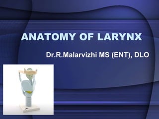
- 1. ANATOMY OF LARYNX Dr.R.Malarvizhi MS (ENT), DLO
- 2. The VOICE BOX • Lies in front of hypopharynx • Opp 3-6th cervical vertebra • Moves vertically and anteroposterior direction during swallowing
- 3. Embrology
- 4. Laryngeal framework • Cartilages • Muscles • Membranes • Joints
- 5. Laryngeal Cartilages Paired • Arytenoid • Cuneiform/Wrisberg • Corniculate / Santorini • Epiglottis • Thyroid • Cricoid
- 6. Laryngeal Cartilages Hyaline • Thyroid • Cricoid • Most of Arytenoid Fibroelastic • Epiglottis • Corniculate • Cuneiform • Tip of Arytenoid
- 7. Thyroid Cartilage • Largest • 2 alae and 2 pairs of superior and inferior cornua • Two alae meet anteriorly forming an angle – Male 90 ° – Female 120° • Vocal folds are attached to the middle of thyroid angle
- 8. Cricoid Cartilage • ONLY complete ring • Anteriorly narrow arch and posteriorly broad lamina
- 9. Arytenoid Cartilage • Paired • Pyramidal shape • Base articulates with cricoid cartilage • Muscular process attached with intrinsic laryngeal muscles • Vocal process attachment to vocal fold • Apex supports corniculate cartilage
- 10. Epiglottis cartilage • Leaf shaped • Yellow, elastic • Forms anterior wall of larynx • Covers inlet • Attached to hyoid by hyoepiglottic ligament – infra hyoid and suprahyoid epiglottis • Petiole
- 11. Corniculate and Cuneiform • Paired • Corniculate- articulates with apex of arytenoid • Cuneiform – rod shaped, situated in aryepiglottic fold, passive support to fold
- 12. Joints • Cricoarytenoid joint- – Synovial – Base of arytenoid, a facet, upper border of cricoid lamina – 2 movements- • Rotatory- arytenoid cartilage moves around vertical axis- abducting and adducting vocal fold • Gliding – one arytenoid glides towards or away from other- closing or opening glottis • Cricothyroid joint – Synovial, – Inferior cornua of thyroid cartilage with facet on cricoid cartilage
- 13. Laryngeal Membranes Extrinsic • Thyrohyoid- – pierced by superior laryngeal vessles – internal laryngeal nerve • Cricothyroid • Cricotracheal Intrinsic • Cricovocal (CONUS ELASTICUS) • Quandrangular
- 16. Muscles of Larynx Intrinsic • Act on vocal folds – Abductors • Posterior Cricoarytenoid – Adductors • Lateral Cricoarytenoid • Interarytenoid (Transverse) • thyroarytenoid – Tensors • Cricothyroid • Vocalis • Act on laryngeal inlet – Openers • Thyroarytenoid (Thyroepiglottic) – Closers • Interarytenoid (Oblique) • Aryepiglottic (posterior oblique part of interarytenoid) Extrinsic • Elevator – Primary (THYROID) • Stylopharyngeus • Salpingopharyngeus • Palatopharyngeus • Thyrohyoid – Secondary (HYOID) • Mylohyoid • Digastric • Stylohyoid • Geniohyoid • Depressors – Sternohyoid – Sternothyroid – Omohyoid
- 22. Cavity of Larynx • Starts at inlet communicating with pharynx • Ends lower borders of cricoid cartilage continuous with lumen of trachea – Inlet – Vestibule – Ventricle – Subglottic space – Vestibular and vocal folds – Rima glottidis
- 23. • Inlet – Oblique opening – Anteriorly by free margin of epiglottis, aryepiglottic folds laterally and posteriorly by interarytenoid fold • Vestibule – Between laryngeal inlet and vestibular folds – Anteriorly by posterior surface of epiglottis, laterally by aryepiglottic folds and posteriorly by mucous membrane over arytenoids
- 24. • Ventricle (Sinus of larynx) – Deep elliptical space – Between vestibular and vocal folds – Extending short distance above and lateral to vestibular folds – Saccule – diverticulum of mucous membrane starting from anterior part of ventricular cavity and extends between vestibular folds and lamina of thyroid cartilage (oil can of larynx) • Laryngocele
- 25. • Subglottic space – Extends from vocal fold to lower border of cricoid cartilage • Rima glottidis – Glottis proper; elongated space between vocal folds anteriorly & vocal process and base of arytenoids posteriorly – Anteroposteriorly • 24 mm in Males • 16 mm in Females – Narrowest part – Size and shape varies – Antr 2/3rd membranous cords and Postr 1/3rd by vocal process of arytenoids
- 29. VOCAL FOLDS • Vocal folds (True) – Pearly sharp bands – Middle of thyroid angle to vocal process of arytenoids – Vocal ligament – true upper edge of cricovocal membr covered by closely bound mucous membrane with scanty subepithelial connective tissue – Multilayered
- 30. VOCAL FOLDS continued • 5 layers (medial to lateral) – Stratified squamous epithelium – Superficial layer- Lamina propria – Middle layer- Lamina propria – Deep layer- Lamina propria – Vocalis muscle
- 31. • Vestibular folds (False) – Fold of mucous membr extending anteroposteriorly across laryngeal cavity – Vestibular ligament, few fibres of thyroarytenoid muscle and mucous glands
- 33. Mucous Membrane of Larynx • Lines larynx • Loosely attached except over posterior surface of epiglottis, vocal folds and corniculate and cuneiform cartilages • Ciliated columnar epithelium – Except over vocal folds and upper part of vestibule- Stratified squamous • Mucous glands – All over – More over posterior surf epiglottis, postr part of aryepiglottic folds and saccules – NONE OVER VOCAL FOLDS
- 34. Lymphatic Drainage • Vocal folds / glottis- nil • Supraglottic- drained by lymphatics piercing thyrohyoid membrane- reach upper deep cervical lymph nodes • Infraglottic – drained by lymphatics piercing cricothyroid membrane – pretracheal and prelaryngeal nodes- lower deep cervical nodes and mediastinal nodes
- 35. Nerve supply • Motor – All by Recurrent laryngeal nerve – Except CRICOTHYROID- External branch of superior laryngeal nerve • Sensory – vocal folds • Above – internal branch of superior laryngeal nerve • Below – recurrent laryngeal nerve
- 36. Nerve supply • Recurrent laryngeal nerve – Right – from VAGUS, level of subclavian artery hooks around it and then ascends b/w trachea and esophagus – Left from VAGUS, level arch of aorta in mediatinum, loops around it and ascends in neck of tracheo- osophageal groove • Longer course
- 37. Nerve supply • Superior laryngeal nerve – Inferior ganglion of VAGUS – Descends behind ICA and at level of greater cornua of hyoid divides 2 branches • External • Internal
- 38. Galen’s Anastamoses • Recurrent laryngeal nerve and Superior laryngeal nerve give off branches and anastamose with each other –Galen’s anastamoses
- 39. SPACES • Pre- epiglottic space – Boyer – Filled with fat , areolar tissue and some lymphatics – Anteriorly- upper part of thyroid cartilage – Superiorly- hyoepiglottic ligt – Posteriorly – infrahyoid epiglottis and quadrangular membrane – Laterlly – continuous paraglottic space
- 40. Spaces • Paraglottic Space – Tucker – Continuous with Boyer’s space – Anteriorly and laterally- thyroid cartilage – Supero medially- quadrangular membrane – Inferomedially – conus elasticus – Posteriorly- mucous membrane over pyriform fossa
- 41. Spaces • Reinke’s space – Potential space between epithelium of vocal fold and vocal ligt – Superficial layer of lamina propria – Bound anteriorly by Broyles’ ligt ; posteriorly by arytenoid cartilage and inferiorly by vocal ligt
- 42. Blood supply • Superior thyroid artery (branch of ECA) • Inferior thyroid artery (branch of Thyrocervical trunk)
- 43. SUBDIVISIONS OF LARYNX • Supraglottis – From infrahyoid portion of epiglottis to vocal folds • Glottis – Vocal folds, anterior and posterior commissures • Subglottis – Below vocal folds to lower border of cricoid cartilage (5mm )
- 46. Physiology • Protection of lower airways • Phonation • Respiration • Fixation of chest
- 47. Theories of phonation Myoelastic Aerodynamic theory Cover body theory