Glomus anatomy n intro
•
4 likes•876 views

a small intro to the big topic of Glomus in Otology

Recommended
Recommended
More Related Content
What's hot
What's hot (20)
Similar to Glomus anatomy n intro
Similar to Glomus anatomy n intro (20)
More from Malarvizhi R
More from Malarvizhi R (20)
Recently uploaded
Recently uploaded (20)
Glomus anatomy n intro
- 2. Intro • Valentin 1840 ▫ Swellings along tympanic nerve • Guild 1941 ▫ Classical description of “glomus jugularis”
- 3. • Glomus is a collection of ganglionic tissue within the Temporal bone in close relation with jugular bulb. • Arising from paraganglionic glomic tissue , distributed along parasympathetic nerves. • Composed of cells that are sensitive changes in arterial oxygen, CO2 & pH.
- 4. • Misnomer ▫ Glomus means a small circumscribed histological structure in which arterioles connect directly with veins • Synonyms ▫ Paraganglioma ▫ Chemodectoma ▫ Tympanic body tumors ▫ Receptoma ▫ Non-chromaffin paraganglioma ▫ Glomerocytoma
- 5. • Histologically BENIGN • Slow growing • Clinically morbid • Internal Carotid Artery • Hereditary • Female 4-6X more • Infants to older age group , commonly in 50s &60s
- 6. • Neural crest origin closely associated anatomically with autonomic ganglia • Diffuse NeuroEndocrine System ▫ ‘chief cells’ ▫ 40 different types of other cells ▫ APUD amine precursor uptake & decarboxylation Necessary step in biosynthesis of neurotransmitters Neuropeptides, catecholamines,neurohormaones , neurotransmitters, hormones and parahormones Functional tumors- 1-3%
- 7. • Paraganglioma of temporal bone are located along Jacobson’s nerve and are symmetric • Innervated by cranial nerves IX &X and supplied Ascending Pharyngeal Artery • Multicentric, Bilateral • Nonfamilial – 10% multicentric • Familial – 25-50% multicentric
- 8. • From middle ear promontory- GLOMUS TYMPANICUM • From jugular foramen – GLOMUS JUGULARE • From perineurium of vagus extending towards jugular foramen- GLOMUS VAGALE
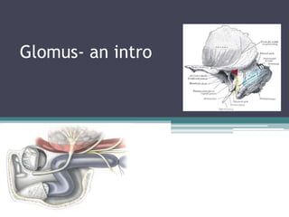
- 9. • In Temporal bone Glomus bodies are in close relation to ▫ Tympanic branch of Glossopharyngeal nerve ▫ Auricular branch of vagus nerve ▫ Adventitita of jugular bulb ▫ Promontory
- 10. Relevant Anatomy • Jugular Bulb • Middle ear cavity • Vagus nerve • Carotid body • Jugular foramen
- 13. Jugular foramen • 2nd largest skull base foramen • Asymmetrical • Vital structures- vascular and neural • Lateral part sigmoid shaped tunnel • Medial part canal in cephalocaudal direction
- 14. • Located behind carotid canal • Bounded ▫ anteriorly by petrous portion of temporal bone ▫ Posteriorly by occipital bone
- 15. • Delineated by jugular spine • Smaller anteromedial ‘pars nervosa’ • Larger posterolateral ‘ pars vascularis’
- 16. • Is large elongated with long axis forwards and medially • Posterior end of hollowed to form JUGULAR FOSSA lodging the superior bulb of internal jugular vein • Fossa large on right • Lateral wall pierced by minute canal, mastoid canaliculus • Medial end possess jugular notch, apex of which opening leading to cochlear canaliculus • Tympanic canaliculus opens on or near thin edge of bone between jugular fossa and lower end of carotid canal
- 17. Structures passing • Anterior part ▫ Inferior petrosal sinus ▫ Meningeal branch of ascending pharyngeal artery • Middle part ▫ IX,X & XI cranial verves • Posterior part ▫ Internal jugular vein ▫ Meningeal branch of Occipital artery Inferior ganglion of glossopharyngeal nerve is lodged in the glossopharyngeal notch near medial end
- 18. Infra Temporal Fossa • The infratemporal fossa lies below the middle cranial fossa, between the ramus of the mandible and the lateral wall of the pharynx • The 'roof' is the infratemporal area of the skull base, which comprises the greater wing of the sphenoid with a small triangular contribution posteriorly from the squamous temporal bone. • .
- 19. • It has ▫ no anatomical floor and continues down into the neck. ▫ Anteriorly lies the posterior wall of the maxilla with the pterygomaxillary and inferior orbital fissures; • Posteriorly, it is bounded by the carotid sheath and styloid apparatus. • The fossa is limited ▫ medially by the medial pterygoid muscle and interpterygoid fascia, and ▫ laterally by the mandible. • The contents of the fossa are the ▫ lateral and medial pterygoid muscles, the maxillary artery and its branches, ▫ the pterygoid venous plexus and maxillary veins and the ▫ branches of the mandibular nerve
- 22. Spread • Growth is by direct extension along lines of least resistance or by ischemic necrosis • Multiple pathways lead intracranially • Incidence of malignant transformation 4%
- 24. • Glomus jugulare, tend to spread around the venous sinuses surrounding the jugular bulb. • Sometimes collectively referred to as the “danger zone,” the venous sinuses most commonly involved include ▫ the inferior petrosal sinus, ▫ internal jugular vein, and ▫ the sigmoid sinus
- 25. • Extend toward and beyond the protympanum, hypotympanum, mesotympanum, or intracranial cavity • From protympanum – ▫ peritubal cell tracts toward the petrous apex, ▫ the carotid canal toward the middle cranial fossa, or ▫ the Eustachian tube toward the nasopharynx
- 26. • From hypotympanum - luminal invasion of either ▫ the sigmoid sinus or ▫ internal jugular vein or ▫ spread toward the inferior petrosal sinus or ▫ neural foramina at the skull base ▫ Carotid sheath lends access to neck
- 27. • From mesotympanum -similar spread as cholesteatoma (antrum and epitympanum or the facial recess, sinus tympani, or mastoid air cells) • Laterally through the tympanic membrane-- external auditory canal, • Medially through the round window and erode the cochlea and internal auditory canal