![International Journal of Trend in Scientific Research and Development @ www.ijtsrd.com eISSN: 2456-6470
@ IJTSRD | Unique Paper ID – IJTSRD53851 | Volume – 7 | Issue – 1 | January-February 2023 Page 1241
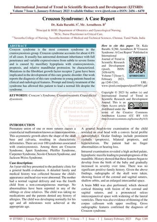
Figure 1: crouzon syndrome Brain MRI
Discussion:
Crouzon syndrome is an inherited disorder, also
known as brachial arch syndrome. This syndrome
affects the embryonic first humeral arch, the
progenitor of the maxilla and mandible. In its early
stages, the syndrome was called "craniofacial
dysostosis". This disorder is characterized by three
features: craniosynostosis, midfacial hypoplasia, and
proptosis. Premature closure of cranial sutures causes
abnormal growth of the skull, affecting growth and
development of the orbital and maxillary complexes.
The cause is due to mutations in the fibroblast growth
factor receptor 2 (FGFR2) gene, which maps to
chromosomal locus 10q25-10q26, although FGFR-2
(Crouzon syndrome) and FGFR3 (Crouzon syndrome
with hyperepidermal hyperplasia) Locus
heterogeneity due to causative mutations is shown.
nigricans) in various patients. The most common
ocular abnormalities are shallow orbit, proptosis,
orbital schisis, strabismus, papilledema, optic nerve
atrophy, exposure keratitis, and decreased vision.
Rarely, nystagmus, iris coloboma, aniridia,
anisocoria, microcornea, macrocornea, cataract,
ectopia lentis, blue sclera, glaucoma, and dislocation
have also occurred.
Treatment is multidisciplinary and multistage surgery
is recommended. Early craniectomy with frontal
advancement is most commonly indicated to prevent
or treat increased intracranial pressure. If necessary,
midface replacement and maxillofacial surgery can be
performed to provide adequate orbital volume, reduce
proptosis, and correct the occlusion to proper
functional position. Prognosis is limited to
malformations depends on the severity.
Conclusion:
Early detection, patient education and timely
intervention are critical steps in the management of
this syndrome. Early initiation of a multidisciplinary
approach improves survival in children with Crouzon
syndrome and also helps subjects overcome social
stigma, as subjects with Crouzon syndrome have a
near-normal lifespan.
Conflict of Interest
None
Funding
None
Consent for publication
Informed consent was obtained from the parents of
the patients to publish this case in medical journal.
References:
[1] Cohen MM jr, Krelborg S. Birth prevalence
studies of the Crouzon’s Syndrome.
Comparison of direct and indirect methods.
Clin Genet 1992; 41:12-5.
[2] Vivek Padmanabhan, Amitha M. Hegde,
Kavitha Rao. Crouzon’s Syndrome: A review
of literature and case report. Contemporary
Clinical Dentistry; Jul-Sep 2011; Vol 2; Issue
3; 211 – 214.
[3] Ahmed I, Afzal A. Diagnosis and evaluation of
Crouzon syndrome. J Coll Physicians Surg Pak
2009; 19(5):318-20.
[4] Bowling EL, Burstein FD. Crouzon syndrome.
Optometry 2006; 77:217-22.
[5] Posnick JC, Ruiz RL. The craniofacial
dysostosis syndromes: current surgical thinking
and future directions. Cleft Palate Craniofac J.
2000; 37:433.](data:image/gif;base64,R0lGODlhAQABAIAAAAAAAP///yH5BAEAAAAALAAAAAABAAEAAAIBRAA7)
Recommended
More Related Content
Similar to Crouzon Syndrome A Case Report
Similar to Crouzon Syndrome A Case Report (15)
More from ijtsrd
More from ijtsrd (20)
Recently uploaded
Recently uploaded (20)
Crouzon Syndrome A Case Report
- 1. International Journal of Trend in Scientific Research and Development (IJTSRD) Volume 7 Issue 1, January-February 2023 Available Online: www.ijtsrd.com e-ISSN: 2456 – 6470 @ IJTSRD | Unique Paper ID – IJTSRD53851 | Volume – 7 | Issue – 1 | January-February 2023 Page 1240 Crouzon Syndrome: A Case Report Dr. Kala Barathi. S1 , Mr. Azrudheen. B2 1 Principal & HOD, Department of Obstetrics and Gynecological Nursing, 2 M.Sc, Nurse Practitioner in Critical Care, 1,2 Saveetha College of Nursing, Saveetha Institute of Medical and Technical Sciences, Chennai, Tamil Nadu, India ABSTRACT Crouzon syndrome is the most common syndrome in the craniosynostosis group. Crouzon syndrome accounts for about 4.8% of all cases. It usually has autosomal dominant inheritance with full penetrance and variable expressiveness from subtle to severe forms and is caused by maxillary hypoplasia with craniosynostosis, proptosis, and relative mandibular protrusion. be characterized. Mutations in the fibroblast growth factor receptor 2 gene have been implicated in the development of this rare genetic disorder. Our work reports the diagnosis of this rare syndrome in young patients based on clinical and radiological features. Prompt and timely treatment of the syndrome has allowed this patient to lead a normal life despite the syndrome. KEYWORDS: Crouzon’s Syndrome, Craniosynostosis, Craniofacial dysostosis How to cite this paper: Dr. Kala Barathi. S | Mr. Azrudheen. B "Crouzon Syndrome: A Case Report" Published in International Journal of Trend in Scientific Research and Development (ijtsrd), ISSN: 2456-6470, Volume-7 | Issue-1, February 2023, pp.1240-1241, URL: www.ijtsrd.com/papers/ijtsrd53851.pdf Copyright © 2023 by author (s) and International Journal of Trend in Scientific Research and Development Journal. This is an Open Access article distributed under the terms of the Creative Commons Attribution License (CC BY 4.0) (http://creativecommons.org/licenses/by/4.0) INTRODUCTION Premature union of one or more sutures causes a craniofacial malformation known as craniosynostosis. This asymmetric growth alters the shape of the skull base and calvaria, resulting in characteristic deformities. There are over 100 syndromes associated with craniosynostosis. Among them are Crouzon Syndrome, Apert Syndrome, Pfeiffer Syndrome, Carpenter Syndrome, Sessle-Chotzen Syndrome, and Jackson-Weiss Syndrome. Case description: An 1year-old boy presented to the pediatric clinic for a general health evaluation. A detailed family and medical history was collected because the child's appearance and head size were abnormal. The mother had a normal vaginal delivery and was the fourth child from a non-consanguineous marriage. No abnormalities have been reported in any of the siblings or close relatives. The child was taking no medications and the parents denied any history of allergies. The child was developing normally for his age and all milestones were achieved at the appropriate age. A general head-to-toe examination of the child revealed an oval head with a convex facial profile (acrocephaly). Ocular findings include prominent eyeballs (proptosis), bilateral proptosis, and hypertelorism. The patient had no finger abnormalities or hearing loss. Intraoral examination revealed a high arched palate, hypoplastic maxilla leading to pseudoprognosis of the mandible. History showed that these features began to develop from the birth of the baby and gradually increased in severity over time. There was no significant positive family history. Given the above findings, radiographs of the skull were taken, showing fusion of the coronal and sagittal sutures, shallow orbits, and an enlarged intercanthal space. A brain MRI was also performed, which showed cortical thinning with fusion of the coronal and sagittal sutures, and a pronounced sulcus interconjunctival space with widening of the lateral ventricles. There was also evidence of thinning of the corpus callosum with upper swelling. Gross radiographic features confirmed the diagnosis of Crouzon syndrome. IJTSRD53851
- 2. International Journal of Trend in Scientific Research and Development @ www.ijtsrd.com eISSN: 2456-6470 @ IJTSRD | Unique Paper ID – IJTSRD53851 | Volume – 7 | Issue – 1 | January-February 2023 Page 1241 Figure 1: crouzon syndrome Brain MRI Discussion: Crouzon syndrome is an inherited disorder, also known as brachial arch syndrome. This syndrome affects the embryonic first humeral arch, the progenitor of the maxilla and mandible. In its early stages, the syndrome was called "craniofacial dysostosis". This disorder is characterized by three features: craniosynostosis, midfacial hypoplasia, and proptosis. Premature closure of cranial sutures causes abnormal growth of the skull, affecting growth and development of the orbital and maxillary complexes. The cause is due to mutations in the fibroblast growth factor receptor 2 (FGFR2) gene, which maps to chromosomal locus 10q25-10q26, although FGFR-2 (Crouzon syndrome) and FGFR3 (Crouzon syndrome with hyperepidermal hyperplasia) Locus heterogeneity due to causative mutations is shown. nigricans) in various patients. The most common ocular abnormalities are shallow orbit, proptosis, orbital schisis, strabismus, papilledema, optic nerve atrophy, exposure keratitis, and decreased vision. Rarely, nystagmus, iris coloboma, aniridia, anisocoria, microcornea, macrocornea, cataract, ectopia lentis, blue sclera, glaucoma, and dislocation have also occurred. Treatment is multidisciplinary and multistage surgery is recommended. Early craniectomy with frontal advancement is most commonly indicated to prevent or treat increased intracranial pressure. If necessary, midface replacement and maxillofacial surgery can be performed to provide adequate orbital volume, reduce proptosis, and correct the occlusion to proper functional position. Prognosis is limited to malformations depends on the severity. Conclusion: Early detection, patient education and timely intervention are critical steps in the management of this syndrome. Early initiation of a multidisciplinary approach improves survival in children with Crouzon syndrome and also helps subjects overcome social stigma, as subjects with Crouzon syndrome have a near-normal lifespan. Conflict of Interest None Funding None Consent for publication Informed consent was obtained from the parents of the patients to publish this case in medical journal. References: [1] Cohen MM jr, Krelborg S. Birth prevalence studies of the Crouzon’s Syndrome. Comparison of direct and indirect methods. Clin Genet 1992; 41:12-5. [2] Vivek Padmanabhan, Amitha M. Hegde, Kavitha Rao. Crouzon’s Syndrome: A review of literature and case report. Contemporary Clinical Dentistry; Jul-Sep 2011; Vol 2; Issue 3; 211 – 214. [3] Ahmed I, Afzal A. Diagnosis and evaluation of Crouzon syndrome. J Coll Physicians Surg Pak 2009; 19(5):318-20. [4] Bowling EL, Burstein FD. Crouzon syndrome. Optometry 2006; 77:217-22. [5] Posnick JC, Ruiz RL. The craniofacial dysostosis syndromes: current surgical thinking and future directions. Cleft Palate Craniofac J. 2000; 37:433.