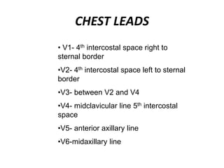
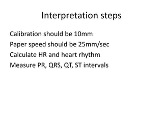
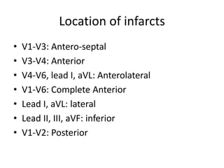
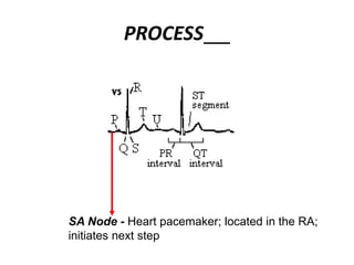
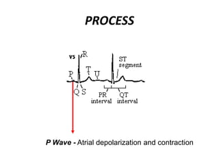
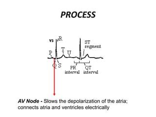
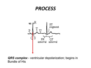
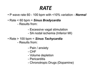
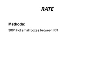
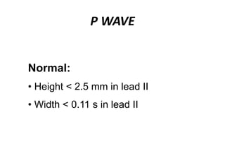
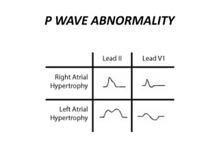
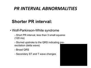
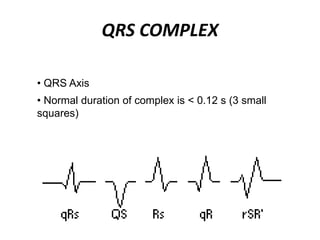
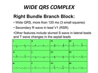
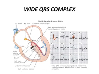
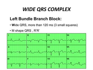
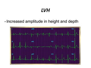
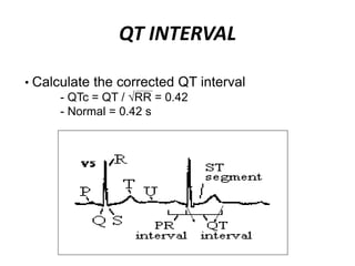
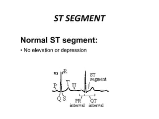
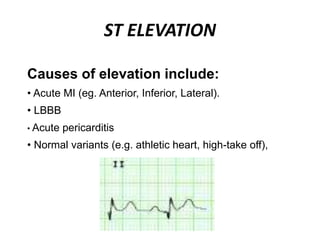
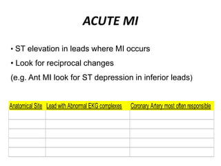
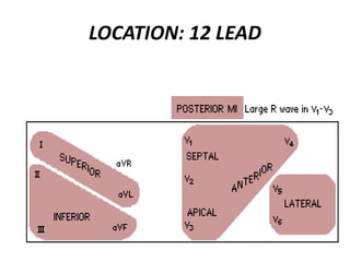
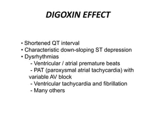
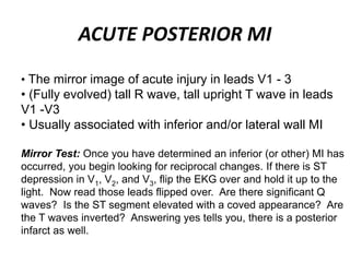
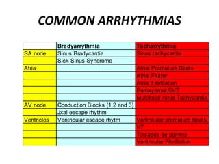
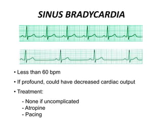
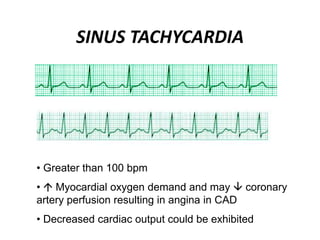
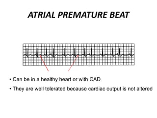
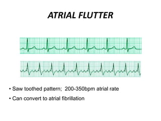
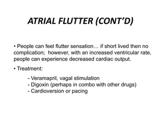
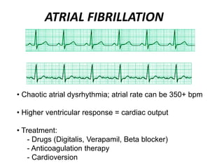
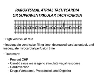
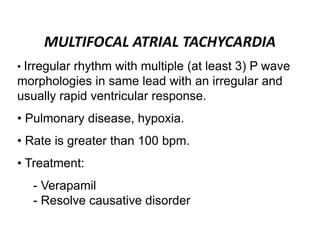
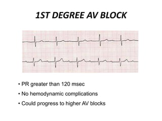
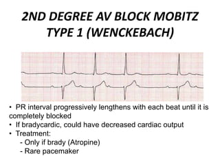
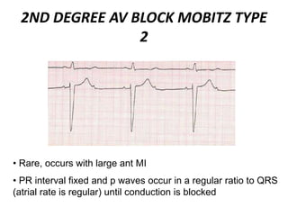
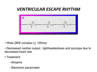
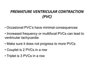
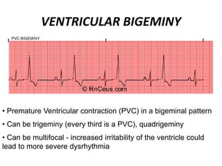
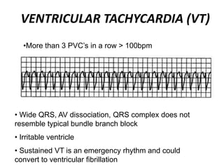
This document provides information on electrocardiograms (ECGs), including ECG paper layout, lead placement, interpretation steps, and how to locate infarcts based on ECG findings. Common arrhythmias are summarized, including definitions and treatments for sinus bradycardia, atrial fibrillation, heart blocks, premature ventricular contractions, ventricular tachycardia, and more. Overall health impacts and treatments are discussed for various rhythms.
2. ECG paper
• Vertical dark lines- 5mm apart
• Vertical light lines- 1mm apart
• Horizontal dark lines-5mm apart
• Horizontal light lines- 1mm apart
• Large square- 5 X 5 mm
• Small square- 1 X 1 mm
3. LEAD PLACEMENT
• Limb Leads - 6 in all
- I, II, III, aVL, aVR, aVF
• Chest leads - 6 in all
-V1,V2,V3,V4,V5,V6
Standard 12-lead ECG:
5. CHEST LEADS
• V1- 4th intercostal space right to
sternal border
•V2- 4th intercostal space left to sternal
border
•V3- between V2 and V4
•V4- midclavicular line 5th intercostal
space
•V5- anterior axillary line
•V6-midaxillary line
7. Location of infarcts
• V1-V3: Antero-septal
• V3-V4: Anterior
• V4-V6, lead I, aVL: Anterolateral
• V1-V6: Complete Anterior
• Lead I, aVL: lateral
• Lead II, III, aVF: inferior
• V1-V2: Posterior
14. VENTRICULAR DEPOLARIZATION
• Q Wave - 1st downward wave of the
complex
• R Wave - 1st upward wave of the complex
• S Wave - downward wave preceded by an
upward wave
22. P WAVE ABNORMALITIES
Right atrial hypertrophy:
• A P wave in lead II taller then 2.5 mm
(2.5 small squares)
• The P wave is usually pointed
23.
24. P WAVE ABNORMALITIES
Left atrial abnormality (dilatation
or hypertrophy):
• M shaped P wave in lead II
• Prominent terminal negative component to
P wave in lead V1
26. PR INTERVAL ABNORMALITIES
Shorter PR interval:
• Wolf-Parkinson-White syndrome
- Short PR interval, less than 3 small squares
(120 ms)
- Slurred upstroke to the QRS indicating pre-
excitation (delta wave)
- Broad QRS
- Secondary ST and T wave changes
28. QRS COMPLEX
• QRS Axis
• Normal duration of complex is < 0.12 s (3 small
squares)
29. WIDE QRS COMPLEX
Right Bundle Branch Block:
• Wide QRS, more than 120 ms (3 small squares)
• Secondary R wave in lead V1 (RSR)
•Other features include slurred S wave in lateral leads
and T wave changes in the septal leads
33. WIDE QRS
- Small or absent P waves
- Atrial fibrillation
- Wide QRS
- Shortened or absent ST segment
- Wide, tall and tented T waves
- Ventricular fibrillation
Hyperkalemia:
• Changes that can be seen:
35. PATHOLOGICAL Q WAVES
• Q waves > 1mm
• Their depth > 25% of the height of the QRS
• Q waves in V6 and aVL (not pathological…small)
• Look for anatomical site, ignore aVR
Anatomical Site Lead with Abnormal EKG complexes Coronary Artery most often responsible
Inferior II, III, aVf RCA
Antero Septal V1-V2 LAD
Antero Apical V3-V4 LAD (distal)
Antero Lateral V5-V6, I, aVL CFX
Posterior V1-V2 (Tall R, Not Q) RCA
37. NON Q WAVE MI
• Not all MIs develop Q waves (up to 1/3 never do or
they develop and resolve)
• WHY?
• Infarct was not complete (transmural)
• Infarct occurred in a electrically “silent” area of
the heart, where an EKG cannot record the injury
• Acute Infarct (Q waves will eventually appear)
39. LEFT VENTRICULAR HYPERTROPHY
(LVH)
Sokolow + Lyon (Am Heart J, 1949;37:161)
S in V1+ R in V5 or V6 > 35 mm
Cornell criteria (Circulation, 1987;3: 565-72)
S in V3 + R in aVL > 28 mm in men
S in V3 + R in aVL > 20 mm in women
Framingham criteria (Circulation,1990; 81:815-820)
R in aVL > 11mm, R in V4-6 > 25mm
S in V1-3 > 25 mm,
S in V1 or V2 + R in V5 or V6 > 35 mm,
R in I + S in III > 25 mm
44. ST ELEVATION
Causes of elevation include:
• Acute MI (eg. Anterior, Inferior, Lateral).
• LBBB
• Acute pericarditis
• Normal variants (e.g. athletic heart, high-take off),
45. ACUTE MI
• ST elevation in leads where MI occurs
• Look for reciprocal changes
(e.g. Ant MI look for ST depression in inferior leads)
Anatomical Site Lead with Abnormal EKG complexes Coronary Artery most often responsible
Inferior II, III, aVf RCA
Antero Septal V1-V2 LAD
Antero Apical V3-V4 LAD (distal)
Antero Lateral V5-V6, I, aVL CFX
Posterior V1-V2 (Tall R, Not Q) RCA
48. ST DEPRESSION
Causes of depression include:
• Myocardial ischemia
• Digoxin Effect
• Ventricular Hypertrophy
• Acute Posterior MI
• Pulmonary Embolus
• LBBB
49. DIGOXIN EFFECT
• Shortened QT interval
• Characteristic down-sloping ST depression
• Dysrhythmias
- Ventricular / atrial premature beats
- PAT (paroxysmal atrial tachycardia) with
variable AV block
- Ventricular tachycardia and fibrillation
- Many others
50. ACUTE POSTERIOR MI
• The mirror image of acute injury in leads V1 - 3
• (Fully evolved) tall R wave, tall upright T wave in leads
V1 -V3
• Usually associated with inferior and/or lateral wall MI
Mirror Test: Once you have determined an inferior (or other) MI has
occurred, you begin looking for reciprocal changes. If there is ST
depression in V1, V2, and V3, flip the EKG over and hold it up to the
light. Now read those leads flipped over. Are there significant Q
waves? Is the ST segment elevated with a coved appearance? Are
the T waves inverted? Answering yes tells you, there is a posterior
infarct as well.
51. ST DEPRESSION
In diagnosis with ischemia:
• Looking for at least 1mm (1 square)
• This can be
1. Upsloping
2. Horizontal (can be combined w/ 1 or 3)
3. Downsloping
54. SMALL, FLATTENED OR INVERTED T
WAVES
Causes are plenty:
• Ischemia, age, race, hyperventilation, anxiety
• LVH, drugs, pericarditis, I-V conduction delay (RBBB),
• Electrolyte disturbances
• The most important thing to consider is INVERTED T waves
associated with Ischemia
56. SINUS BRADYCARDIA
• Less than 60 bpm
• If profound, could have decreased cardiac output
• Treatment:
- None if uncomplicated
- Atropine
- Pacing
57. SINUS TACHYCARDIA
• Greater than 100 bpm
• Myocardial oxygen demand and may coronary
artery perfusion resulting in angina in CAD
• Decreased cardiac output could be exhibited
58. SSS (SICK SINUS SYNDROME)
• Deceased cardiac output, related to
periods of excessive bradycardia, AV block
and/or tachycardia
• Treatment:
- Pacemaker
- Anti coagulation therapy
59. ATRIAL PREMATURE BEAT
• Can be in a healthy heart or with CAD
• They are well tolerated because cardiac output is not altered
60. ATRIAL FLUTTER
• Saw toothed pattern; 200-350bpm atrial rate
• Can convert to atrial fibrillation
61. ATRIAL FLUTTER (CONT’D)
• People can feel flutter sensation… if short lived then no
complication; however, with an increased ventricular rate,
people can experience decreased cardiac output.
• Treatment:
- Veramapril, vagal stimulation
- Digoxin (perhaps in combo with other drugs)
- Cardioversion or pacing
63. PAROXYSMAL ATRIAL TACHYCARDIA
OR SUPRAVENTRICULAR TACHYCARDIA
• High ventricular rate
• Inadequate ventricular filling time, decreased cardiac output, and
inadequate myocardial perfusion time
• Treatment:
- Prevent CHF
- Carotid sinus massage to stimulate vagal response
- Cardioversion
- Drugs (Verapamil, Propranolol, and Digoxin)
64. MULTIFOCAL ATRIAL TACHYCARDIA
• Irregular rhythm with multiple (at least 3) P wave
morphologies in same lead with an irregular and
usually rapid ventricular response.
• Pulmonary disease, hypoxia.
• Rate is greater than 100 bpm.
• Treatment:
- Verapamil
- Resolve causative disorder
65. 1ST DEGREE AV BLOCK
• PR greater than 120 msec
• No hemodynamic complications
• Could progress to higher AV blocks
66. 2ND DEGREE AV BLOCK MOBITZ
TYPE 1 (WENCKEBACH)
• PR interval progressively lengthens with each beat until it is
completely blocked
• If bradycardic, could have decreased cardiac output
• Treatment:
- Only if brady (Atropine)
- Rare pacemaker
67. 2ND DEGREE AV BLOCK MOBITZ TYPE
2
• Rare, occurs with large ant MI
• PR interval fixed and p waves occur in a regular ratio to QRS
(atrial rate is regular) until conduction is blocked
68. • Symptoms of decreased cardiac output occur with
slowing ventricular rate
- Could progress to complete block
• Treatment:
- Atropine initially
- Permanent pacemaker
2ND DEGREE AV BLOCK MOBITZ
TYPE 2 (CONT’D)
69. 3RD DEGREE AV BLOCK (COMPLETE)
• Atria and ventricles are independent of each other;
no relationship present
• Symptoms could include lightheadedness or syncope from
decreased rate
71. JUNCTIONAL ESCAPE RHYTHM
• QRS complexes are not preceded by normal P
waves, because the impulse originate below the SA
node
• Can cause a missed P wave, inverted or in the
QRS
72. VENTRICULAR ESCAPE RHYTHM
• Wide QRS complex (> 120ms)
• Decreased cardiac output , lightheadedness and syncope due to
decreased heart rate
• Treatment:
- Atropine
- Electronic pacemaker
73. PREMATURE VENTRICULAR CONTRACTION
(PVC)
• Occasional PVC’s have minimal consequences
• Increased frequency or multifocal PVCs can lead to
ventricular tachycardia
• Make sure it does not progress to more PVCs
• Couplet is 2 PVCs in a row
• Triplet is 3 PVCs in a row
74. VENTRICULAR BIGEMINY
• Premature Ventricular contraction (PVC) in a bigeminal pattern
• Can be trigeminy (every third is a PVC), quadrigeminy
• Can be multifocal - increased irritability of the ventricle could
lead to more severe dysrhythmia
75. VENTRICULAR TACHYCARDIA (VT)
•More than 3 PVC’s in a row > 100bpm
• Wide QRS, AV dissociation, QRS complex does not
resemble typical bundle branch block
• Irritable ventricle
• Sustained VT is an emergency rhythm and could
convert to ventricular fibrillation
76. VT
• Decreased cardiac output , irritable ventricle
• Treatment:
- Cardioversion
- Lidocaine or Procainamide to get NSR
- Emergent care
- Long term care: ICD (implantable
cardioverter defibrillator)
77. TORSADES DE POINTES
• Form of VT – “twisting of the points”
• People with prolonged QT interval are susceptible
78. VENTRICULAR FIBRILLATION
• Chaotic activity of the ventricles
• No effective cardiac output or coronary perfusion
• Associated with severe myocardial ischemia.
• Life-threatening - death occurs within 4 min.
• Treatment:
- Immediate defibrillation
- CPR
- Lidocaine, bretylium. epinephrine
80. How to read the ECG
– Look at the whole tracing.
Rhythm: Is there a P wave before each QRS complex?
− Yes: sinus rhythm No: AV junctional or heart block
Rate: Count boxes; use caliper, ruler
PR interval: Normal - 0.20 sec. or less
QRS complex: Skinny (0.10 sec. or less) or broad (BBB or
ventricular)
ST segment: Isoelectric (normal), elevated or depressed
T wave: Upright, flat or inverted
Interpretation: Normal or abnormal.
− Is the rhythm dangerous?