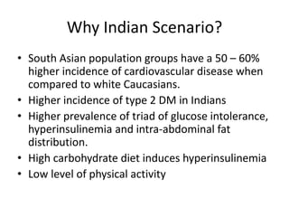
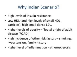
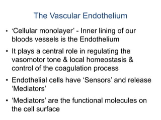
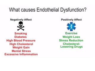
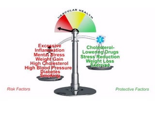
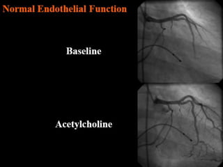
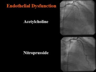
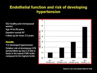
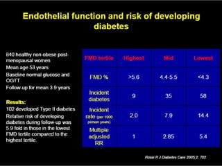
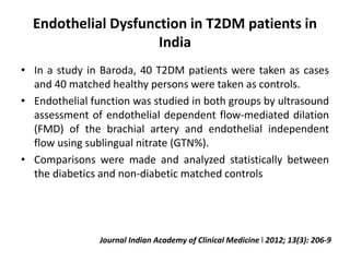
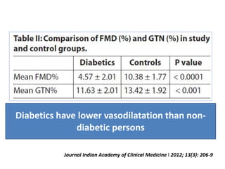
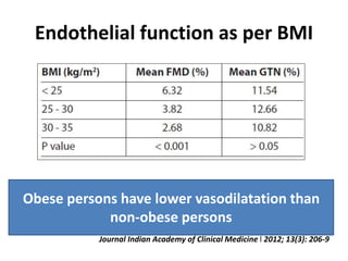
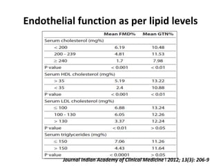
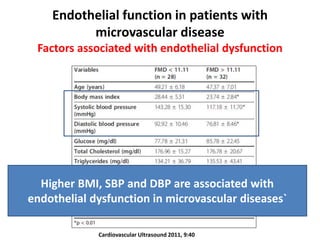
ENDOTHELIAL DYSFUNCTION IN
THE INDIAN SCENARIO
HeartSense Team
www.heartsense.in
Topics
• Why Indian Scenario?
• Describing endothelial dysfunction
• Measures of endothelial dysfunction
• Clinical relevance
• What the future holds...
Why Indian Scenario?
• South Asian population groups have a 50 – 60%
higher incidence of cardiovascular disease when
compared to white Caucasians.
• Higher incidence of type 2 DM in Indians
• Higher prevalence of triad of glucose intolerance,
hyperinsulinemia and intra-abdominal fat
distribution.
• High carbohydrate diet induces hyperinsulinemia
• Low level of physical activity
Why Indian Scenario?
• High levels of insulin resistance
• Low HDL (and high levels of small HDL
particles), high small dense LDL.
• Higher levels of obesity – ‘foetal origin of adult
disease (FOAD)’
• High incidence of other risk factors – smoking,
hypertension, family history
• Higher level of inflammation - atherosclerosis
• ‘Cellular monolayer’ - Inner lining of our
bloods vessels is the Endothelium
• It plays a central role in regulating the
vasomotor tone & local homeostasis &
control of the coagulation process
• Endothelial cells have ‘Sensors’ and release
‘Mediators’
• ‘Mediators’ are the functional molecules on
the cell surface
The Vascular Endothelium
(S)
The vascular endothelium serves multiple functions:
1) It regulates fluid and molecule traffic between blood and tissues
2) It is an anti-coagulant surface
3) It contributes to vascular homeostasis and repair
4) It plays a vital role in vascular tone and blood flow regulation ***
Endothelial cells serve multiple functions.
Assessing this function is the most practical way of measuring
endothelial function.
Molecular mechanisms of Endothelial Functions
In Health Dysfunction
Vasodilation Vasoconstriction
NO, PGI2, EDHF, BK,
C-NP
ROS, ET-1, TxA2, A-II
Thrombolysis Thrombosis
Platelet Disaggregation
NO, PGI2
Adhesion Molecules
CAMs, Selectins
Antiproliferation
NO, PGI2, TGF-, Hep
Growth Factors
ET-1, A-II, PDGF, bFGF, ILGF,
Interleukins
Lipolysis Inflammation
ROS, NF-B
PAI-1, TF, Tx-A2
tPA, Protein C, TF-I,
vonWF
LPL Vogel R
Coronary
Heart
Disease
Endothelial
Dysfunction
NO
↑Inflammation
↑Thrombosis
“Response-to-Injury” Hypothesis
Genes
Coronary
Risk
Factors
 O2 Endothelial Cells and
 H2O2 Vascular Smooth Muscle
Oxidative Stress: Endothelial
Dysfunction and CAD/Renal Risk
Factors
Endothelial Dysfunction
Apoptosis
Vasoconstriction
Leukocyte
adhesion
Lipid
deposition
ThrombosisVSMC
growth
Hypertension
SmokingDiabetes LDL Homocysteine Estrogen
deficiency
Endothelial and Vascular Dysfunction:
A “Barometer of Cardiovascular Risk”
• Marker of the inherent atherosclerotic risk
• An integrated index of both the overall CV risk
factor burden and the sum of all vasculo-
protective factors in an individual.
Aging Diet
Smoking Inactivity
Diabetes ↑ Cholesterol
↑ Blood Pressure Oxidative Stress
Genetics Medications
Clinical Methods for Assessing
Endothelium-Dependent Dilation
Coronary Arteries
• Epicardial Artery Diameter
 with ACh
• CBF  with ACh
• Epicardial Artery Diameter
 with Adenosine
Forearm
• Brachial Artery Diameter 
with Arterial Occlusion
• Forearm Blood Flow with
ACh
BASELINE POST OCCLUSION
Brachial Artery Ultrasound with FMD
http://www.cvphysiology.com/Blood%20Flow/BF006.htm
Reactive Hyperemia
Reactive hyperemia is the transient increase in organ blood flow that occurs following a
brief period of ischemia (e.g., arterial occlusion).
The left panel shows the effects of a 2 min arterial occlusion on blood flow. In this example, blood flow goes to zero
during arterial occlusion. When the occlusion is released, blood flow rapidly increases (i.e., hyperemia occurs) that lasts
for several minutes. The hyperemia occurs because during the period of occlusion, tissue hypoxia and a build up of
vasodilator metabolites (e.g., adenosine) dilate arterioles and decrease vascular resistance. Then when perfusion
pressure is restored (i.e., occlusion released), flow becomes elevated because of the reduced vascular resistance. During
the hyperemia, the tissue becomes reoxygenated and vasodilator metabolites are washed out of the tissue. This causes
the resistance vessels to regain their normal vascular tone, thereby returning flow to control.
Brachial Artery Flow-Mediated Vasodilation
Baseline 5 Minutes Post-Occlusion
Blood Pressure Cuff
Occlusion – 1 Minute
Release
3.1 mm 3.6 mm
Takese B, Am J Cardiol 1998:82:1535
Comparison of Brachial and Coronary
Flow-Mediated Vasodilation
Brachial artery FMD represents the
endothelial function of coronary artery
Effect of ACh-Induced Forearm Vasodilation* on 32-
Month CVE’s (%) in 225 Never Treated Hypertensive
Subjects
(* Relative Flow Increase)
0
1
2
3
4
5
6
7
8
Tertile 1 Tertile 2 Tertile 3
ACh FBF
%CVE's
Lower Ach induced vasodilatation is
associated with higher CV events
Perticone F et al, Circulation 2001;104:191
Murakami T et al. J Am Coll Cardiol 2001;37:294A
CVE’s over 4 Years in 480 Patients with Suspected
CAD According to Brachial Artery FMD
0%
5%
10%
15%
20%
25%
<4% 4%-8% >8%
CHD Events
CV Events
Endothelial dysfunction: Indian Scenario
• India has high prevalence of diseases associated with
endothelial dysfunction: CAD, DM, Hypertension, etc.
• Risk factors of endothelial dysfunction like smoking,
dyslipidemia are also very common in Indian
population.
• There are relatively less studies in Indian population
for direct assessment of endothelial function
Endothelial Dysfunction in T2DM patients in
India
• In a study in Baroda, 40 T2DM patients were taken as cases
and 40 matched healthy persons were taken as controls.
• Endothelial function was studied in both groups by ultrasound
assessment of endothelial dependent flow-mediated dilation
(FMD) of the brachial artery and endothelial independent
flow using sublingual nitrate (GTN%).
• Comparisons were made and analyzed statistically between
the diabetics and non-diabetic matched controls
Journal Indian Academy of Clinical Medicine l 2012; 13(3): 206-9
Diabetics have lower vasodilatation than non-
diabetic persons
Journal Indian Academy of Clinical Medicine l 2012; 13(3): 206-9
Endothelial function as per BMI
Obese persons have lower vasodilatation than
non-obese persons
Journal Indian Academy of Clinical Medicine l 2012; 13(3): 206-9
Endothelial function as per lipid levels
Journal Indian Academy of Clinical Medicine l 2012; 13(3): 206-9
Conclusion
• Diabetics have worse endothelial functions than
non diabetics
• Endothelial function was related to dyslipidemia
and obesity
Journal Indian Academy of Clinical Medicine l 2012; 13(3): 206-9
Endothelial function in patients with
microvascular disease
• Cardiac syndrome X (Csx) : presence of angina-like
chest pain, a positive stress test and angiographically
normal coronaries, considered as a microvascular
CAD
• In a study in Hyderabad, the brachial artery FMD was
measure in both endothelium dependent/
independent vasodilatation by high resolution
ultrasound in 30 cardiac syndrome X patients and
matched with 30 healthy control subjects
Cardiovascular Ultrasound 2011, 9:40
Endothelial function in patients with
microvascular disease
Results:
• Significantly decreased flow mediated dilatation was
observed in patients when compared to control (9.42
vs 21.11, p < 0.01)
• Total, 46% of subjects had endothelial dysfunction
and of them, Csx subjects had higher prevalence
(76% vs 16% p < 0.01) than control subjects.
Cardiovascular Ultrasound 2011, 9:40
Endothelial function in patients with
microvascular disease
Factors associated with endothelial dysfunction
Cardiovascular Ultrasound 2011, 9:40
Higher BMI, SBP and DBP are associated with
endothelial dysfunction in microvascular diseases`
Endothelial Senescence in premature
CAD patients in India
• Objective: To evaluate role of vascular senescence in
premature CAD (PCAD) by comparing numerical status and
senescence of circulating endothelial progenitor cells (EPCs) in
PCAD patients to controls.
• EPCs were measured by flow cytometry in 57 patients with
CAD, and 57 controls without evidence of CAD, recruited from
random patients ≤ 50 years at AIIMS, New Delhi.
• EPC senescence as determined by telomere length (EPC-TL)
and telomerase activity (EPC-TA) was studied by real time
polymerase chain reaction (PCR) and PCR– ELISA respectively.
Vemparala et al. BMC Cardiovascular Disorders 2013,
13:104
Endothelial Senescence in premature
CAD patients in India
Vemparala et al. BMC Cardiovascular Disorders 2013, 13:104
*Adjusted for age, sex, BMI, smoking and medication
Endothelial Senescence in premature
CAD patients in India
Conclusion
• There is an association between increased
endothelial cell senescence with PCAD in young
patients from India.
• This suggests that early accelerated vascular cell
senescence may play an important mechanistic role
in CAD epidemic in developing countries like India
Vemparala et al. BMC Cardiovascular Disorders 2013, 13:104
What does all this mean?
• Endothelial dysfunction is a precursor to most
cardiovascular risk factors and CVD.
• Methods to alter endothelial dysfunction can
help modify incidence of risk factors
• In vitro EPC modification and injection of
‘potent’ EPCs into coronary arteries is the way
forward.
Available research

Endothelial dysfunction in indian scenario

  • 1.
    ENDOTHELIAL DYSFUNCTION IN THEINDIAN SCENARIO HeartSense Team www.heartsense.in
  • 2.
    Topics • Why IndianScenario? • Describing endothelial dysfunction • Measures of endothelial dysfunction • Clinical relevance • What the future holds...
  • 3.
    Why Indian Scenario? •South Asian population groups have a 50 – 60% higher incidence of cardiovascular disease when compared to white Caucasians. • Higher incidence of type 2 DM in Indians • Higher prevalence of triad of glucose intolerance, hyperinsulinemia and intra-abdominal fat distribution. • High carbohydrate diet induces hyperinsulinemia • Low level of physical activity
  • 4.
    Why Indian Scenario? •High levels of insulin resistance • Low HDL (and high levels of small HDL particles), high small dense LDL. • Higher levels of obesity – ‘foetal origin of adult disease (FOAD)’ • High incidence of other risk factors – smoking, hypertension, family history • Higher level of inflammation - atherosclerosis
  • 5.
    • ‘Cellular monolayer’- Inner lining of our bloods vessels is the Endothelium • It plays a central role in regulating the vasomotor tone & local homeostasis & control of the coagulation process • Endothelial cells have ‘Sensors’ and release ‘Mediators’ • ‘Mediators’ are the functional molecules on the cell surface The Vascular Endothelium
  • 7.
    (S) The vascular endotheliumserves multiple functions: 1) It regulates fluid and molecule traffic between blood and tissues 2) It is an anti-coagulant surface 3) It contributes to vascular homeostasis and repair 4) It plays a vital role in vascular tone and blood flow regulation *** Endothelial cells serve multiple functions. Assessing this function is the most practical way of measuring endothelial function.
  • 9.
    Molecular mechanisms ofEndothelial Functions In Health Dysfunction Vasodilation Vasoconstriction NO, PGI2, EDHF, BK, C-NP ROS, ET-1, TxA2, A-II Thrombolysis Thrombosis Platelet Disaggregation NO, PGI2 Adhesion Molecules CAMs, Selectins Antiproliferation NO, PGI2, TGF-, Hep Growth Factors ET-1, A-II, PDGF, bFGF, ILGF, Interleukins Lipolysis Inflammation ROS, NF-B PAI-1, TF, Tx-A2 tPA, Protein C, TF-I, vonWF LPL Vogel R
  • 10.
  • 11.
     O2 EndothelialCells and  H2O2 Vascular Smooth Muscle Oxidative Stress: Endothelial Dysfunction and CAD/Renal Risk Factors Endothelial Dysfunction Apoptosis Vasoconstriction Leukocyte adhesion Lipid deposition ThrombosisVSMC growth Hypertension SmokingDiabetes LDL Homocysteine Estrogen deficiency
  • 13.
    Endothelial and VascularDysfunction: A “Barometer of Cardiovascular Risk” • Marker of the inherent atherosclerotic risk • An integrated index of both the overall CV risk factor burden and the sum of all vasculo- protective factors in an individual. Aging Diet Smoking Inactivity Diabetes ↑ Cholesterol ↑ Blood Pressure Oxidative Stress Genetics Medications
  • 17.
    Clinical Methods forAssessing Endothelium-Dependent Dilation Coronary Arteries • Epicardial Artery Diameter  with ACh • CBF  with ACh • Epicardial Artery Diameter  with Adenosine Forearm • Brachial Artery Diameter  with Arterial Occlusion • Forearm Blood Flow with ACh
  • 18.
    BASELINE POST OCCLUSION BrachialArtery Ultrasound with FMD
  • 19.
    http://www.cvphysiology.com/Blood%20Flow/BF006.htm Reactive Hyperemia Reactive hyperemiais the transient increase in organ blood flow that occurs following a brief period of ischemia (e.g., arterial occlusion). The left panel shows the effects of a 2 min arterial occlusion on blood flow. In this example, blood flow goes to zero during arterial occlusion. When the occlusion is released, blood flow rapidly increases (i.e., hyperemia occurs) that lasts for several minutes. The hyperemia occurs because during the period of occlusion, tissue hypoxia and a build up of vasodilator metabolites (e.g., adenosine) dilate arterioles and decrease vascular resistance. Then when perfusion pressure is restored (i.e., occlusion released), flow becomes elevated because of the reduced vascular resistance. During the hyperemia, the tissue becomes reoxygenated and vasodilator metabolites are washed out of the tissue. This causes the resistance vessels to regain their normal vascular tone, thereby returning flow to control.
  • 21.
    Brachial Artery Flow-MediatedVasodilation Baseline 5 Minutes Post-Occlusion Blood Pressure Cuff Occlusion – 1 Minute Release 3.1 mm 3.6 mm
  • 22.
    Takese B, AmJ Cardiol 1998:82:1535 Comparison of Brachial and Coronary Flow-Mediated Vasodilation Brachial artery FMD represents the endothelial function of coronary artery
  • 23.
    Effect of ACh-InducedForearm Vasodilation* on 32- Month CVE’s (%) in 225 Never Treated Hypertensive Subjects (* Relative Flow Increase) 0 1 2 3 4 5 6 7 8 Tertile 1 Tertile 2 Tertile 3 ACh FBF %CVE's Lower Ach induced vasodilatation is associated with higher CV events Perticone F et al, Circulation 2001;104:191
  • 24.
    Murakami T etal. J Am Coll Cardiol 2001;37:294A CVE’s over 4 Years in 480 Patients with Suspected CAD According to Brachial Artery FMD 0% 5% 10% 15% 20% 25% <4% 4%-8% >8% CHD Events CV Events
  • 29.
    Endothelial dysfunction: IndianScenario • India has high prevalence of diseases associated with endothelial dysfunction: CAD, DM, Hypertension, etc. • Risk factors of endothelial dysfunction like smoking, dyslipidemia are also very common in Indian population. • There are relatively less studies in Indian population for direct assessment of endothelial function
  • 30.
    Endothelial Dysfunction inT2DM patients in India • In a study in Baroda, 40 T2DM patients were taken as cases and 40 matched healthy persons were taken as controls. • Endothelial function was studied in both groups by ultrasound assessment of endothelial dependent flow-mediated dilation (FMD) of the brachial artery and endothelial independent flow using sublingual nitrate (GTN%). • Comparisons were made and analyzed statistically between the diabetics and non-diabetic matched controls Journal Indian Academy of Clinical Medicine l 2012; 13(3): 206-9
  • 31.
    Diabetics have lowervasodilatation than non- diabetic persons Journal Indian Academy of Clinical Medicine l 2012; 13(3): 206-9
  • 32.
    Endothelial function asper BMI Obese persons have lower vasodilatation than non-obese persons Journal Indian Academy of Clinical Medicine l 2012; 13(3): 206-9
  • 33.
    Endothelial function asper lipid levels Journal Indian Academy of Clinical Medicine l 2012; 13(3): 206-9
  • 34.
    Conclusion • Diabetics haveworse endothelial functions than non diabetics • Endothelial function was related to dyslipidemia and obesity Journal Indian Academy of Clinical Medicine l 2012; 13(3): 206-9
  • 35.
    Endothelial function inpatients with microvascular disease • Cardiac syndrome X (Csx) : presence of angina-like chest pain, a positive stress test and angiographically normal coronaries, considered as a microvascular CAD • In a study in Hyderabad, the brachial artery FMD was measure in both endothelium dependent/ independent vasodilatation by high resolution ultrasound in 30 cardiac syndrome X patients and matched with 30 healthy control subjects Cardiovascular Ultrasound 2011, 9:40
  • 36.
    Endothelial function inpatients with microvascular disease Results: • Significantly decreased flow mediated dilatation was observed in patients when compared to control (9.42 vs 21.11, p < 0.01) • Total, 46% of subjects had endothelial dysfunction and of them, Csx subjects had higher prevalence (76% vs 16% p < 0.01) than control subjects. Cardiovascular Ultrasound 2011, 9:40
  • 37.
    Endothelial function inpatients with microvascular disease Factors associated with endothelial dysfunction Cardiovascular Ultrasound 2011, 9:40 Higher BMI, SBP and DBP are associated with endothelial dysfunction in microvascular diseases`
  • 38.
    Endothelial Senescence inpremature CAD patients in India • Objective: To evaluate role of vascular senescence in premature CAD (PCAD) by comparing numerical status and senescence of circulating endothelial progenitor cells (EPCs) in PCAD patients to controls. • EPCs were measured by flow cytometry in 57 patients with CAD, and 57 controls without evidence of CAD, recruited from random patients ≤ 50 years at AIIMS, New Delhi. • EPC senescence as determined by telomere length (EPC-TL) and telomerase activity (EPC-TA) was studied by real time polymerase chain reaction (PCR) and PCR– ELISA respectively. Vemparala et al. BMC Cardiovascular Disorders 2013, 13:104
  • 39.
    Endothelial Senescence inpremature CAD patients in India Vemparala et al. BMC Cardiovascular Disorders 2013, 13:104 *Adjusted for age, sex, BMI, smoking and medication
  • 40.
    Endothelial Senescence inpremature CAD patients in India Conclusion • There is an association between increased endothelial cell senescence with PCAD in young patients from India. • This suggests that early accelerated vascular cell senescence may play an important mechanistic role in CAD epidemic in developing countries like India Vemparala et al. BMC Cardiovascular Disorders 2013, 13:104
  • 41.
    What does allthis mean? • Endothelial dysfunction is a precursor to most cardiovascular risk factors and CVD. • Methods to alter endothelial dysfunction can help modify incidence of risk factors • In vitro EPC modification and injection of ‘potent’ EPCs into coronary arteries is the way forward.
  • 42.