Chest x
•
1 like•696 views

The document summarizes several common chest x-ray abnormalities seen with pleural disease: 1. Pleural effusions appear as a collection of fluid in the pleural space that obscures structures like the costophrenic angle. A large left pleural effusion is seen with an underlying bronchogenic carcinoma in one example. 2. Pneumothoraces form when air is trapped in the pleural space, usually from trauma or underlying lung disease. A left pneumothorax is seen due to a rib fracture in one example. 3. Asbestos exposure can lead to benign calcified pleural plaques appearing on chest x-rays as irregular, well-defined shadows similar to holly

Recommended
Recommended
More Related Content
What's hot
What's hot (20)
Viewers also liked
Viewers also liked (12)
Similar to Chest x
Similar to Chest x (20)
More from Bs. Nhữ Thu Hà
More from Bs. Nhữ Thu Hà (20)
Recently uploaded
Recently uploaded (20)
Chest x
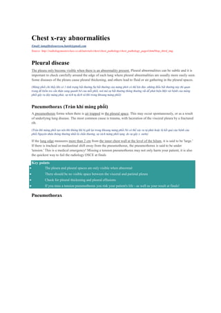
- 1. Chest x-ray abnormalities Email: iamgiftedsoareyou.hatok@gmail.com Source: http://radiologymasterclass.co.uk/tutorials/chest/chest_pathology/chest_pathology_page4.html#top_third_img Pleural disease The pleura only become visible when there is an abnormality present. Pleural abnormalities can be subtle and it is important to check carefully around the edge of each lung where pleural abnormalities are usually more easily seen. Some diseases of the pleura cause pleural thickening, and others lead to fluid or air gathering in the pleural spaces. (Màng phổi chỉ thấy khi có 1 tình trạng bất thường.Sự bất thường của màng phởi có thể kín đáo ,những điều bất thường này thì quan trọng để kiểm tra cẩn thận xung quanh bờ của mỗi phổi, nơi mà sự bất thường thông thường rất dễ phát hiện.Một vài bệnh của màng phổi gây ra dày màng phải, sự tích tụ dịch và khí trong khoang màng phổi) Pneumothorax (Tràn khí màng phổi) A pneumothorax forms when there is air trapped in the pleural space. This may occur spontaneously, or as a result of underlying lung disease. The most common cause is trauma, with laceration of the visceral pleura by a fractured rib. (Tràn khí màng phổi tạo nên khi không khí bị giữ lại trong khoang màng phổi.Nó có thể xảy ra tự phát hoặc là kết quả của bệnh của phổi.Nguyên nhân thông thường nhất là chấn thương, sự rách màng phổi tạng do sự gãy x .sườn) If the lung edge measures more than 2 cm from the inner chest wall at the level of the hilum, it is said to be 'large.' If there is tracheal or mediastinal shift away from the pneumothorax, the pneumothorax is said to be under 'tension.' This is a medical emergency! Missing a tension pneumothorax may not only harm your patient, it is also the quickest way to fail the radiology OSCE at finals Key points The pleura and pleural spaces are only visible when abnormal There should be no visible space between the visceral and parietal pleura Check for pleural thickening and pleural effusions If you miss a tension pneumothorax you risk your patient's life - as well as your result at finals! Pneumothorax
- 2. Air in pleural space - Pneumothorax Visible pleural edge (blue line) Lung markings not visible beyond this edge ( “lung marking” không thấy ở bên ngoài bờ phổi) Clinical information Fall from height - trauma to chest Diagnosis Left pneumothorax due to a rib fracture (arrowhead) The trachea and mediastinal structures are not displaced so there is no 'tension' Đặt vấn đề: 1.”Lung marking” là gì? Lung tissue is air-filled and appears black on Chest X-ray. The lung markings are a reflection of other structures within the lungs that show-up as white on the X-ray. These other structures are made up primarily of blood vessels, but also bronchial walls, lymphatics and some fibrotic tissue that accompanies vessels and bronchi. Lung markings are thus, normal X-ray findings 2.Phân biệt 2 tình trạng “large” và “tension”? Pleural thickening Pleural thickening is best seen at the lung edges where the pleura runs tangentially to the x-ray beam. (Sự dày màng phổi nhìn thấy rõ nhất tại rìa phổi, nơi mà màng phổi tiếp tuyến với chùm tia X) Unilateral pleural thickening
- 3. Unilateral pleural thickening Peripheral shadowing on the right Loss of right lung volume Shadowing over the whole right lung due to circumferential pleural thickening Clinical information History of asbestos exposure(tiếp xúc với amiang) Diagnosis Malignant mesothelioma - a disease caused by exposure to asbestos Differential Empyema*( the presence of pus in a body cavity,especially the pleural cavity) Pleural metastases*([Greek, from methistanai, to change : meta-, meta- + histanai, to cause to stand, place; see stā- in Indo-European roots.]) Pleural effusions* * Do not cause volume loss Asbestos plaques Calcified asbestos related pleural plaques have a characteristic appearance, and are generally considered to be benign. They are irregular, well-defined, and classically said to look like holly leaves. holly leaves Asbestos related pleural plaques
- 4. Asbestos related pleural plaques Bilateral well defined irregular shadows that are as dense as the bones Peripheral pleural thickening Clinical information Chronic mild shortness of breath (thở ngắn nông) Retired dock worker (người đóng tàu đã về hưu)with clear history of asbestos exposure Diagnosis Bilateral calcified asbestos related pleural plaques Pleural effusions A pleural effusion is a collection of fluid in the pleural space. Fluid gathers in the lowest part of the chest, according to the patient's position. If the patient is upright when the x-ray is taken, a pleural effusion will obscure the costophrenic angle and hemidiaphragm. If a patient is supine a pleural effusion layers along the posterior aspect of the chest cavity and becomes difficult to see on a chest x-ray. Costophrenic angle: In anatomy, the costophrenic angles are the places where the diaphragm (-phrenic) meets the ribs (costo-). Comparison between a normal costophrenic angle on the patient's right, and an obscured costophrenic angle (circled) on the patient's left, due to pneumonia with parapneumonic effusion. Pleural effusions(tràn dịch màng phổi)
- 5. Đặt vấn đề: 1. “Meniscus sign”? Pleural effusions The left lower zone is uniformly white At the top of this white area there is a concave surface - meniscus sign The left heart border, costophrenic angle and hemidiaphragm are obscured Slight blunting of the right costophrenic angle indicates a small pleural effusion on that side Clinical information Life long smoker Weight loss and increasing shortness of breath Diagnosis Large left pleural effusion Underlying bronchogenic carcinoma MmmmM« Previous Tutorials Next »