Bangalore Call Girls Marathahalli 📞 9907093804 High Profile Service 100% Safe
Electroretinography
1. 26/04/2010
ELECTRORETINOGRAPHY
Dr Frank FAMOSE
DVM, CES Ophtalmologie
Ekaterinburg May 2010
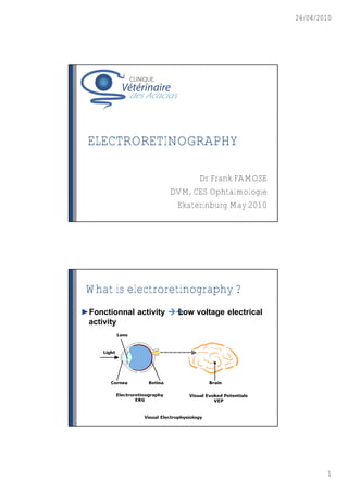
What is electroretinography ?
►Fonctionnal activity
Low voltage electrical
activity
1
2. 26/04/2010
History
►1849 : Dipolar activity of eye (Dubois-Raymond)
►1865 : Electrical activity of retina (Holmgren)
►1924 : First recordings of human ERG (L•wenstein)
►1933 : ERG components (Granit)
►1967 : ERG applications in Vet ophthalmology (Rubin)
2
5. 26/04/2010
General morphology of ERG
c c
a-wave : receptor level Other waves :
b-wave : post-receptor level Oscillatory potentials (OP)
c-wave : pre-receptor level Early Receptor Potential (ERP)
The components of ERG
►From Granit’s work : 3 components
Pigmentary epithelium
Ganglionar cells
Photoreceptors
►Intra-retinal potentials
a-wave : Photoreceptors
b-wave : bipolar cells depolarisation
c-wave : pigmentary epithelium
5
6. 26/04/2010
Electroretinography material
►Light stimulation
Intensity
Spectral composition
Spatial repartition
Repetition frequency
►Filters
Neutral
Coloured
Electroretinography material
►Electrodes
Active electrode
Reference electrode
Ground electrode
6
7. 26/04/2010
Electroretinography material
►Collecting and
recording data
Amplification
Filtration
Computer
13
Variations in components of ERG :
photoreceptors
►Rods ►Cones
Low light intensity High light intensity
Low frequency High frequency (>30Hz)
Rhodopsine 2 spectral sensitivities
Saturation process No saturation
14
7
8. 26/04/2010
Variations in components of ERG :
intensity of light stimulation
►Standardisation
►Effects of increased
intensity
►Spectral
composition
►Time frequency
15
Variations in components of ERG :
environmental light conditions
►Scotopic conditions ►Photopic conditions
(darkness) (daylight conditions)
Rods in dim flash Saturation of rods
Cones in strong flash Specific stimulation
stimulation of cones
Dim flash of blue light Strong flash of achromatic light
in darkness conditions in darkness conditions
Stimulation of rods only Saturation of rods
Specific evaluation of cones
8
9. 26/04/2010
Variations in components of ERG :
anesthesia
►Necessity of
anesthesia
►Effects of inhalation
anesthaetics
►Effects of hypoxia
Variations in components of ERG :
the patient
►Pupillary size :
mydriasis
►Age : attenuation
►Ocular opacities :
various effects
9
10. 26/04/2010
ERG protocols
►Goals of the ERG
Selective stimulation
►Animal preparation
Mydriatics
General anesthesia
►Electrodes fixation
Active electrode
Reference electrode
Ground electrode
ERG protocols : ISCEV
►Only white stimulation
►Darkness adaptation
(20 min)
►Dim stimulation in
scotopic conditions Rods stimulation
►Photopic stimulation in
photopic conditions Rods and cones
►Photopic stimulation in stimulation
30 hz frequency Cones stimulation
10
11. 26/04/2010
ERG protocols : coloured
stimulations
►Darkness adaptation
(20 min)
►Dim blue stimulation in
scotopic conditions Rods stimulation
►Photopic red stimulation in
photopic conditions Cones stimulation
►Photopic white stimulation
in 30 hz frequency Cones stimulation
ERG protocols : « mix » protocol
►Room light adaptation
(2 hours)
►Photopic white
stimulation Cones stimulation
►Darkness adaptation
(20 min)
►Scotopic blue Rods stimulation
stimulation in darkness
►Photopic stimulation in
30 hz frequency Cones stimulation
11
12. 26/04/2010
ERG : other techniques
►Multifocal ERG
►Pattern ERG
For Human
and laboratory use
ERG analysis
►Waves amplitude
►Time of culmination
No reference value !!
12
13. 26/04/2010
ERG analysis
One month later…
Clinical indications of ERG
►Preoperative ►Clinical evaluation of
evaluation of retina retina in pathologic
(cataract surgery) conditions
►Amaurosis ►Early diagnosis of
retinal conditions
13