Approach to a patient with ST segment abnormality in ECG
•Download as PPSX, PDF•
4 likes•1,047 views

Case: A 45 years old presented with chest discomfort and excessive sweating for last 2 hours. He was diabetic, smoker and dyslipidemic. His pulse 68b/min and BP-130/80 mm Hg. In emergency department he had the following ECG. Case: A 25 years old gentleman presented with chest pain and fever .He was normotensive, non-smoker and non-diabetic. His pulse 128b/min and BP-130/80 mm Hg. Troponin I was normal. Case: A 54 years old lady presented with chest discomfort and excessive sweating for last 4 hours. She was diabetic, hypertensive and dyslipidemic. Her pulse 62b/min and BP-160/90 mm Hg. In emergency department she had the following ECG.

Recommended
More Related Content
What's hot
What's hot (20)
Similar to Approach to a patient with ST segment abnormality in ECG
Similar to Approach to a patient with ST segment abnormality in ECG (20)
More from PROFESSOR DR. MD. TOUFIQUR RAHMAN
More from PROFESSOR DR. MD. TOUFIQUR RAHMAN (20)
Recently uploaded
Recently uploaded (20)
Approach to a patient with ST segment abnormality in ECG
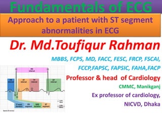
- 1. Fundamentals of ECG Approach to a patient with ST segment abnormalities in ECG Dr. Md.Toufiqur Rahman MBBS, FCPS, MD, FACC, FESC, FRCP, FSCAI, FCCP,FAPSC, FAPSIC, FAHA,FACP Professor & head of Cardiology CMMC, Manikganj Ex professor of cardiology, NICVD, Dhaka
- 2. Fundamentals of ECG ST segment
- 3. drtoufiq1971@gmail.com ST segment Professor Dr Md Toufiqur Rahman Fundamentals of ECG Case: A 45 years old presented with chest discomfort and excessive sweating for last 2 hours. He was diabetic, smoker and dyslipidemic. His pulse 68b/min and BP-130/80 mm Hg. In emergency department he had the following ECG. Figure: ST segment elevation in V2-V6, Lead 1 and aVL suggesting ( Extensive anterior wall myocardial infarction) and ST segment depression in inferior leads (II, III, aVF).
- 4. drtoufiq1971@gmail.com ST segment Professor Dr Md Toufiqur Rahman Fundamentals of ECG Case: A 25 years old gentleman presented with chest pain and fever .He was normotensive, non-smoker and non-diabetic. His pulse 128b/min and BP-130/80 mm Hg. Troponin I was normal. Figure: ECG showing Wide spread ST segment elevation in lead 1, II, III, aVF, aVL, V4-V6 suggestive of acute Pericarditis.
- 5. drtoufiq1971@gmail.com ST segment Professor Dr Md Toufiqur Rahman Fundamentals of ECG Case: A 54 years old lady presented with chest discomfort and excessive sweating for last 4 hours. She was diabetic, hypertensive and dyslipidemic. Her pulse 62b/min and BP-160/90 mm Hg. In emergency department she had the following ECG. Figure: ST segment elevation in inferior leads (II, III, aVF) suggestive of inferior myocardial infarction and there is reciprocal ST segment depression in lead 1, aVL.
- 6. drtoufiq1971@gmail.com ST segment Professor Dr Md Toufiqur Rahman Fundamentals of ECG Case: A 23 years old gentleman presented with occasional chest discomfort. He was smoker, normotensive and non-diabetic. He had the following ECG. His Echocardiogram was normal and troponin I level was normal. ECG showing characteristic ST segment elevation in V1-V3 suggestive of Benign early repolarization (BER).
- 7. drtoufiq1971@gmail.com ST segment Professor Dr Md Toufiqur Rahman Fundamentals of ECG Case: A 33 years old gentleman presented with occasional chest discomfort, dizziness and several episodes of syncope. He had an ejection systolic murmur in precordium, BP-95/60 mm Hg. Echocardiogram showed bicuspid aortic valve and aortic valve gradient was 123 mm Hg. He had the following ECG. Figure: ECG showing ST segment elevation in lead 1, aVL, V1-V4 with with specific pattern of LBBB. There is prolonged PR interval suggestive of presence of first degree AV block.
- 8. drtoufiq1971@gmail.com ST segment Professor Dr Md Toufiqur Rahman Fundamentals of ECG Case: A 43 years old lady presented with headache for 2 months. Her BP-160/100 mm Hg. Her echocardiogram showed concentric left ventricular hypertrophy. She had the following ECG. Figure: S wave in V1/V2+ R in V5/V6 more than 35 mm suggestive of Left ventricular hypertrophy. In V1-V2 there is deep s wave with ST segment elevation.
- 9. drtoufiq1971@gmail.com ST segment Professor Dr Md Toufiqur Rahman o The ST segment is the flat, isoelectric section of the ECG between the end of the S wave (the J point) and the beginning of the T wave. o The ST Segment represents the interval between ventricular depolarization and repolarization. o The most important cause of ST segment abnormality (elevation or depression) is myocardial ischaemia or infarction. Fundamentals of ECG
- 10. drtoufiq1971@gmail.com ST segment Professor Dr Md Toufiqur Rahman Fundamentals of ECG
- 11. drtoufiq1971@gmail.com ST segment Professor Dr Md Toufiqur Rahman Causes of ST Segment Elevation Acute myocardial infarction Coronary vasospasm (Printzmetal’s angina) Pericarditis Benign early repolarization Left bundle branch block Left ventricular hypertrophy Ventricular aneurysm Brugada syndrome Ventricular paced rhythm Raised intracranial pressure Takotsubo Cardiomyopathy Fundamentals of ECG
- 12. drtoufiq1971@gmail.com ST segment Professor Dr Md Toufiqur Rahman Morphology of the Elevated ST segment Myocardial Infarction Acute STEMI may produce ST elevation with either concave, convex or obliquely straight morphology. Fundamentals of ECG
- 13. drtoufiq1971@gmail.com ST segment Professor Dr Md Toufiqur Rahman Morphology of the Elevated ST segment Fundamentals of ECG
- 14. drtoufiq1971@gmail.com ST segment Professor Dr Md Toufiqur Rahman Morphology of the Elevated ST segment Fundamentals of ECG
- 15. drtoufiq1971@gmail.com ST segment Professor Dr Md Toufiqur Rahman Morphology of the Elevated ST segment Patterns of ST Elevation Acute ST elevation myocardial infarction (STEMI) ST segment elevation and Q-wave formation in contiguous leads. Septal (V1-2) Anterior (V3-4) Lateral (I + aVL, V5-6) Inferior (II, III, aVF) Right ventricular (V1, V4R) Posterior (V7-9) Fundamentals of ECG
- 16. drtoufiq1971@gmail.com ST segment Professor Dr Md Toufiqur Rahman Morphology of the Elevated ST segment Acute ST elevation myocardial infarction (STEMI) There is usually reciprocal ST depression in the electrically opposite leads. For example, STE in the high lateral leads I + aVL typically produces reciprocal ST depression in lead III Fundamentals of ECG
- 17. drtoufiq1971@gmail.com ST segment Professor Dr Md Toufiqur Rahman Coronary Vasospasm (Prinzmetal’s angina) • This causes a pattern of ST elevation that is very similar to acute STEMI — i.e. localised ST elevation with reciprocal ST depression occurring during episodes of chest pain. • However, unlike acute STEMI the ECG changes are transient, reversible with vasodilators and not usually associated with myocardial necrosis. • It may be impossible to differentiate these two conditions based on the ECG alone. Fundamentals of ECG
- 18. drtoufiq1971@gmail.com ST segment Professor Dr Md Toufiqur Rahman Pericarditis Acute Pericarditis causes widespread concave (“saddleback”) ST segment elevation with PR segment depression in multiple leads, typically involving I, II, III, aVF, aVL, and V2-6. Spodick’s sign was first described by David H. Spodick in 1974 as a downward sloping TP segment with specificity for acute pericarditis. Fundamentals of ECG
- 19. drtoufiq1971@gmail.com ST segment Professor Dr Md Toufiqur Rahman Pericarditis Concave “saddleback” ST elevation in leads I, II, III, aVF, V5-6 with depressed PR segments. There is reciprocal ST depression and PR elevation in leads aVR and V1. Fundamentals of ECG
- 20. drtoufiq1971@gmail.com ST segment Professor Dr Md Toufiqur Rahman Benign Early Repolarization Benign Early Repolarization (BER) causes mild ST elevation with tall T-waves mainly in the precordial leads. BER is a normal variant commonly seen in young, healthy patients. There is often notching of the J-point — the “fish- hook” pattern. The ST changes may be more prominent at slower heart rates and disappear in the presence of tachycardia. Fundamentals of ECG
- 21. drtoufiq1971@gmail.com ST segment Professor Dr Md Toufiqur Rahman Benign Early Repolarization There is slight concave ST elevation in the precordial and inferior leads with notching of the J-point (the “fish-hook” pattern) Fundamentals of ECG
- 22. drtoufiq1971@gmail.com ST segment Professor Dr Md Toufiqur Rahman Left Bundle Branch Block (LBBB) • In Left bundle branch block (LBBB), the ST segments and T waves show “appropriate discordance” — i.e. they are directed opposite to the main vector of the QRS complex. • This produces ST elevation and upright T waves in leads with a negative QRS complex (dominant S wave), while producing ST depression and T wave inversion in leads with a positive QRS complex (dominant R wave). Fundamentals of ECG
- 23. drtoufiq1971@gmail.com ST segment Professor Dr Md Toufiqur Rahman Left Bundle Branch Block (LBBB) ST elevation in leads with deep S waves — most apparent in V1-3. ST depression in leads with tall R waves — most apparent in I and aVL. Fundamentals of ECG
- 24. drtoufiq1971@gmail.com ST segment Professor Dr Md Toufiqur Rahman Left Ventricular Hypertrophy (LVH) Left Ventricular Hypertrophy (LVH) causes a similar pattern of repolarization abnormalities as LBBB, with ST elevation in the leads with deep S-waves (usually V1-3) and ST depression/T-wave inversion in the leads with tall R waves (I, aVL, V5-6). Fundamentals of ECG
- 25. drtoufiq1971@gmail.com ST segment Professor Dr Md Toufiqur Rahman Left Ventricular Hypertrophy (LVH) • Deep S waves with ST elevation in V1-3 • ST depression and T-wave inversion in the lateral leads V5-6 • there is also right axis deviation, which is unusual for LVH and may be due to associated left posterior fascicular block Fundamentals of ECG
- 26. drtoufiq1971@gmail.com ST segment Professor Dr Md Toufiqur Rahman Ventricular Aneurysm • Ventricular Aneurysm – residual ST elevation and deep Q waves seen in patients with previous myocardial infarction. • It is associated with extensive myocardial damage and paradoxical movement of the left ventricular wall during systole. Fundamentals of ECG
- 27. drtoufiq1971@gmail.com ST segment Professor Dr Md Toufiqur Rahman Ventricular Aneurysm • ST elevation with deep Q waves and inverted T waves in V1-3. • This pattern suggests the presence of a left ventricular aneurysm due to a prior anteroseptal MI. Fundamentals of ECG
- 28. drtoufiq1971@gmail.com ST segment Professor Dr Md Toufiqur Rahman Brugada Syndrome • Brugada Syndrome is an inherited channelopathy (a disease of myocardial sodium channels) that leads to paroxysmal ventricular arrhythmias and sudden cardiac death in young patients. • The tell-tale sign on the resting ECG is the “Brugada sign” — ST elevation and partial RBBB in V1-2 with a “coved” morphology. Fundamentals of ECG
- 29. drtoufiq1971@gmail.com ST segment Professor Dr Md Toufiqur Rahman Brugada Syndrome • ST elevation and partial RBBB in V1-2 with a coved morphology — the “Brugada sign”. Fundamentals of ECG
- 30. drtoufiq1971@gmail.com ST segment Professor Dr Md Toufiqur Rahman Ventricular Paced Rhythm Ventricular pacing (with a pacing wire in the right ventricle) causes ST segment abnormalities identical to that seen in LBBB. There is appropriate discordance, with the ST segment and T wave directed opposite to the main vector of the QRS complex. Fundamentals of ECG
- 31. drtoufiq1971@gmail.com ST segment Professor Dr Md Toufiqur Rahman Ventricular Paced Rhythm There is appropriate discordance, with the ST segment and T wave directed opposite to the main vector of the QRS complex. Fundamentals of ECG
- 32. drtoufiq1971@gmail.com ST segment Professor Dr Md Toufiqur Rahman Raised Intracranial Pressure • Raised Intracranial Pressure (ICP) (e.g. due to intracranial haemorrhage, traumatic brain injury) may cause ST elevation or depression that simulates myocardial ischaemia or pericarditis. • More commonly, raised ICP is associated with widespread, deep T-wave inversions (“cerebral T waves“). Fundamentals of ECG
- 33. drtoufiq1971@gmail.com ST segment Professor Dr Md Toufiqur Rahman Raised Intracranial Pressure Widespread ST elevation with concave (pericarditis-like) morphology in a patient with severe traumatic brain injury Fundamentals of ECG
- 34. drtoufiq1971@gmail.com ST segment Professor Dr Md Toufiqur Rahman Takotsubo Cardiomyopathy o Takotsubo Cardiomyopathy, A STEMI mimic producing ischaemic chest pain, ECG changes +/- elevated cardiac enzymes with characteristic regional wall motion abnormalities on echocardiography. o Typically occurs in the context of severe emotional distress (“broken heart syndrome“). Commonly associated with new ECG changes (ST elevation or T wave inversion) or moderate troponin rise. Fundamentals of ECG
- 35. drtoufiq1971@gmail.com ST segment Professor Dr Md Toufiqur Rahman Takotsubo Cardiomyopathy Fundamentals of ECG
- 36. drtoufiq1971@gmail.com ST segment Professor Dr Md Toufiqur Rahman Less Common Causes of ST segment Elevation Pulmonary embolism and acute cor pulmonale (usually in lead III) Acute aortic dissection (classically causes inferior STEMI due to RCA dissection) Hyperkalaemia Sodium-channel blocking drugs (secondary to QRS widening) J-waves (hypothermia, hypercalcaemia) Following electrical cardioversion Others: Cardiac tumour, myocarditis, pancreas or gallbladder disease Fundamentals of ECG
- 37. drtoufiq1971@gmail.com ST segment Professor Dr Md Toufiqur Rahman Transient ST elevation after DC cardioversion from VF Fundamentals of ECG
- 38. drtoufiq1971@gmail.com ST segment Professor Dr Md Toufiqur Rahman J waves in hypothermia simulating ST elevation Fundamentals of ECG
- 39. drtoufiq1971@gmail.com ST segment Professor Dr Md Toufiqur Rahman Causes of ST Depression Myocardial ischaemia / NSTEMI Reciprocal change in STEMI Posterior MI Digoxin effect Hypokalaemia Supraventricular tachycardia Right bundle branch block Right ventricular hypertrophy Left bundle branch block Left ventricular hypertrophy Ventricular paced rhythm Fundamentals of ECG
- 40. drtoufiq1971@gmail.com ST segment Professor Dr Md Toufiqur Rahman Morphology of ST Depression ST depression can be either upsloping, downsloping, or horizontal. Horizontal or downsloping ST depression ≥ 0.5 mm at the J-point in ≥ 2 contiguous leads indicates myocardial ischaemia (according to the 2007 Task Force Criteria). Upsloping ST depression in the precordial leads with prominent De Winter T waves is highly specific for occlusion of the LAD. Fundamentals of ECG
- 41. drtoufiq1971@gmail.com ST segment Professor Dr Md Toufiqur Rahman Morphology of ST Depression Reciprocal change has a morphology that resembles “upside down” ST elevation and is seen in leads electrically opposite to the site of infarction. Posterior MI manifests as horizontal ST depression in V1-3 and is associated with upright T waves and tall R waves. Fundamentals of ECG
- 42. drtoufiq1971@gmail.com ST segment Professor Dr Md Toufiqur Rahman Morphology of ST Depression Fundamentals of ECG
- 43. drtoufiq1971@gmail.com ST segment Professor Dr Md Toufiqur Rahman ST segment morphology in myocardial ischaemia Fundamentals of ECG
- 44. drtoufiq1971@gmail.com ST segment Professor Dr Md Toufiqur Rahman Morphology of ST Depression Reciprocal change Fundamentals of ECG
- 45. drtoufiq1971@gmail.com ST segment Professor Dr Md Toufiqur Rahman Morphology of ST Depression ST segment morphology in posterior MI Fundamentals of ECG
- 46. drtoufiq1971@gmail.com ST segment Professor Dr Md Toufiqur Rahman Morphology of ST Depression Myocardial Ischaemia ST depression due to subendocardial ischaemia may be present in a variable number of leads and with variable morphology. It is often most prominent in the left precordial leads V4-6 plus leads I, II and aVL. Widespread ST depression with ST elevation in aVR is seen in left main coronary artery occlusion and severe triple vessel disease. Fundamentals of ECG
- 47. drtoufiq1971@gmail.com ST segment Professor Dr Md Toufiqur Rahman Morphology of ST Depression Myocardial Ischaemia ST depression localised to the inferior or high lateral leads is more likely to represent reciprocal change than subendocardial ischaemia Fundamentals of ECG
- 48. drtoufiq1971@gmail.com ST segment Professor Dr Md Toufiqur Rahman Morphology of ST Depression Reciprocal Change • ST elevation during acute STEMI is associated with simultaneous ST depression in the electrically opposite leads. • Inferior STEMI produces reciprocal ST depression in aVL (± lead I). • Lateral or anterolateral STEMI produces reciprocal ST depression in III and aVF (± lead II). • Reciprocal ST depression in V1-3 occurs with posterior infarction Fundamentals of ECG
- 49. drtoufiq1971@gmail.com ST segment Professor Dr Md Toufiqur Rahman Morphology of ST Depression Reciprocal Change Reciprocal ST depression in aVL with inferior STEMI Fundamentals of ECG
- 50. drtoufiq1971@gmail.com ST segment Professor Dr Md Toufiqur Rahman Morphology of ST Depression Reciprocal Change Reciprocal ST depression in III and aVF with high lateral STEMI Fundamentals of ECG
- 51. drtoufiq1971@gmail.com ST segment Professor Dr Md Toufiqur Rahman Posterior Myocardial Infarction Acute posterior STEMI causes ST depression in the anterior leads V1- 3, along with dominant R waves (“Q-wave equivalent”) and upright T waves. There is ST elevation in the posterior leads V7-9. Fundamentals of ECG
- 52. drtoufiq1971@gmail.com ST segment Professor Dr Md Toufiqur Rahman De Winter T Waves De Winter T waves: a pattern of up-sloping ST depression with symmetrically peaked T waves in the precordial leads is considered to be a STEMI equivalent, and is highly specific for an acute occlusion of the LAD. Fundamentals of ECG
- 53. drtoufiq1971@gmail.com ST segment Professor Dr Md Toufiqur Rahman Digoxin Effect Digoxin Effect: Treatment with digoxin causes down sloping ST depression with a “sagging” morphology, reminiscent of Salvador Dali’s moustache. Fundamentals of ECG
- 54. drtoufiq1971@gmail.com ST segment Professor Dr Md Toufiqur Rahman Hypokalaemia Hypokalaemia causes widespread downsloping ST depression with T-wave flattening/inversion, prominent U waves and a prolonged QU interval. Fundamentals of ECG
- 55. drtoufiq1971@gmail.com ST segment Professor Dr Md Toufiqur Rahman Right ventricular hypertrophy (RVH) Right ventricular hypertrophy (RVH) causes ST depression and T-wave inversion in the right precordial leads V1-3. Fundamentals of ECG
- 56. drtoufiq1971@gmail.com ST segment Professor Dr Md Toufiqur Rahman Right Bundle Branch Block (RBBB) Right Bundle Branch Block (RBBB) may produce a similar pattern of repolarisation abnormalities to RVH, with ST depression and T wave inversion in V1-3. Fundamentals of ECG
- 57. drtoufiq1971@gmail.com ST segment Professor Dr Md Toufiqur Rahman Supraventricular tachycardia (SVT) Fundamentals of ECG
- 58. drtoufiq1971@gmail.com ST segment Professor Dr Md Toufiqur Rahman Supraventricular tachycardia (SVT) Supraventricular tachycardia (e.g. AVNRT) typically causes widespread horizontal ST depression, most prominent in the left precordial leads (V4-6). This rate-related ST depression does not necessarily indicate the presence of myocardial ischaemia, provided that it resolves with treatment. Fundamentals of ECG