
Recommended
More Related Content
What's hot
What's hot (20)
Similar to Electron microscope
Similar to Electron microscope (20)
Recently uploaded
Recently uploaded (20)
Electron microscope
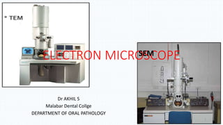
- 1. ELECTRON MICROSCOPE Dr AKHIL S Malabar Dental Collge DEPARTMENT OF ORAL PATHOLOGY SEM 1
- 2. The Central player - (e) • The electron “e” is an elementary particle • Also called corpuscle • carries a negative charge. • the electron was discovered by J. J. Thompson in 1897 • e is a constituent of the atom • 1000 times smaller than a hydrogen atom. • the mass of the electron 1/1836 of that of a proton. 2
- 3. Scheme of electron-matter interactions arising from the impact of an electron beam onto a specimen. A signal below the specimen is observable if the thickness is small enough to allow some electrons to pass through 3
- 4. Elastic Electron Interactions • no energy is transferred from the electron to the sample. • These signals are mainly exploited in - Transmission Electron Microscopy and - Electron diffraction methods. 4
- 5. Inelastic Electron Interactions - Energy is transferred from the electrons to the specimen - The energy transferred can cause different signals such as - X-rays, - Auger electrons - secondary electrons, - plasmons, - phonons, - UV quanta or cathodoluminescence. • Used in Analytical Electron Microscopy … SEM 5
- 6. What is Electron Microscopy? • Electron microscopy is a diagnostic tool with diversified combination of techniques …… • that offer unique possibilities to gain insights into - structure, - topology, - morphology, and - composition of a material. 6
- 7. What is an Electron Microscope ? • A special type of microscope having a high resolution of images, able to magnify objects in nanometres, which are formed by controlled use of electrons in vacuum captured on a phosphorescent screen 7
- 8. Why were the EMs invented? • To study objects of < 0.2 micrometer • For analysis of sub cellular structures • Intra cellular pathogens - viruses • Cell metabolism • Study of minute structures in the nature Greater resolving power of the EMs than light microscope • An EM can magnify structures from 100 – 250000 times than light microscopy 8
- 9. The novelty of EMs from others • Beam of Electrons …… instead of a beam of light • Electro-magnetic lens ………..instead of Ground glass lenses • Cylindrical Vacuum column - Electrons should travel in vacuum to avoid collisions with air molecules that cause scattering of electrons distorting the image 9
- 10. 10
- 11. Types of electron microscope 1. Transmission electron microscopy : 2. Scanning electron microscopy: 11
- 12. SEM 12
- 13. Transmission Electron Microscope Principle • TEM is the direct counterpart of Light microscope • Involves passage of high velocity electron beam through specimen, thin enough to transmit 50% of the electrons • Transmitted electrons – focused by lens systems to form a 2 dimensional magnified image 13
- 14. Analogy between LM & TEM • Arrangement & function of their components 1. Illuminating system – source & condenser 2. Imaging system – lenses to produce magnified image – objective & projector 3. Image translating system – Final image is viewed 14
- 15. THE LIGHT MICROSCOPE v THE ELECTRON MICROSCOPE VacuumAir-filledInterior MagnetsGlassLenses High voltage (50kV) tungsten filament Tungsten or quartz halogen lamp Radiation source X 5,00,000x1000 – x1500 Maximum magnification 0.14nm Fine detail app. 200nm or 0.2micron Maximum resolving power Electrons app. 4nm Visible light 390nm (red) – 760nm Electromagnetic spectrum used ELECTRON MICROSCOPELIGHT MICROSCOPEFEATURE © 2007 Paul Billiet ODWS Focus Lens is movable Rigidly fixed, adjust lens currents 15
- 16. THE LIGHT MICROSCOPE v THE ELECTRON MICROSCOPE Copper gridGlass slideSupport Heavy metalsWater soluble dyesStains Ultramicrotome Slices - 50nm Parts of cells visible Microtome slices - 20 000nm Whole cells visible Sectioning ResinWaxEmbedding Glutaraldehyde,OsO4formaldehydeFixation ELECTRON MICROSCOPE LIGHT MICROSCOPEFEATURE © 2007 Paul Billiet ODWS Focussing screen Human eye (retina), photographic film Fluorescent screen, photographic film 16
- 18. Electron source • The electron source consists of a cathode and an anode. • The cathode is a tungsten filament which emits electrons when being heated. • A negative cap confines the electrons into a loosely focused beam. • The beam is then accelerated towards the specimen by the positive anode. 18
- 19. Electromagnetic lens system • The system allows electrons within a small energy range to pass through, so the electrons in the electron beam will have a well-defined energy. • 1. Magnetic Lens: Circular electro- magnets capable of generating a precise circular magnetic field. The field acts like an optical lens to focus the electrons. • 2. Aperture: A thin disk with a small (2-100 micrometers) circular through-hole. It is used to restrict the electron beam and filter out unwanted electrons before hitting the specimen. 19
- 20. The Vacuum System • The electron beam must be generated in and traverse through the microscope column under a high vacuum condition. • The presence of air molecules will result in the collision and scattering of the electrons from their path. • In the electron microscope the vacuum is maintained by a series of highly efficient vacuum pumps. • THE VACUUM FACTOR: Biological material must be properly fixed and preserved 20
- 21. Sample holder • The sample holder is a platform equipped with a mechanical arm for holding the specimen and controlling its position. 21
- 22. Imaging system • The imaging system consists of another electromagnetic lens system and a screen. • The electromagnetic lens - two lens, one for refocusing the electrons after they pass through the specimen, and the other for enlarging the image and projecting it onto the screen. • The screen has a phosphorescent plate which glows when being hit by electrons. 22
- 23. Image Formation in the TEM • The basis of image formation in the TEM is the scattering of electrons. • The scattering results in a shadow on the viewing screen or photographic film. • Material with high atomic numbers will cause more scattering and produce a deep shadow. Such material is termed "electron dense" and has high image contrast. • Biological material has low electron density and is known generally as "electron transparent". Hence, an inherent low contrast image is formed. • BIOLOGICAL MATERIAL must, therefore, be STAINED with heavy metal salts. 23
- 24. • ELECTRON SOURCE • ELECTROMAGNETIC LENS SYSTEM • SAMPLE HOLDER • IMAGING SYSTEM. 24
- 25. THE SCANNING ELECTRON MICROSCOPE • To directly visualize the surface topography of solid unsectioned specimens. • Probe scans the specimen in square raster pattern. • The first scanning electron microscope (SEM) debuted in 1938 ( Von Ardenne) with the first commercial instruments around 1965. • Differs from TEM in construction & operational modes • TEM – information is obtained from transmitted electrons • SEM – majority is obtained from secondary, backscattered electrons & from X-rays. 25
- 26. 26
- 27. 27
- 28. 28
- 29. • After the impingement of the primary electrons on the specimens, secondary electrons as well as other forms of radiation are emitted. • But only the secondary electrons will be collected by the signal detector. • In the detector these electrons strike a scintillator and the light produced is converted to electric signals by a photomultiplier. • The electric signal is then amplified and displayed on the cathode ray tube (CRT). 29
- 30. • In the SEM the electron beam is rapidly scanned back and forth in an orderly pattern across the specimen surface. • It is a composite of many individual image spots similar to the image formed on the TV screen. • The SEM has a specimen stage that allows the specimen to move freely so that the surface of the specimen can be viewed from all angles. 30
- 31. The focused electron beam is moved from one pixel to another. At every pixel, the beam stays for a defined time and generates a signal (e.g.secondary electrons) which are detected, amplified and displayed on a computer screen 31
- 32. • A smaller area is scanned with the same number of pixels. • The scanned pixels are smaller Image magnification in SEM 32
- 33. 33
- 34. TEM vs SEM TEM SEM 5 lenses – C1, C2, objective, 2 projector 3 lenses – 2 condensor, 1 objective High accelerating voltage - penetration low accelerating voltage Not complicated Specimen Stage – complicated X & y axis X,Y,Z-axis, tilting, rotating 34
- 35. • Diseases of kidney • Metabolic storage diseases • Respiratory tract biopsies • Skeletal muscle diseases • Infectious agents • Cutaneous diseases • Peripheral nerve biopsies • Epithelial tumors • Mesothelioma • Melanoma • Hematopoietic and lymphopoietic tumors • Soft-tissue tumors • Central nervous system tumors • Small round cell tumors Non tumor biopsies Tumor diagnosis 35
- 36. A, Adenovirus, an icosahedral nonenveloped DNA virus with fibers. B, Epstein-Barr virus, an icosahedral enveloped DNA virus. C, Rotavirus, a nonenveloped, wheel-like, RNA virus. D, Paramyxovirus, a spherical enveloped RNA virus. RNA is seen spilling out of the disrupted virus 36
- 37. Squamous cell carcinoma Well differentiated squamous cell carcinoma Abundant cytokeratin filaments. Frequent desmosomes between cells. 37
- 38. Well differentiated squamous cell carcinoma- frequent desmosomes 38
- 39. Disadvantages 1. EM is not economical- stable high voltage supply, vaccum system etc 2. Findings unlikely to influence treatment, IHC & LM together are confirmatory . 3. Tissue preparation is tough 4. Only a small proportion of neoplasm can be studied 5. Misinterpretation of non- neoplastic elements belonging to the tumor 39
- 40. Conclusion Currently the use of EM is limited for the expense and lack of surgical pathologists to interpret EM findings . Still it provides unique insight into the structure of some tumors and renal pathologies. So better to use it selectively in study and diagnosis of human diseases and research areas and correlating the findings with LM findings and IHC results. 40
- 41. References • Theory and practices of histopathological techniques, John D Bancroft, 4th edition;. • Robbins and Cotran, PATHOLOGIC BASIS OF DISEASE, 8th edition. • Cellular pathology technique, C.F.A. Culling, 4th edition;pg . • Gail Stewart .Microscopes. Kidhaven Press,1 2002:23=65. • Shar Levine, Leslie Johnstone The Ultimate Guide to Your Microscope. Sterling Publishing Company, Inc.,3 2008: 41
- 42. • Kenneth Rainis & Bruce Russell Guide to Micro life .Microscope and micro life. Watts 2 2001:11-76. • Elizabeth M. Slayter .Light and Electron Microscopy. Cambridge University Press, 1: 30-Oct-1992. • Various internet sources. 42