SlideShare a Scribd company logo
1 of 133
Download to read offline
BACTERIOLOGY, VIROLOGY AND
CULTURE MEDIA
Presented by
Dr. Shireen Singh
• CONTENTS
1. Bacteriology
• Introduction
• Microscopy
• Study of bacteria
• Background
• Gram Stain
• Bacteria – size, shape, structure.
• Bacterial growth
• Microbiology of periodontal disease
2. Virology
• Introduction
• Morphology
• Oral viral infections of adults
3. Culture media
• Introduction
• Need for culture media
• Requirements
• Types of media
• Culture methods
• References
Robert Koch
(1843-1910)
Father of Medical
Microbiology
 Developed Pure culture
techniques
 Used Potato slices
First found individual bacterial
colonies growing with different
appearance.
 Microscopic examination
revealed cells within a single
colony were similar pure culture
may be isolated from a diseased
organ or normally sterile site of
the body
INTRODUCTION
• Microorganisms are living structures of microscopical size.
• Organisms originally classified under plant and animal
kingdoms. This classification proved unsatisfactory, therefore,
a third kingdom Protista was formed.
Protista
Prokaryotes Eukaryotes
MICROSCOPY
Done for following purposes:
The following types of microscopes are being employed
a) Optimal or Light Microscope
b) Phase Contrast Microscope
c) Dark Field Microscope
d) Fluorescent Microscope
e) Electron Microscope
f) Interference Microscope
Magnification of an object Maximization of resolution
Optimization of the contrast between structures,
organisms and background.
A. Optical or light
microscope
The compound light microscope
consists of three sets of lenses
• the condenser focuses light onto
the specimen to give optimum
illumination
• the objective provides a magnified
and inverted image of the specimen
• the eyepiece adds further
magnification
 Equipped with objective lenses of
low power(X10), high power (X40)
and oil immersion (X100).
Immersion oils have a refractive index
similar to glass thus , use of these
oils permit more light to be
incorporated in the image resulting
in improving the resolution power.
Visualization of bacteria requires the use
of immersion oil with X100 objective.
This combination results in resolution of
0.2 microns.
B. Phase contrast microscope
• Contrast enhancing optical technique that can be utilized to
produce high contrast images of transparent specimens(living
cells,microorganisms,thin tissue slices,subcellular particles).
• It employs an optical mechanism to translate minute variations
in phase into corresponding changes in amplitude.
• The living cells can be examined in their natural state without
previously being killed, fixed and stained.
• A special optical system(special , condensor and objective
lens) converts difference in phase into difference in intensity
of light producing light and dark contrast of the image.
C. Dark field microscope
Positive dark-field examination.
Treponemes are recognizable by their
characteristic corkscrew shape.
Lighting system is modified to reach the
specimen from the sides only. This is
accomplished through the use of a special
condenser that both blocks direct light rays
and deflects light off a mirror on the side of
the condenser at an oblique angle. This
creates a “dark field” that contrasts against
the highlighted edge of the specimens and
results when the oblique rays are reflected
from the edge of the specimen upward into
the objective of the microscope.
 Resolution by dark-field microscopy is
quite high. this technique has been
particularly useful for observing organisms
such as
Treponema pallidum(smaller than 0.2 mm)
D. Fluorescent Microscope
• The specimens are exposed to a
light of shorter wavelength
(ultraviolet light),
which results in emission of longer
wavelength visible light.
• Bacteria stained with fluorescent
dye(auramine, rhodamine) become visible as brighter glowing
objects in a darker background.
 Employed for detection of antigen (direct fluorescent
antibody technique ) and antibodies (indirect fluorescent
antibody methods).
Fluorescence photomicrograph. A rod-shaped
bacterium tagged with a fluorescent marker
E. Electron Microscope
• Resolution of the electron
microscope is due to the fact
that electrons have a much
shorter wavelength than the
photons of white light.
• Wavelength of electrons is
0.005 nm as compared to
500 nm with visible light.
• The resolving power of an
electron microscope may be
as low as 1–2 nm, enabling
us to see viruses.
Two types of electron microscopes in general use: the transmission electron
microscope (TEM), which has many features in common with the light
microscope, and the scanning electron microscope (SEM).
TEM SEM
Study of Bacteria
A. Unstained(wet) preparations
Unstained preparations are examined mainly for bacterial motility and
for demonstration of spirochetes.
B. Stained preparations
Structural detail of bacteria cant be seen under light microscope due to
lack of contrast. Hence it is necessary to use staining methods to
produce color contrast.
Common staining techniques
Simple stains Negative staining
Impregnation
methods Differential stains
Differential Stains
• These stains impart different colors to different bacteria or
bacterial structures. The most commonly employed differential
stains are the Gram stain, the Acid-fast stain and the Albert
stain.
GRAM STAIN
• The Gram stain was devised by the Danish
physician, Hans Christian Gram, while
working in Berlin in 1883. He later
published this procedure in 1884.
First Paper on Gram Staining
Dr. Gram in his paper described how he
was able to visualize what we now call
Staphylococcus, Streptococcus, Bacillus,
and Clostridia in various histological
sections. Interestingly, Dr. Gram did not
actually use safranin as a counter stain in
the original procedure (Gram negative cells
would be colorless). He instead
recommended using Bismarck brown as a
counter stain to enable tissue cell nuclei to
be visualized.
Definition
A method of staining bacteria using a violet stain. The gram staining
characteristics (denoted as positive or negative). A heat fixed bacterial
smear is stained with crystal violet (methyl violet), treated with 3%
iodine/potassium iodide solution, washed with alcohol and counterstained.
The method differentiates bacteria into two main classes, gram-positive
and gram-negative.
• Gram (+)
– accept gram stain
– have simpler cell walls with large amounts of peptidoglycan
• Gram (-)
– do not stain
– have more complex cell walls with less peptidoglycan
– cell walls contain lipopolysaccharides
– are more likely to be pathogenic (cause disease)
– more resistant to antibiotics
Difference in cell walls
PRINCIPLES
• Based on the composition of the cell wall Gram staining uses
crystal violet to stain cell walls, iodine as a mordant, and a fuchsin
or safranin counter stain to mark all bacteria. Gram status is
important in medicine; the presence or absence of a cell wall will
change the bacterium's susceptibility to some antibiotics.
• Gram-positive bacteria
- resist decolourization and retain the color of primary stain. i.e.
dark blue or violet.
• Gram negative bacteria
- are decolorized by acetone/alcohol and therefore, take counter
stain and appear red.
Gram (+) and (-) Bacteria
Four Major Steps
in Gram Staining
Applying a primary stain (crystal violet)or
Methyl violet to a heat-fixed smear of a
bacterial culture
Addition of a mordant (Gram's iodine)
Rapid decolorization with alcohol or
acetone(10-30s)
Counterstaining with Safranin or basic
fuchsin(30s)
STEP 1 Here, crystal violet dye is applied to
the slide
STEP 2 The slide is washed with water
for 10seconds
STEP 3 Gram iodide is applied & then
washed with top water
STEP 4 Washed off under top water
STEP 5 Slide is being decolorized using ethanol &
acetone
STEP 6 Counter stain safranin applied for 30s
importing pink color to gram negative bacteria
How the Gram Stain Work?
The Gram reaction is based on the structure of the bacterial cell
wall.
I. The Gram positive cells have a more acidic protoplasm
which accounts for retaining the basic dye. The dark purple
crystal violet stain is retained by the thick layer of
peptidoglycan which forms the outer layer of the cell.
II. In Gram-negative bacteria, the thin peptidoglycan layer in
the periplasm does not retain the dark stain, and the pink
safranin counter stains the peptidoglycan layer.
ACID-FAST STAIN(ZIEHL-NEELSEN
STAIN)
 Ziehl-Neelsen staining is used to stain species of Mycobacterium
tuberculosis that do not stain with the standard laboratory staining
procedures like Gram staining.
 The stains used are the red colored Carbol fuchsin that stains the
bacteria and a counter stain like Methylene blue or Malachite
green.
BACKGROUND
The Three Domain System or Super-Kingdom
• Proposed by Woese 1970.
• Under this system, organisms are classified into three domains and six
kingdoms.
 The domains are Archaea, Bacteria and Eukarya.
 The kingdoms are Archaebacteria(ancient), Eubacteria(true),Protista,
Fungi, Plantae and Animalia.
• The current system groups the organisms based on the differences in
ribosomal RNA structure.
What are Prokaryotes and Eukaryotes?
• Prokaryotes- Bacteria and blue green algae belong to this group.
Bacteria are unicellular without any true branching except
Actinomycetales. They don’t contain chlorophyll except for blue
green algae.
• Eukaryotes- Fungi, algae( except blue green algae),protozoa and
slime moulds are included in this group.
STRUCTURE Prokaryotes Eukaryotes
Nucleus
Nuclear membrane Absent Present
Nucleolus Absent Present
Chromosome One More than one
Division By binary fusion By mitosis
Cytoplasm
Mitochondria, Golgi
apparatus,Lysosomes,Endoplasmic
reticulum
All are absent All are present
SIZE OF BACTERIA
• Unit of measurement in bacteriology is the micron
(micrometre, µm)
• Bacteria of medical importance
- 0.2 – 1.5 µm in diameter
- 2 – 5 µm in length
Morphology of bacteria
Depending on the shape of bacteria, they are classified as:
• Cocci – spherical/ oval shaped major groups
• Bacilli – rod shaped
• Spirochetes – flexible spiral forms
• Actinomycetes – branching filamentous bacteria
• Mycoplasmas – lack cell wall
• Rickettsiae
& Chlamydiae- small obligate parasites
ARRANGEMENT OF BACTERIA
Cocci in pair- Diplococcus Cocci in chain-Streptococci
Cocci in cluster - Staphylococci
Group of four-Tetrad
Sarcina – groups of eight
COCCI
Coccobacilli Bacilli in chains
Comma shaped Chinese letter
pattern
BACILLI
BACTERIAL CELL
The bacterial structures may be divided
into two categories:
I. Essential structures
II. Other structures
I. Essential structures
• Nucleoid (nuclear material)
• Cytoplasm
• Cytoplasmic membrane
• Cell wall
II. Other structures
• Capsule
• Flagella
• Pilli (Fimbriae)
• Spores
Nucleoid (nuclear material)
Nucleoid
• Only contains a single
chromosome.
• Has no nuclear membrane.
• Has no nucleolus.
Genome –
- single, circular double stranded
deoxyribonucleic acid (DNA)
- Haploid
- Divides by binary fission
It is a closed circular thread about 1
mm long when straightened.
Nucleoid of Bacillus cereus(2500x)
Cytoplasm
• The bacterial cytoplasm is a
colloidal system containing a
variety of organic and
inorganic solutes in a viscous
watery solution.
• Acts as a semi permeable
membrane: controls the
inflow and outflow of
metabolites
• Accounts for 30% of the dry
weight of bacterial cell.
• Composed of 60% protein, 20-
30% lipids and 10-20%
carbohydrate.
• Contains ribosomes,
mesosomes, vacuoles and
inclusions granules.
• Not containing endoplasmic reticulum.
• Not containing mitochondria.
Cytoplasm
Cytoplasm
Cytosol is the major component of the cytoplasm; it
is the liquid portion of the cytoplasm typically
composed of water, salts and organic molecules.
Cell membrane
The cell membrane is a typical
phospholipid bilayer that contains
the following constituents:
a. Cytochromes and enzymes
involved in electron transport
and oxidative phosphorylation.
b. Carrier lipids, enzymes, and
penicillin-binding proteins
(PBP) involved in cell wall
biosynthesis.
c. Enzymes involved in
phospholipid synthesis and DNA
replication.
d. Chemoreceptor.
Ribosomes • Centres of protein synthesis.
• Ribosomes are a prominent particle
of the cell. They are present in both
eukaryotic and prokaryotic cell.
• 10-20nm size with a sedimentation
constant of 70 S(Svedberg units).
Composed
of
Ribosomal
RNA(rRNA)
Ribosomal
proteins
Inclusion granules
Inclusion granules are insoluable, osmotically inert, round,
neutral polymers in the cytoplasm of bacteria. They act as food
reserves and may contain organic substances such as starch,
glycogen or lipid. They consist of
• Volutin granules (polymetaphosphate)
• Lipid granules
• Polysaccharide granules (starch or glycogen)
• Granules of sulphur
Mesosomes(Chondroids)
Functions
• Principal sites of respiratory
enzymes
• Coordinate nuclear &
Cytoplasmic division during
binary fission
 More prominent in Gram +ve
bacteria
 Two types- septal and lateral.
The septal mesosome is
attached to bacterial
chromosome and involved in
DNA segregation.
Vesicular, multilaminated structures formed as invaginations of plasma
membrane.
CELL WALL
• It refers to that portion of the cell
envelope that is external to the
cytoplasmic membrane and internal to
the capsule or glycocalyx. Tough and
rigid structure, surrounding the
bacterium like a shell.
1. Confers shape and rigidity
2. 10 - 25 nm thick
3. Weighs about 20-25% of dry
weight of the cell.
4. Average thickness is 0.15-0.5
μm.
5. Young and rapidly growing
bacteria has thin cell wall but
old and slowly dividing
bacteria has thick cell wall.
Composed of N-acetyl Muramic acid and N-
acetyl Glucosamine back bones cross linked
with peptide chain and pentaglycine bridge.
• Components of cell wall of Gram positive bacteria
1. Peptidoglycan
2. Teichoic and teichuronic acid
3. Polysaccharides
• Components of cell wall of Gram negative bacteria
1. Peptidoglycan
2. Lipoprotein
3. Phospholipids
4. Lipopolysaccharide
The Gram-positive cell wall is composed of a thick, multilayered
peptidoglycan sheath outside of the cytoplasmic membrane.
Teichoic acids are linked to and embedded in the peptidoglycan,
and lipoteichoic acids extend into the cytoplasmic membrane
The Gram-negative cell wall is composed of an outer membrane linked to
thin, mainly single-layered peptidoglycan by lipoproteins. The
peptidoglycan is located within the periplasmic space that is created
between the outer and inner membranes. The outer membrane includes
porins, which allow the passage of small hydrophilic molecules across the
membrane, and lipopolysaccharide molecules that extend into
extracellular space.
FUNCTIONS
1. Accounts for the shape of the cell.
2. Provides protection to the cell against osmotic
damage.
3. Confers rigidity upon bacteria.
4. Provides staining characteristics to the bacterium
5. Takes part in cell division.
6. Possesses target site for antibiotics, lysozymes and
bacteriophages. It carries bacterial antigens that are
important in virulence and immunity.
Additional Organelles
1. Capsule & Slime layer –
– Viscous layer secreted around the cell
wall.
– Polysaccharide / polypeptide in nature
a) Capsule – sharply defined structure,
antigenic in nature
• Protects bacteria from lytic enzymes
• Inhibits phagocytosis
• Stained by negative staining using
India Ink
• Can be demonstrated by Quellung
reaction.
b) Slime layer – loose undemarcated
secretion which is easily washed off
from cell envelope. All bacteria have at
least a thin slime layer.
(contd)..
2. Flagella –
Filamentous protein structures attached
to the cell surface that provide the swimming
movement for most motile prokaryotes.
 Size: 3-20μm in length and 0.01-0.013μm in diameter. It is composed
of protein named as flagellin.
It is the organ of locomotion in bacterial cell and
consists of thee parts.
-The filament
-The hook
- The basal body
The basal body and hook are embedded in the cell surface while the
filament is free on the surface of bacterial cell.
Electron Micrograph of
Vibrio cholera
 Their presence in bacterial cell is detected by
-Hanging drop preparation.
-Swarming phenomenon on surface of plate agar .
-Motility media
-Special staining methods
-Silver impregnation methods
-Dark –field microscopy
-Electron microscopy
Structure of Prokaryotic Flagella
Structures of
Movement
(contd)..
3. Fimbriae/ Pili –
 Thin, hair like appendages on the
surface of many Gram-negative bacteria
 10-20µm long, acts as organs of
adhesion (attachment) - allowing bacteria to colonize
environmental surfaces or cells and resist flushing
 Made up of proteins called pilins.
 Pili can be of two types –
 Common pili – short & abundant
 Sex pili - small number (one to six), very long pili, helps in
conjugation (process of transfer of DNA)
Electron micrograph of Fimbriae of
Neisseria gonorrhoeaea
(contd)..
4. Spores –
Resting cells which are capable
of surviving under adverse
environmental conditions like
heat, drying, freezing, action of
toxic chemicals and radiation.
• Bacterial spore is smooth
walled and oval or spherical
in shape.
• It does not take up ordinary
stains. It looks like areas of
high refractivity under light
microscope.
Spores are detected by
 Simple staining methods
 Special staining methods
Bacterial Growth
Bacterial growth refers to an increase in bacterial cell numbers
(multiplication).
 It results from bacterial reproduction due to binary fission,
which may be characterized by a parameter called
generation time (the average time required for cell numbers
to double).
 It usually occurs asynchronously (i.e., all cells do not divide
at precisely the same moment)
Cell Growth and Binary Fission
• In a growing rod-shaped cell,
elongation continues until the
cell divides into two new cells.
This process is called binary
fission (“binary” to express the
fact that two cells have arisen
from one).
• In a growing culture of a rod-
shaped bacterium such as
Escherichia coli, cells elongate
to approximately twice their
original length and then form a
partition that constricts the cell
into two daughter cells.
• The partition is called a septum and
results from the inward growth of
the Cytoplasmic membrane and cell
wall from opposing directions;
septum formation continues until the
two daughter cells are pinched off.
• Generation time is the time taken
for the size of a bacterial population
to double. Bacteria grow by taking
nutrients and incorporate them into
cellular components; then bacteria
divide into two equal daughter cells
and double the number.
• The generation time in most of the
medically important bacteria is
about 20minutes. In Mycobacterium
tuberculosis it is about 20 hours &
in lepra bacilli, it is 20 days.
Bacterial growth curve
• The bacterial growth curve involves the inoculation
of bacteria from a saturated culture into fresh liquid
media. It is unique for a particular nutritional
environment.
• Illustrated in a plot of logarithmic number of the
number of bacteria versus time; the generation time is
determined by observing the time necessary for the
cells to double in number during the log phase of
growth.
The growth curve has four
phases
Growth curve
Lag Phase
• When a microbial culture is inoculated into a fresh medium,
growth usually begins only after a period of time called the lag
phase.
• There is increase in the size of cells but there is no appreciable
increase in numbers.
• During the end of the lag phase, the bacteria have maximum
cell size.
Log or Exponential phase
• During the exponential phase of growth each cell
divides to form two cells, each of which also divides
to form two more cells, and so on.
• Cells in exponential growth are typically in their
healthiest state and hence are most desirable for
studies of their enzymes or other cell components.
• The cells in this phase are smaller in size and stain
uniformly.
Stationary Phase
• In the stationary phase, there is no net increase or
decrease in cell number and thus the growth rate of the
population is zero. Although the population may not
grow during the stationary phase, many cell functions
can continue, including energy metabolism and
biosynthetic processes.
• Some cells may even divide during the stationary phase
but no net increase in cell number occurs. This is
because some cells in the population grow, whereas
others die, the two processes balancing each other out.
This is a phenomenon called cryptic growth.
Phase of Decline
• After a period of stationary phase, the bacterial population
decreases due to the death of cells.
• The decline phase starts due to exhaustion of nutrients,
accumulation of toxic products and autolytic enzymes.
• There is decline in viable count and not in total count.
MICROBIOLOGY OF
PERIODONTAL DISEASES
Periodontal disease is a chronic bacterial infection
characterized by persistent inflammation, connective
tissue breakdown and alveolar bone destruction.
(Yamamoto et al., 2011)
• Gingivitis is a reversible dental plaque induced
inflammation of the gingiva.
• Periodontitis, which is bacterially induced, can be
defined as a chronic inflammatory disease initiated by
dental plaque bio film and perpetuated by a deregulated
immune response (Suvan et al., 2011)
usually accompanied by gingivitis resulting in
irreversible destruction of the supporting tissues
surrounding the tooth, including the alveolar bone.
(Yamamoto et al., 2011)
It is estimated that more than 700 different
species are capable of colonizing the adult
mouth and that any individual typically
harbors 150 or more different species.
Koch's Postulates(1876)
1.The micro-organism should be found in every case of the
disease and under conditions which explain the
pathological changes and clinical features.
2.It should be possible to isolate the causative agent in pure
culture from the lesion.
3.When such pure culture is inoculated into appropriate
laboratory animal, the lesion of the disease should be
reproduced.
4.It should be possible to re isolate the bacterium in pure culture
from the lesion produced in the experimental animal.
Socransky’s Postulates (1992)
1. The organism must be found in relatively high numbers in
proximity to the periodontal lesion
2. The organism must be absent or present in much smaller
numbers in periodontally healthy subjects or in subjects with
other forms of periodontal disease.
3. The organism must have high levels of serum, salivary and
gingival crevicular fluid antibody developed against it in
periodontally diseased subjects.
4. The organism must be found to produce virulence factors in
vitro which can be correlated with clinical histopathology.
5. The organism must mimic similar pathogenic properties in an
appropriate animal model.
Socransky et al. (1998) examined over 13,000 sub
gingival plaque samples from 185 adults, and identified
six specific microbial groups of bacterial species.
Microorganisms associated with
Specific Periodontal disease
Possible etiologic agents of
periodontal disease
• Aggregatibacter actinomycetemcomitans
• Porphyromonas gingivalis
• Tannerella forsythia (Bacteroides forsythus)
• Treponema denticola
• Prevotella intermedia
• Fusobacterium nucleatum
• Eikenella corrodens
• Campylobacter rectus (Wolinella recta)
• Peptostreptococcus micros
• Streptococcus intermedius
AGGREGATIBACTER
ACTINOMYCETEMCOMITANS
• Gram negative coccobacillus, facultative non motile,
rod-shaped.
• Non sporulating, non-motile and non-branching
bacteria
• The genus name Actinobacillus refers to
actin- star shaped
bacillus- rod shaped
History
• First recognized as periodontal pathogen by Newman et al.,
1976.
• It was originally named
- Bacterium actinomycetem comitans (Klinger 1912)
- Bacterium comitans (Lieske 1921)
- Actinobacillus actinomycetemcomitans (Topley &
Wilson1929)
- Haemophilus actinomycetemcomitans (Potts 1985)
Aggregatibacter a member of family Pasteurellaceae,
has been recognized as one of the key pathogens in
periodontitis. (Slots et
al. 1990)
• Small, non motile, G-ve, saccharolytic, round ended rod.
(Haffajee and Socransky 1994)
• Forms small convex colonies with a star shaped center when
grown on blood agar.
• Its presence in the periodontal pocket is associated with
preadolescent, localized juvenile now known as localized
aggressive periodontitis (LAP) (Newman et al. 1976) and
advanced adult aggressive periodontal disease.
PORPHYROMONAS GINGIVALIS
• It is a G-ve, anaerobic, non motile, asaccharolytic rod that
usually exhibit coccal to short rod morphologies.
• Group of “black pigmented Bacteroides”
• Possesses a capsule which protects against
phagocytosis,fimbriae (for adhesion)
and vesicles & produces a number of virulence factors:
- proteases (for destruction of immunoglobulins and the
complement factors, and degradation of host-cell collagenase
inhibitors)
- collagenase
- hemolysin
- endotoxin
- fatty acids
• Form brown to black colonies on blood agar
plates.
• P.gingivalis has been shown to induce elevated
systemic and local immune responses in
subjects with various forms of periodontitis.
(Haffaje & Socransky 1994)
VIROLOGY
Introduction to Virology
A virus is an obligate intracellular
infective agent containing only one
type of nucleic acid (DNA or RNA)
as their genome.
 Have no metabolic activity outside
the living cells.
 Do not possess a cellular
organization and lack the enzymes
necessary for protein and nucleic
acid synthesis.
 Multiply by complex process and not
by binary fission.
Morphology of virus
MORPHOLOGY OF VIRUS
A. SIZE
Viruses are much smaller in size than other organisms.
The extracellular infectious virus particle is called
the virion.
Size ranges from 20 to 300 nm in diameter.
The most direct method for measuring virus size is
electron microscopy.
B. STRUCTURE
Viruses consist of Nucleic acid
core(genome) surrounded
by a protein coat. The NA is
the genome that contains the
information necessary for
virus multiplication; the
protein is arranged around
the genome in the form of a
shell that is the capsid. The
shell + the NA is the
Nucleocapsid.
Virions
o Infectious particle composed
of either RNA or DNA that is
encased in a protein coat called
a capsid.
o They are either naked or
enveloped, depending on
whether the capsid is
surrounded by a lipoprotein
envelope.
o Virions replicate only in living
cells and therefore are obligate
intracellular parasites.
o They cannot be observed with
a light microscope.
Capsid
Composed of structural units called capsomers, which are aggregates
of viral- specific polypeptides.
1. They are classified as helical, icosahedral (a 20-sided polygon),
or complex; used as a criterion for viral classification.
2. Serves four functions:
a. Protects the viral genome.
b. Is the site of receptors necessary for naked viruses to initiate
infection.
c. Stimulates antibody production.
d. Is the site of antigenic determinants important in some serologic
tests.
3. The structure is composed of the nucleic acid genome and the capsid
proteins is called the nucleocapsid.
C. SYMMETRY
Three types of symmetry are determined by the arrangement of
capsid around the nucleic acid core
• Icosahedral symmetry is the one in which the capsomers are
made up of 20 triangular faces (five at the top, five at the
bottom, and ten around the middle) that form a symmetric
figure (an icosahedron). An icosahedron has three axes of
symmetry: fivefold, three fold, and twofold. This type of
symmetry is found in papova,picorna,adenovirus(all naked or
enveloped) and herpes.
• In the Helical symmetry, the nucleic acid and the capsomers
are wound together to form a helical or spiral tube.
• Some viruses do not show ether icosahedral or helical
symmetry due to the complexity of their structures. These are
referred to have Complex symmetry. E.g. Poxvirus.
Envelopes
 The viral envelope surrounds the nucleocapsid of enveloped
viruses and is composed of viral specific glycoproteins and
host-cell-derived lipids and lipoproteins.
 It contains molecules that are necessary for enveloped viruses
to initiate infection, act as a stimulus for antibody production,
and serve as antigens in serologic tests; it also forms the basis
of ether sensitivity of a virus.
D. Shape of virus
The shape of virus particles is determined by the
arrangement of the repeating subunits that form the
protein coat (capsid) of the virus.
Oral viral infections of adults
• Viral diseases of the oral mucosa and the perioral
region are often encountered in dental practice, but
have received only limited research interest.
• Viruses are important ulcerogenic and tumorigenic
agents of the human mouth.
• The finding of an abundance of mammalian viruses
in periodontitis lesions may suggest a role for viruses
in more oral diseases than previously recognized.
Virus Viral genome Enveloped
virus
Characteristics Disease association
Herpesviruses Double-
stranded DNA
Yes
HSV-1 Latency in sensory ganglia.
Causes orolabial disease.
Herpetic gingivostomatitis, recurrent orolabial lesion,
encephalitis, neonatal infections
HSV-2 Latency in sensory ganglia.
Causes genital and newborn
infections
Genital infection, aseptic meningitis, sacral autonomic
nervous system dysfunction
Varicella–
zoster virus
Latency in sensory ganglia. Varicella (chickenpox), herpes zoster (shingles), CNS
involvement, pneumonia, secondary bacterial infections
&death.
Epstein–Barr
virus
Identified initially in 1964
from African Burkitt
lymphoma. Infects
epithelial cells with a
cytolytic infection and B
lymphocytes with a latent
infection
Infectious mononucleosis, hairy leukoplakia of the tongue,
Burkitt lymphoma, B lymphoproliferative disease, Hodgkin!s
lymphoma, nasopharyngeal carcinoma, gastric carcinoma,
parotid carcinoma .
HSV-6 Cell tropism for T
lymphocytes and neural
cells. Frequently shed in the
saliva of healthy donors
Roseola infantum, meningitis, encephalitis, possibly multiple
sclerosis
HSV-7 Latency in macrophages
and T lymphocytes.
Frequently shed in the
saliva of healthy donors
Exanthema subitum, macular-papular rashes, transplant-
recipient pathogen
HSV-8 Six genetic subtypes with
marked clustering to
geographical areas. B
lymphocytes and
monocytes serve as
Kaposi’s sarcoma, multicentric Castleman disease, primary
effusion lymphoma, mononucleosis-like illness, aplastic
anemia
Oral viral infections of adults
Periodontology 2000, Vol. 49, 2009
Virus Viral genome Enveloped virus Characteristics Disease association
Papilloma viruses Double-
stranded DNA
No Epithelial cell
proliferation with
specificity in the ano-
genital area, urethra,
skin, larynx, tracheo-
bronchial and oral
mucosa
Genital and cutaneous warts, cervical and
anogenital cancers, condylomata
acuminate(sexually transmitted diease),
recurrent respiratory papillomatosis
Picornaviruses Single-stranded
RNA
No
Coxsackievirus Coxsackie virus A16 is
closely related to
Enterovirus-71, and both
belong to a discrete
subgroup of type A
enteroviruses that are
prominently associated
with hand, foot and
mouth disease
Uncomplicated hand, foot and mouth disease
herpangina , myocarditis, infectious type 1
diabetes , atherosclerosis
Echovirus Some echovirus
replication occurs in the
nasopharynx
Meningitis, pericarditis, myocarditis,
herpangina
Enterovirus Enterovirus-71 was first
isolated in 1969 from a
child with encephalitis.
Can cause large
epidemics of acute
disease. Mutates readily
Hand, foot and mouth disease , herpangina,
poliomyelitis-like illness, meningoencephalitis,
acute pulmonary edema
Oral viral infections of adults
Periodontology 2000, Vol. 49, 2009
Herpes viruses
• Infection that accompanies primary herpetic gingivostomatitis is usually
subclinical in early childhood & only a small percentage of patients
develop an acute primary infection.
• Usually occurs in older children
• Consists of
- High temperature (102-104°F)
- Anorexia and listlessness
- headache
-Tender regional lymphadenopathy
- vesiculo-ulcerative eruption on the peri-oral skin, vermilion or any intra-
oral mucosal surface.
• Abrupt onset
• Gingivitis (most striking feature, with markedly swollen, erythematous,
friable gums.)
• Perioral skin involvement due to contamination with infected saliva
• The vesicles are 2 to 3 mm in diameter, rupture,
leaving painful ulcers that heal without scarring after
seven to ten days.
• Gingivae are swollen and reddish due to a general
inflammation.
Vesicles that break down to form clusters of
small round or irregular superficial ulcers with
a yellowish base and a red margin.
Lesions presented as multiple
ulcerations on the lip and tongue
Human immunodeficiency virus
• Etiologic agent of Acquired immune deficiency syndrome(AIDS)
• The lesions may be present in up to 50% of people with HIV
infection & in up to 80% of those with a diagnosis of AIDS.
• The lesions are clearly visible and can be diagnosed reliably from
the clinical features alone.
• The lesions parallel the decline in number of CD4 cells and an
increase in viral load and are independent indicators of disease
progression.
• Disease progression is characterized by an increase prevalence of
oral candidiasis, oral hairy leukoplakia, ulcerative periodontal
disease and xerostomia.
Classification of orofacial lesions
associated with HIV/AIDS in adults
Stomatologija, Baltic Dental and Maxillofacial Journal, 2015, Vol. 17
LINEAR GINGIVAL ERYTHEMA
• a distinct fiery red band along the margin of the gingiva, most
frequently found in anterior teeth, accompanied in some cases
by bleeding and discomfort.
• Possible etiology –Candida dubliniensis
• It manifests in immuno compromised patients with CD4+ T
lymphocyte counts
<200 cells/mm3.
Linear gingival erythema in 49 year old female
with AIDS, CD4 count 115.
NECROTISING ULCERATIVE DISEASE
• Necrotising ulcerative disease (NUD) is sub classified as
necrotising ulcerative gingivitis (NUG) and necrotising
ulcerative periodontitis (NUP) that appear to be different
stages of the same disease.
(NUG) (NUP)
escorted by bleeding, extremely sharp
pain, ulcerated gingival papillae, rapid
and extensive soft tissue necrosis and
advanced loss of periodontal
attachment frequently leading to bone
exposure and crater shaped defects.
characterized by rapid onset
and acute painful
inflammation of gingiva with
rapid destruction of soft
tissues.
• NUD is usually followed by fever, malaise, halitosis and
lympthadenopathia.
• NUD is more common among immuno compromised patients,
particularly if they have psychological/motivational problems, poor
nutrition, and use tobacco or other drugs.
Necrotizing ulcerative disease in 17
year old immunocompetent female
HIV infection and periodontal
diseases: an overview of the
post-HAART era
HIV infection and periodontal diseases
M Mataftsi et al
CULTURE MEDIA
INTRODUCTION
• Definition
Culture is the term given to microorganisms that are cultivated in
the lab for the purpose of identifying and studying them.
Medium is the term given to the combination of ingredients that
will support the growth and cultivation of microorganisms by
providing all the essential nutrients required for the growth
(that is, multiplication) in order to cultivate these
microorganisms in large numbers to study them.
Inoculum is the suspension of microorganisms.
Inoculation is the introduction of microbes into culture medium.
Need for Culture Media
It is usually essential to obtain a culture by
growing the organism in an artificial medium.
If more than one species or type of organism
are present each requires to be carefully
separated or isolated in pure culture.
Several organism need the determination of
Antibiotic sensitivity pattern for optimal
antibiotic selection.
Essential requirements in culture
media
Any culture medium must contains:
-A source of energy
-Sources of carbon, nitrogen, sulfur, phosphorus
-Minerals
-Vitamins and growth factors
-Water
Types of media
I. Based on physical state
a) solid medium
b) liquid medium
c) semi solid medium
II. Based on nutritional factors
a) simple medium
b) complex medium
c) synthetic or defined medium
d) Special media
Special media
– Enriched media
– Enrichment media
– Selective media
– Differential media
– Indicator media
– Transport media
– Sugar media
III.Based on Oxygen requirement
- Aerobic media
- Anaerobic media
Simple media / basal media
Nutrient broth(NB) is an example of simple medium.
 May be in liquid form:
- NB consists of peptone +meat extract 1% + NaCl.
 May be in a solid form
- NB + 2-3% agar = Nutrient agar
Simplest & routinely employed medium for diagnostic
purposes
Complex media
• Media other than basal media.
• They have added ingredients.
• Provide special nutrients
Synthetic or defined media
• Media prepared from pure chemical substances and
its exact composition is known
• Eg: peptone water – 1% peptone + 0.5% NaCl in
water
SPECIAL MEDIA
1. Enriched media
• Substances like blood, serum, egg are added to the
basal medium.
• Used to grow bacteria that are exacting in their
nutritional needs.
• Eg: Blood agar, Chocolate agar
Blood agar Chocolate agar
2. Enrichment media
• Liquid media used to isolate pathogens from a mixed
culture.
• Media is incorporated with inhibitory substances to
suppress the unwanted organism.
• Eg:
– Selenite ‘F’ Broth – for the isolation of
Salmonella, Shigella
– Alkaline Peptone Water – for growingVibrio
cholerae
3. Selective media
The inhibitory substance is added to a solid media.
- Used to isolate a particular bacteria from specimens
where mixed bacterial flora is expected.
Examples
Deoxycholate citrate agar(DCA) : for enteric
bacilli(Salmonella,Shigella)
Bile salt agar(BSA) : Vibrio cholerae
4. Differential media
A media which has substances incorporated in it
enabling it to distinguish between bacteria.
Eg: Mac Conkey’s medium
– Peptone
– Lactose
– Agar
– Neutral red
– Taurocholate
• Distinguish between lactose fermenters(LF) & non lactose
fermenters(NLF) by forming pink colored colonies in LF &
colorless or pale pink colonies in NLF.
5. Indicator media
• These media contain an
indicator which changes
its color when a
bacterium grows in
them.
• Eg:
– Blood agar
– Mac Conkey’s medium
– Wilson & Blair medium
6. Transport media
• Media used for transporting the samples.
• Delicate organisms may not survive the
time taken for transporting the specimen
without a transport media.
• Eg:
– Stuart’s medium – non nutrient
soft agar gel containing a reducing agent
– Buffered glycerol saline – enteric
bacilli.
7. Sugar media
• Media containing any fermentable substance helpful
for identification of bacteria.
• Eg: glucose, sucrose, lactose, starch etc.
• Media consists of 1% of the sugar in peptone water.
• Contain a small tube (Durham’s tube) for the
detection of gas by the bacteria.
ANAEROBIC MEDIA
• Used for cultivation of anaerobic bacteria eg:
a. Cooked meat broth(CMB)
b. Thioglycollate broth.
Cooked meat broth(CMB)
• Known as Robertson’s cooked meat(RCM) medium.
Contains nutrient broth & pieces of fat free minced
cooked meat of ox heart.
Procedure
Before inoculation, the medium is boiled in water bath
at 80°C for 30 min to make it oxygen free. For strict
anaerobiosis the surface of CMB medium is covered
with a layer of sterile liquid paraffin.
CULTURE METHODS
 Culture methods employed depend on the purpose for
which they are intended.
 The indications for culture are:
– To isolate bacteria in pure cultures.
– To demonstrate their properties.
– To obtain sufficient growth for the preparation of
antigens and for other tests.
– For bacteriophage & bacteriocin susceptibility.
– To determine sensitivity to antibiotics.
– To estimate viable counts.
– Maintain stock cultures.
Culture methods include:
1. Streak culture
2. Lawn culture
3. Stroke culture
4. Stab culture
5. Pour plate method
6. Liquid culture
7. Anaerobic culture methods
STREAK CULTURE (Surface plating)
It is a routine method employed
for bacterial isolation in pure
culture. A platinum or nichrome
wire loop is used. This loop is first
sterilized in the Bunsen flame by
making it red hot and cooled by
touching an uninoculated part of
the medium.
Primary Streak (shown in yellow)
 Hold the plate on one hand &
inoculated loop on the other.
 Dip your inoculating loop into
a broth culture, or touch it to
the material you want to
spread -an isolated colony or
swab an area for which you
want to quantify the microbial
species present.
 Go back and forth a number of
times in a small area of the
Petrie plate. The goal is to
spread your material
completely over this initial
area of the plate
Secondary Streak (shown in Blue)
 Sterilize inoculating loop
& squelch it in an unused
area of the agar before
streaking to cool it.
 Pick up the plate and
rotate it 1/4 of a turn to
your left.
 Run the loop through the
previous streak 2-3 times,
then draw it along 1/3 of
the remaining plate.
Tertiary Streak (Orange)
• Rotate the plate another
1/4 turn and sterilize your
inoculating loop or take a
fresh, sterile stick or
swab. Squelch a heated
inoculated loop in an
unused area of the agar
before streaking to cool it.
• Run the loop through the
previous, secondary streak
2-3 times, and draw the
streak over a remaining
1/3 of the plate.
Quaternary Streak (Black)
• Rotate the plate another 1/4
turn and sterilize the
inoculating loop. Squelch a
heated inoculated loop in an
unused area of the agar before
streaking to cool it.
• Run the loop through the
previous tertiary streak 2 times
and draw over the remaining
free space in the plate, being
careful not to contact the
primary streak (yellow).
• Incubate the plate as needed,
and check 18-24 hours later for
growth.
References
1. Textbook of microbiology -CP Baveja
2. Essential microbiology - Stuart Hogg
3. Todar's Online Textbook of Bacteriology
4. Medical Microbiology (Twenty-Sixth Edition)
5. Periodontal Microbiology
6. Microbiology of Periodontal Diseases. A Review
7. Microbial etiology of periodontal disease – mini review; Ljiljana Kesic et al
8. Actinobacillus actinomycetemcomitans in human periodontal disease: a cross-
sectional microbiological investigation Jorgen Slots Sept. 1980
9. Human viruses in periodontitis JØRGEN SLOTS Periodontology 2000
2010;53:89–110
10. Introduction to Modern Virology N. J. Dimmock A. J. Easton K. N. Leppard
Sixth Edition
11. Oral viral infections of adults Jørgen Slots Periodontology 2000 2009;49:60–
86.
THANK YOU
bacteriology-171227075624.pdf

More Related Content

Similar to bacteriology-171227075624.pdf

lab.Technique Second Term Lectures.pdf
lab.Technique Second Term Lectures.pdflab.Technique Second Term Lectures.pdf
lab.Technique Second Term Lectures.pdfbiko333
 
Observing microorganism9 06 (1)
Observing microorganism9 06 (1)Observing microorganism9 06 (1)
Observing microorganism9 06 (1)WBarfie1
 
diagnosticmicrobiology-140523042250-phpapp01.pdf
diagnosticmicrobiology-140523042250-phpapp01.pdfdiagnosticmicrobiology-140523042250-phpapp01.pdf
diagnosticmicrobiology-140523042250-phpapp01.pdfFatima Fasih
 
Diagnostic microbiology.
Diagnostic microbiology.Diagnostic microbiology.
Diagnostic microbiology.DCROWN
 
Mici 1100 sept_08_lectures_1-5
Mici 1100 sept_08_lectures_1-5Mici 1100 sept_08_lectures_1-5
Mici 1100 sept_08_lectures_1-5Star Reddy
 
Patel college of pharmacy m sandeep mewada.ppt.pptm
Patel college of pharmacy  m sandeep mewada.ppt.pptmPatel college of pharmacy  m sandeep mewada.ppt.pptm
Patel college of pharmacy m sandeep mewada.ppt.pptmSANDEEP MEWADA
 
Identification of microbes
Identification of microbesIdentification of microbes
Identification of microbesDr. sreeremya S
 
morphphysioofbacteriadr-180131090251.pdf
morphphysioofbacteriadr-180131090251.pdfmorphphysioofbacteriadr-180131090251.pdf
morphphysioofbacteriadr-180131090251.pdfdawitg2
 
Chapter 2 Staining.pptx
Chapter 2 Staining.pptxChapter 2 Staining.pptx
Chapter 2 Staining.pptxdrcmhabibullah
 
Introduction to Bacteriology, Morphology & staining
Introduction to Bacteriology, Morphology & stainingIntroduction to Bacteriology, Morphology & staining
Introduction to Bacteriology, Morphology & stainingJNMC AMU, Aligarh, India
 
identification of bacteria- lecture 7.pptx
identification of bacteria- lecture 7.pptxidentification of bacteria- lecture 7.pptx
identification of bacteria- lecture 7.pptxOsmanAli92
 
INTRODUCTION TO MICRO LAB, STAINING TECHNIQUES & MORPHOLOGY OF BACTERIA
INTRODUCTION TO MICRO LAB, STAINING TECHNIQUES & MORPHOLOGY OF BACTERIAINTRODUCTION TO MICRO LAB, STAINING TECHNIQUES & MORPHOLOGY OF BACTERIA
INTRODUCTION TO MICRO LAB, STAINING TECHNIQUES & MORPHOLOGY OF BACTERIADrBhavikapatel
 

Similar to bacteriology-171227075624.pdf (20)

lab.Technique Second Term Lectures.pdf
lab.Technique Second Term Lectures.pdflab.Technique Second Term Lectures.pdf
lab.Technique Second Term Lectures.pdf
 
bacteria .pdf
bacteria .pdfbacteria .pdf
bacteria .pdf
 
Observing microorganism9 06 (1)
Observing microorganism9 06 (1)Observing microorganism9 06 (1)
Observing microorganism9 06 (1)
 
diagnosticmicrobiology-140523042250-phpapp01.pdf
diagnosticmicrobiology-140523042250-phpapp01.pdfdiagnosticmicrobiology-140523042250-phpapp01.pdf
diagnosticmicrobiology-140523042250-phpapp01.pdf
 
Diagnostic microbiology.
Diagnostic microbiology.Diagnostic microbiology.
Diagnostic microbiology.
 
Mici 1100 sept_08_lectures_1-5
Mici 1100 sept_08_lectures_1-5Mici 1100 sept_08_lectures_1-5
Mici 1100 sept_08_lectures_1-5
 
Patel college of pharmacy m sandeep mewada.ppt.pptm
Patel college of pharmacy  m sandeep mewada.ppt.pptmPatel college of pharmacy  m sandeep mewada.ppt.pptm
Patel college of pharmacy m sandeep mewada.ppt.pptm
 
Bacteraial staianing.pptx
Bacteraial staianing.pptxBacteraial staianing.pptx
Bacteraial staianing.pptx
 
Identification of microbes
Identification of microbesIdentification of microbes
Identification of microbes
 
Bacteria 1
Bacteria 1  Bacteria 1
Bacteria 1
 
microbiology
microbiologymicrobiology
microbiology
 
morphphysioofbacteriadr-180131090251.pdf
morphphysioofbacteriadr-180131090251.pdfmorphphysioofbacteriadr-180131090251.pdf
morphphysioofbacteriadr-180131090251.pdf
 
Chapter 2 Staining.pptx
Chapter 2 Staining.pptxChapter 2 Staining.pptx
Chapter 2 Staining.pptx
 
Introduction to Bacteriology, Morphology & staining
Introduction to Bacteriology, Morphology & stainingIntroduction to Bacteriology, Morphology & staining
Introduction to Bacteriology, Morphology & staining
 
bacteriology (1).pptx
bacteriology (1).pptxbacteriology (1).pptx
bacteriology (1).pptx
 
Identification of bacteria
Identification of bacteriaIdentification of bacteria
Identification of bacteria
 
Staining Methods.pdf
Staining Methods.pdfStaining Methods.pdf
Staining Methods.pdf
 
Staining techniques
Staining techniquesStaining techniques
Staining techniques
 
identification of bacteria- lecture 7.pptx
identification of bacteria- lecture 7.pptxidentification of bacteria- lecture 7.pptx
identification of bacteria- lecture 7.pptx
 
INTRODUCTION TO MICRO LAB, STAINING TECHNIQUES & MORPHOLOGY OF BACTERIA
INTRODUCTION TO MICRO LAB, STAINING TECHNIQUES & MORPHOLOGY OF BACTERIAINTRODUCTION TO MICRO LAB, STAINING TECHNIQUES & MORPHOLOGY OF BACTERIA
INTRODUCTION TO MICRO LAB, STAINING TECHNIQUES & MORPHOLOGY OF BACTERIA
 

More from dawitg2

bacterial plant pathogen jeyarajesh-190413122916.pptx
bacterial plant pathogen jeyarajesh-190413122916.pptxbacterial plant pathogen jeyarajesh-190413122916.pptx
bacterial plant pathogen jeyarajesh-190413122916.pptxdawitg2
 
unit1_practical-applications-of-biotechnology.ppt
unit1_practical-applications-of-biotechnology.pptunit1_practical-applications-of-biotechnology.ppt
unit1_practical-applications-of-biotechnology.pptdawitg2
 
Introduction-to-Plant-Cell-Culture-lec1.ppt
Introduction-to-Plant-Cell-Culture-lec1.pptIntroduction-to-Plant-Cell-Culture-lec1.ppt
Introduction-to-Plant-Cell-Culture-lec1.pptdawitg2
 
fertilizers-150111082613-conversion-gate01.pdf
fertilizers-150111082613-conversion-gate01.pdffertilizers-150111082613-conversion-gate01.pdf
fertilizers-150111082613-conversion-gate01.pdfdawitg2
 
sac-301-manuresfertilizersandsoilfertilitymanagement-210105122952.pdf
sac-301-manuresfertilizersandsoilfertilitymanagement-210105122952.pdfsac-301-manuresfertilizersandsoilfertilitymanagement-210105122952.pdf
sac-301-manuresfertilizersandsoilfertilitymanagement-210105122952.pdfdawitg2
 
davindergill 135021014 -170426133338.pdf
davindergill 135021014 -170426133338.pdfdavindergill 135021014 -170426133338.pdf
davindergill 135021014 -170426133338.pdfdawitg2
 
preparationofdifferentagro-chemicaldosesforfield-140203005533-phpapp02.pdf
preparationofdifferentagro-chemicaldosesforfield-140203005533-phpapp02.pdfpreparationofdifferentagro-chemicaldosesforfield-140203005533-phpapp02.pdf
preparationofdifferentagro-chemicaldosesforfield-140203005533-phpapp02.pdfdawitg2
 
pesticide stoeage storage ppwscript.ppt
pesticide stoeage storage  ppwscript.pptpesticide stoeage storage  ppwscript.ppt
pesticide stoeage storage ppwscript.pptdawitg2
 
-pre-cautions--on--seed-storage--1-.pptx
-pre-cautions--on--seed-storage--1-.pptx-pre-cautions--on--seed-storage--1-.pptx
-pre-cautions--on--seed-storage--1-.pptxdawitg2
 
Intgrated pest management 02 _ 06_05.pdf
Intgrated pest management 02 _ 06_05.pdfIntgrated pest management 02 _ 06_05.pdf
Intgrated pest management 02 _ 06_05.pdfdawitg2
 
major potato and tomato disease in ethiopia .pptx
major potato and tomato disease in ethiopia .pptxmajor potato and tomato disease in ethiopia .pptx
major potato and tomato disease in ethiopia .pptxdawitg2
 
pesticide formulation pp602lec1to2-211207073233.pdf
pesticide formulation pp602lec1to2-211207073233.pdfpesticide formulation pp602lec1to2-211207073233.pdf
pesticide formulation pp602lec1to2-211207073233.pdfdawitg2
 
diseasespests-2013-130708184617-phpapp02.pptx
diseasespests-2013-130708184617-phpapp02.pptxdiseasespests-2013-130708184617-phpapp02.pptx
diseasespests-2013-130708184617-phpapp02.pptxdawitg2
 
pesticide pp602lec1to2-211207073233.pptx
pesticide pp602lec1to2-211207073233.pptxpesticide pp602lec1to2-211207073233.pptx
pesticide pp602lec1to2-211207073233.pptxdawitg2
 
agricultaraly important agrochemicals.ppt
agricultaraly important agrochemicals.pptagricultaraly important agrochemicals.ppt
agricultaraly important agrochemicals.pptdawitg2
 
agri pesticide chemistry-180722174627.pdf
agri pesticide chemistry-180722174627.pdfagri pesticide chemistry-180722174627.pdf
agri pesticide chemistry-180722174627.pdfdawitg2
 
372922285 -important Fungal-Nutrition.ppt
372922285 -important Fungal-Nutrition.ppt372922285 -important Fungal-Nutrition.ppt
372922285 -important Fungal-Nutrition.pptdawitg2
 
disease development and pathogenesis-201118142432.pptx
disease development and pathogenesis-201118142432.pptxdisease development and pathogenesis-201118142432.pptx
disease development and pathogenesis-201118142432.pptxdawitg2
 
ppp211lecture8-221211055228-824cf9da.pptx
ppp211lecture8-221211055228-824cf9da.pptxppp211lecture8-221211055228-824cf9da.pptx
ppp211lecture8-221211055228-824cf9da.pptxdawitg2
 
defensemechanismsinplants-180308104711.pptx
defensemechanismsinplants-180308104711.pptxdefensemechanismsinplants-180308104711.pptx
defensemechanismsinplants-180308104711.pptxdawitg2
 

More from dawitg2 (20)

bacterial plant pathogen jeyarajesh-190413122916.pptx
bacterial plant pathogen jeyarajesh-190413122916.pptxbacterial plant pathogen jeyarajesh-190413122916.pptx
bacterial plant pathogen jeyarajesh-190413122916.pptx
 
unit1_practical-applications-of-biotechnology.ppt
unit1_practical-applications-of-biotechnology.pptunit1_practical-applications-of-biotechnology.ppt
unit1_practical-applications-of-biotechnology.ppt
 
Introduction-to-Plant-Cell-Culture-lec1.ppt
Introduction-to-Plant-Cell-Culture-lec1.pptIntroduction-to-Plant-Cell-Culture-lec1.ppt
Introduction-to-Plant-Cell-Culture-lec1.ppt
 
fertilizers-150111082613-conversion-gate01.pdf
fertilizers-150111082613-conversion-gate01.pdffertilizers-150111082613-conversion-gate01.pdf
fertilizers-150111082613-conversion-gate01.pdf
 
sac-301-manuresfertilizersandsoilfertilitymanagement-210105122952.pdf
sac-301-manuresfertilizersandsoilfertilitymanagement-210105122952.pdfsac-301-manuresfertilizersandsoilfertilitymanagement-210105122952.pdf
sac-301-manuresfertilizersandsoilfertilitymanagement-210105122952.pdf
 
davindergill 135021014 -170426133338.pdf
davindergill 135021014 -170426133338.pdfdavindergill 135021014 -170426133338.pdf
davindergill 135021014 -170426133338.pdf
 
preparationofdifferentagro-chemicaldosesforfield-140203005533-phpapp02.pdf
preparationofdifferentagro-chemicaldosesforfield-140203005533-phpapp02.pdfpreparationofdifferentagro-chemicaldosesforfield-140203005533-phpapp02.pdf
preparationofdifferentagro-chemicaldosesforfield-140203005533-phpapp02.pdf
 
pesticide stoeage storage ppwscript.ppt
pesticide stoeage storage  ppwscript.pptpesticide stoeage storage  ppwscript.ppt
pesticide stoeage storage ppwscript.ppt
 
-pre-cautions--on--seed-storage--1-.pptx
-pre-cautions--on--seed-storage--1-.pptx-pre-cautions--on--seed-storage--1-.pptx
-pre-cautions--on--seed-storage--1-.pptx
 
Intgrated pest management 02 _ 06_05.pdf
Intgrated pest management 02 _ 06_05.pdfIntgrated pest management 02 _ 06_05.pdf
Intgrated pest management 02 _ 06_05.pdf
 
major potato and tomato disease in ethiopia .pptx
major potato and tomato disease in ethiopia .pptxmajor potato and tomato disease in ethiopia .pptx
major potato and tomato disease in ethiopia .pptx
 
pesticide formulation pp602lec1to2-211207073233.pdf
pesticide formulation pp602lec1to2-211207073233.pdfpesticide formulation pp602lec1to2-211207073233.pdf
pesticide formulation pp602lec1to2-211207073233.pdf
 
diseasespests-2013-130708184617-phpapp02.pptx
diseasespests-2013-130708184617-phpapp02.pptxdiseasespests-2013-130708184617-phpapp02.pptx
diseasespests-2013-130708184617-phpapp02.pptx
 
pesticide pp602lec1to2-211207073233.pptx
pesticide pp602lec1to2-211207073233.pptxpesticide pp602lec1to2-211207073233.pptx
pesticide pp602lec1to2-211207073233.pptx
 
agricultaraly important agrochemicals.ppt
agricultaraly important agrochemicals.pptagricultaraly important agrochemicals.ppt
agricultaraly important agrochemicals.ppt
 
agri pesticide chemistry-180722174627.pdf
agri pesticide chemistry-180722174627.pdfagri pesticide chemistry-180722174627.pdf
agri pesticide chemistry-180722174627.pdf
 
372922285 -important Fungal-Nutrition.ppt
372922285 -important Fungal-Nutrition.ppt372922285 -important Fungal-Nutrition.ppt
372922285 -important Fungal-Nutrition.ppt
 
disease development and pathogenesis-201118142432.pptx
disease development and pathogenesis-201118142432.pptxdisease development and pathogenesis-201118142432.pptx
disease development and pathogenesis-201118142432.pptx
 
ppp211lecture8-221211055228-824cf9da.pptx
ppp211lecture8-221211055228-824cf9da.pptxppp211lecture8-221211055228-824cf9da.pptx
ppp211lecture8-221211055228-824cf9da.pptx
 
defensemechanismsinplants-180308104711.pptx
defensemechanismsinplants-180308104711.pptxdefensemechanismsinplants-180308104711.pptx
defensemechanismsinplants-180308104711.pptx
 

Recently uploaded

Wellbeing inclusion and digital dystopias.pptx
Wellbeing inclusion and digital dystopias.pptxWellbeing inclusion and digital dystopias.pptx
Wellbeing inclusion and digital dystopias.pptxJisc
 
SOC 101 Demonstration of Learning Presentation
SOC 101 Demonstration of Learning PresentationSOC 101 Demonstration of Learning Presentation
SOC 101 Demonstration of Learning Presentationcamerronhm
 
Tatlong Kwento ni Lola basyang-1.pdf arts
Tatlong Kwento ni Lola basyang-1.pdf artsTatlong Kwento ni Lola basyang-1.pdf arts
Tatlong Kwento ni Lola basyang-1.pdf artsNbelano25
 
Python Notes for mca i year students osmania university.docx
Python Notes for mca i year students osmania university.docxPython Notes for mca i year students osmania university.docx
Python Notes for mca i year students osmania university.docxRamakrishna Reddy Bijjam
 
How to Manage Call for Tendor in Odoo 17
How to Manage Call for Tendor in Odoo 17How to Manage Call for Tendor in Odoo 17
How to Manage Call for Tendor in Odoo 17Celine George
 
How to Add New Custom Addons Path in Odoo 17
How to Add New Custom Addons Path in Odoo 17How to Add New Custom Addons Path in Odoo 17
How to Add New Custom Addons Path in Odoo 17Celine George
 
Beyond_Borders_Understanding_Anime_and_Manga_Fandom_A_Comprehensive_Audience_...
Beyond_Borders_Understanding_Anime_and_Manga_Fandom_A_Comprehensive_Audience_...Beyond_Borders_Understanding_Anime_and_Manga_Fandom_A_Comprehensive_Audience_...
Beyond_Borders_Understanding_Anime_and_Manga_Fandom_A_Comprehensive_Audience_...Pooja Bhuva
 
Exploring_the_Narrative_Style_of_Amitav_Ghoshs_Gun_Island.pptx
Exploring_the_Narrative_Style_of_Amitav_Ghoshs_Gun_Island.pptxExploring_the_Narrative_Style_of_Amitav_Ghoshs_Gun_Island.pptx
Exploring_the_Narrative_Style_of_Amitav_Ghoshs_Gun_Island.pptxPooja Bhuva
 
Interdisciplinary_Insights_Data_Collection_Methods.pptx
Interdisciplinary_Insights_Data_Collection_Methods.pptxInterdisciplinary_Insights_Data_Collection_Methods.pptx
Interdisciplinary_Insights_Data_Collection_Methods.pptxPooja Bhuva
 
Understanding Accommodations and Modifications
Understanding  Accommodations and ModificationsUnderstanding  Accommodations and Modifications
Understanding Accommodations and ModificationsMJDuyan
 
Spellings Wk 4 and Wk 5 for Grade 4 at CAPS
Spellings Wk 4 and Wk 5 for Grade 4 at CAPSSpellings Wk 4 and Wk 5 for Grade 4 at CAPS
Spellings Wk 4 and Wk 5 for Grade 4 at CAPSAnaAcapella
 
Accessible Digital Futures project (20/03/2024)
Accessible Digital Futures project (20/03/2024)Accessible Digital Futures project (20/03/2024)
Accessible Digital Futures project (20/03/2024)Jisc
 
HMCS Max Bernays Pre-Deployment Brief (May 2024).pptx
HMCS Max Bernays Pre-Deployment Brief (May 2024).pptxHMCS Max Bernays Pre-Deployment Brief (May 2024).pptx
HMCS Max Bernays Pre-Deployment Brief (May 2024).pptxEsquimalt MFRC
 
21st_Century_Skills_Framework_Final_Presentation_2.pptx
21st_Century_Skills_Framework_Final_Presentation_2.pptx21st_Century_Skills_Framework_Final_Presentation_2.pptx
21st_Century_Skills_Framework_Final_Presentation_2.pptxJoelynRubio1
 
This PowerPoint helps students to consider the concept of infinity.
This PowerPoint helps students to consider the concept of infinity.This PowerPoint helps students to consider the concept of infinity.
This PowerPoint helps students to consider the concept of infinity.christianmathematics
 
How to Add a Tool Tip to a Field in Odoo 17
How to Add a Tool Tip to a Field in Odoo 17How to Add a Tool Tip to a Field in Odoo 17
How to Add a Tool Tip to a Field in Odoo 17Celine George
 
FICTIONAL SALESMAN/SALESMAN SNSW 2024.pdf
FICTIONAL SALESMAN/SALESMAN SNSW 2024.pdfFICTIONAL SALESMAN/SALESMAN SNSW 2024.pdf
FICTIONAL SALESMAN/SALESMAN SNSW 2024.pdfPondicherry University
 
OSCM Unit 2_Operations Processes & Systems
OSCM Unit 2_Operations Processes & SystemsOSCM Unit 2_Operations Processes & Systems
OSCM Unit 2_Operations Processes & SystemsSandeep D Chaudhary
 
Basic Civil Engineering first year Notes- Chapter 4 Building.pptx
Basic Civil Engineering first year Notes- Chapter 4 Building.pptxBasic Civil Engineering first year Notes- Chapter 4 Building.pptx
Basic Civil Engineering first year Notes- Chapter 4 Building.pptxDenish Jangid
 

Recently uploaded (20)

Wellbeing inclusion and digital dystopias.pptx
Wellbeing inclusion and digital dystopias.pptxWellbeing inclusion and digital dystopias.pptx
Wellbeing inclusion and digital dystopias.pptx
 
SOC 101 Demonstration of Learning Presentation
SOC 101 Demonstration of Learning PresentationSOC 101 Demonstration of Learning Presentation
SOC 101 Demonstration of Learning Presentation
 
Tatlong Kwento ni Lola basyang-1.pdf arts
Tatlong Kwento ni Lola basyang-1.pdf artsTatlong Kwento ni Lola basyang-1.pdf arts
Tatlong Kwento ni Lola basyang-1.pdf arts
 
Python Notes for mca i year students osmania university.docx
Python Notes for mca i year students osmania university.docxPython Notes for mca i year students osmania university.docx
Python Notes for mca i year students osmania university.docx
 
How to Manage Call for Tendor in Odoo 17
How to Manage Call for Tendor in Odoo 17How to Manage Call for Tendor in Odoo 17
How to Manage Call for Tendor in Odoo 17
 
How to Add New Custom Addons Path in Odoo 17
How to Add New Custom Addons Path in Odoo 17How to Add New Custom Addons Path in Odoo 17
How to Add New Custom Addons Path in Odoo 17
 
Beyond_Borders_Understanding_Anime_and_Manga_Fandom_A_Comprehensive_Audience_...
Beyond_Borders_Understanding_Anime_and_Manga_Fandom_A_Comprehensive_Audience_...Beyond_Borders_Understanding_Anime_and_Manga_Fandom_A_Comprehensive_Audience_...
Beyond_Borders_Understanding_Anime_and_Manga_Fandom_A_Comprehensive_Audience_...
 
Exploring_the_Narrative_Style_of_Amitav_Ghoshs_Gun_Island.pptx
Exploring_the_Narrative_Style_of_Amitav_Ghoshs_Gun_Island.pptxExploring_the_Narrative_Style_of_Amitav_Ghoshs_Gun_Island.pptx
Exploring_the_Narrative_Style_of_Amitav_Ghoshs_Gun_Island.pptx
 
Call Girls in Uttam Nagar (delhi) call me [🔝9953056974🔝] escort service 24X7
Call Girls in  Uttam Nagar (delhi) call me [🔝9953056974🔝] escort service 24X7Call Girls in  Uttam Nagar (delhi) call me [🔝9953056974🔝] escort service 24X7
Call Girls in Uttam Nagar (delhi) call me [🔝9953056974🔝] escort service 24X7
 
Interdisciplinary_Insights_Data_Collection_Methods.pptx
Interdisciplinary_Insights_Data_Collection_Methods.pptxInterdisciplinary_Insights_Data_Collection_Methods.pptx
Interdisciplinary_Insights_Data_Collection_Methods.pptx
 
Understanding Accommodations and Modifications
Understanding  Accommodations and ModificationsUnderstanding  Accommodations and Modifications
Understanding Accommodations and Modifications
 
Spellings Wk 4 and Wk 5 for Grade 4 at CAPS
Spellings Wk 4 and Wk 5 for Grade 4 at CAPSSpellings Wk 4 and Wk 5 for Grade 4 at CAPS
Spellings Wk 4 and Wk 5 for Grade 4 at CAPS
 
Accessible Digital Futures project (20/03/2024)
Accessible Digital Futures project (20/03/2024)Accessible Digital Futures project (20/03/2024)
Accessible Digital Futures project (20/03/2024)
 
HMCS Max Bernays Pre-Deployment Brief (May 2024).pptx
HMCS Max Bernays Pre-Deployment Brief (May 2024).pptxHMCS Max Bernays Pre-Deployment Brief (May 2024).pptx
HMCS Max Bernays Pre-Deployment Brief (May 2024).pptx
 
21st_Century_Skills_Framework_Final_Presentation_2.pptx
21st_Century_Skills_Framework_Final_Presentation_2.pptx21st_Century_Skills_Framework_Final_Presentation_2.pptx
21st_Century_Skills_Framework_Final_Presentation_2.pptx
 
This PowerPoint helps students to consider the concept of infinity.
This PowerPoint helps students to consider the concept of infinity.This PowerPoint helps students to consider the concept of infinity.
This PowerPoint helps students to consider the concept of infinity.
 
How to Add a Tool Tip to a Field in Odoo 17
How to Add a Tool Tip to a Field in Odoo 17How to Add a Tool Tip to a Field in Odoo 17
How to Add a Tool Tip to a Field in Odoo 17
 
FICTIONAL SALESMAN/SALESMAN SNSW 2024.pdf
FICTIONAL SALESMAN/SALESMAN SNSW 2024.pdfFICTIONAL SALESMAN/SALESMAN SNSW 2024.pdf
FICTIONAL SALESMAN/SALESMAN SNSW 2024.pdf
 
OSCM Unit 2_Operations Processes & Systems
OSCM Unit 2_Operations Processes & SystemsOSCM Unit 2_Operations Processes & Systems
OSCM Unit 2_Operations Processes & Systems
 
Basic Civil Engineering first year Notes- Chapter 4 Building.pptx
Basic Civil Engineering first year Notes- Chapter 4 Building.pptxBasic Civil Engineering first year Notes- Chapter 4 Building.pptx
Basic Civil Engineering first year Notes- Chapter 4 Building.pptx
 

bacteriology-171227075624.pdf

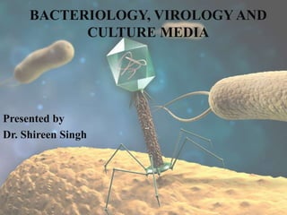
  • 1. BACTERIOLOGY, VIROLOGY AND CULTURE MEDIA Presented by Dr. Shireen Singh
  • 2. • CONTENTS 1. Bacteriology • Introduction • Microscopy • Study of bacteria • Background • Gram Stain • Bacteria – size, shape, structure. • Bacterial growth • Microbiology of periodontal disease 2. Virology • Introduction • Morphology • Oral viral infections of adults
  • 3. 3. Culture media • Introduction • Need for culture media • Requirements • Types of media • Culture methods • References
  • 4. Robert Koch (1843-1910) Father of Medical Microbiology  Developed Pure culture techniques  Used Potato slices First found individual bacterial colonies growing with different appearance.  Microscopic examination revealed cells within a single colony were similar pure culture may be isolated from a diseased organ or normally sterile site of the body
  • 5. INTRODUCTION • Microorganisms are living structures of microscopical size. • Organisms originally classified under plant and animal kingdoms. This classification proved unsatisfactory, therefore, a third kingdom Protista was formed. Protista Prokaryotes Eukaryotes
  • 6. MICROSCOPY Done for following purposes: The following types of microscopes are being employed a) Optimal or Light Microscope b) Phase Contrast Microscope c) Dark Field Microscope d) Fluorescent Microscope e) Electron Microscope f) Interference Microscope Magnification of an object Maximization of resolution Optimization of the contrast between structures, organisms and background.
  • 7. A. Optical or light microscope The compound light microscope consists of three sets of lenses • the condenser focuses light onto the specimen to give optimum illumination • the objective provides a magnified and inverted image of the specimen • the eyepiece adds further magnification  Equipped with objective lenses of low power(X10), high power (X40) and oil immersion (X100). Immersion oils have a refractive index similar to glass thus , use of these oils permit more light to be incorporated in the image resulting in improving the resolution power. Visualization of bacteria requires the use of immersion oil with X100 objective. This combination results in resolution of 0.2 microns.
  • 8. B. Phase contrast microscope • Contrast enhancing optical technique that can be utilized to produce high contrast images of transparent specimens(living cells,microorganisms,thin tissue slices,subcellular particles). • It employs an optical mechanism to translate minute variations in phase into corresponding changes in amplitude. • The living cells can be examined in their natural state without previously being killed, fixed and stained. • A special optical system(special , condensor and objective lens) converts difference in phase into difference in intensity of light producing light and dark contrast of the image.
  • 9.
  • 10. C. Dark field microscope Positive dark-field examination. Treponemes are recognizable by their characteristic corkscrew shape. Lighting system is modified to reach the specimen from the sides only. This is accomplished through the use of a special condenser that both blocks direct light rays and deflects light off a mirror on the side of the condenser at an oblique angle. This creates a “dark field” that contrasts against the highlighted edge of the specimens and results when the oblique rays are reflected from the edge of the specimen upward into the objective of the microscope.  Resolution by dark-field microscopy is quite high. this technique has been particularly useful for observing organisms such as Treponema pallidum(smaller than 0.2 mm)
  • 11. D. Fluorescent Microscope • The specimens are exposed to a light of shorter wavelength (ultraviolet light), which results in emission of longer wavelength visible light. • Bacteria stained with fluorescent dye(auramine, rhodamine) become visible as brighter glowing objects in a darker background.  Employed for detection of antigen (direct fluorescent antibody technique ) and antibodies (indirect fluorescent antibody methods). Fluorescence photomicrograph. A rod-shaped bacterium tagged with a fluorescent marker
  • 12. E. Electron Microscope • Resolution of the electron microscope is due to the fact that electrons have a much shorter wavelength than the photons of white light. • Wavelength of electrons is 0.005 nm as compared to 500 nm with visible light. • The resolving power of an electron microscope may be as low as 1–2 nm, enabling us to see viruses.
  • 13. Two types of electron microscopes in general use: the transmission electron microscope (TEM), which has many features in common with the light microscope, and the scanning electron microscope (SEM). TEM SEM
  • 14. Study of Bacteria A. Unstained(wet) preparations Unstained preparations are examined mainly for bacterial motility and for demonstration of spirochetes. B. Stained preparations Structural detail of bacteria cant be seen under light microscope due to lack of contrast. Hence it is necessary to use staining methods to produce color contrast. Common staining techniques Simple stains Negative staining Impregnation methods Differential stains
  • 15. Differential Stains • These stains impart different colors to different bacteria or bacterial structures. The most commonly employed differential stains are the Gram stain, the Acid-fast stain and the Albert stain.
  • 16. GRAM STAIN • The Gram stain was devised by the Danish physician, Hans Christian Gram, while working in Berlin in 1883. He later published this procedure in 1884. First Paper on Gram Staining Dr. Gram in his paper described how he was able to visualize what we now call Staphylococcus, Streptococcus, Bacillus, and Clostridia in various histological sections. Interestingly, Dr. Gram did not actually use safranin as a counter stain in the original procedure (Gram negative cells would be colorless). He instead recommended using Bismarck brown as a counter stain to enable tissue cell nuclei to be visualized.
  • 17. Definition A method of staining bacteria using a violet stain. The gram staining characteristics (denoted as positive or negative). A heat fixed bacterial smear is stained with crystal violet (methyl violet), treated with 3% iodine/potassium iodide solution, washed with alcohol and counterstained. The method differentiates bacteria into two main classes, gram-positive and gram-negative. • Gram (+) – accept gram stain – have simpler cell walls with large amounts of peptidoglycan • Gram (-) – do not stain – have more complex cell walls with less peptidoglycan – cell walls contain lipopolysaccharides – are more likely to be pathogenic (cause disease) – more resistant to antibiotics
  • 19. PRINCIPLES • Based on the composition of the cell wall Gram staining uses crystal violet to stain cell walls, iodine as a mordant, and a fuchsin or safranin counter stain to mark all bacteria. Gram status is important in medicine; the presence or absence of a cell wall will change the bacterium's susceptibility to some antibiotics. • Gram-positive bacteria - resist decolourization and retain the color of primary stain. i.e. dark blue or violet. • Gram negative bacteria - are decolorized by acetone/alcohol and therefore, take counter stain and appear red.
  • 20. Gram (+) and (-) Bacteria
  • 21.
  • 22. Four Major Steps in Gram Staining Applying a primary stain (crystal violet)or Methyl violet to a heat-fixed smear of a bacterial culture Addition of a mordant (Gram's iodine) Rapid decolorization with alcohol or acetone(10-30s) Counterstaining with Safranin or basic fuchsin(30s)
  • 23. STEP 1 Here, crystal violet dye is applied to the slide STEP 2 The slide is washed with water for 10seconds
  • 24. STEP 3 Gram iodide is applied & then washed with top water STEP 4 Washed off under top water
  • 25. STEP 5 Slide is being decolorized using ethanol & acetone STEP 6 Counter stain safranin applied for 30s importing pink color to gram negative bacteria
  • 26. How the Gram Stain Work? The Gram reaction is based on the structure of the bacterial cell wall. I. The Gram positive cells have a more acidic protoplasm which accounts for retaining the basic dye. The dark purple crystal violet stain is retained by the thick layer of peptidoglycan which forms the outer layer of the cell. II. In Gram-negative bacteria, the thin peptidoglycan layer in the periplasm does not retain the dark stain, and the pink safranin counter stains the peptidoglycan layer.
  • 27. ACID-FAST STAIN(ZIEHL-NEELSEN STAIN)  Ziehl-Neelsen staining is used to stain species of Mycobacterium tuberculosis that do not stain with the standard laboratory staining procedures like Gram staining.  The stains used are the red colored Carbol fuchsin that stains the bacteria and a counter stain like Methylene blue or Malachite green.
  • 28. BACKGROUND The Three Domain System or Super-Kingdom • Proposed by Woese 1970. • Under this system, organisms are classified into three domains and six kingdoms.  The domains are Archaea, Bacteria and Eukarya.  The kingdoms are Archaebacteria(ancient), Eubacteria(true),Protista, Fungi, Plantae and Animalia. • The current system groups the organisms based on the differences in ribosomal RNA structure.
  • 29.
  • 30. What are Prokaryotes and Eukaryotes? • Prokaryotes- Bacteria and blue green algae belong to this group. Bacteria are unicellular without any true branching except Actinomycetales. They don’t contain chlorophyll except for blue green algae. • Eukaryotes- Fungi, algae( except blue green algae),protozoa and slime moulds are included in this group. STRUCTURE Prokaryotes Eukaryotes Nucleus Nuclear membrane Absent Present Nucleolus Absent Present Chromosome One More than one Division By binary fusion By mitosis Cytoplasm Mitochondria, Golgi apparatus,Lysosomes,Endoplasmic reticulum All are absent All are present
  • 31.
  • 32. SIZE OF BACTERIA • Unit of measurement in bacteriology is the micron (micrometre, µm) • Bacteria of medical importance - 0.2 – 1.5 µm in diameter - 2 – 5 µm in length
  • 33. Morphology of bacteria Depending on the shape of bacteria, they are classified as: • Cocci – spherical/ oval shaped major groups • Bacilli – rod shaped • Spirochetes – flexible spiral forms • Actinomycetes – branching filamentous bacteria • Mycoplasmas – lack cell wall • Rickettsiae & Chlamydiae- small obligate parasites
  • 34. ARRANGEMENT OF BACTERIA Cocci in pair- Diplococcus Cocci in chain-Streptococci Cocci in cluster - Staphylococci Group of four-Tetrad Sarcina – groups of eight COCCI
  • 35. Coccobacilli Bacilli in chains Comma shaped Chinese letter pattern BACILLI
  • 37. The bacterial structures may be divided into two categories: I. Essential structures II. Other structures
  • 38. I. Essential structures • Nucleoid (nuclear material) • Cytoplasm • Cytoplasmic membrane • Cell wall II. Other structures • Capsule • Flagella • Pilli (Fimbriae) • Spores
  • 39. Nucleoid (nuclear material) Nucleoid • Only contains a single chromosome. • Has no nuclear membrane. • Has no nucleolus. Genome – - single, circular double stranded deoxyribonucleic acid (DNA) - Haploid - Divides by binary fission It is a closed circular thread about 1 mm long when straightened. Nucleoid of Bacillus cereus(2500x)
  • 40. Cytoplasm • The bacterial cytoplasm is a colloidal system containing a variety of organic and inorganic solutes in a viscous watery solution. • Acts as a semi permeable membrane: controls the inflow and outflow of metabolites • Accounts for 30% of the dry weight of bacterial cell. • Composed of 60% protein, 20- 30% lipids and 10-20% carbohydrate. • Contains ribosomes, mesosomes, vacuoles and inclusions granules. • Not containing endoplasmic reticulum. • Not containing mitochondria. Cytoplasm Cytoplasm Cytosol is the major component of the cytoplasm; it is the liquid portion of the cytoplasm typically composed of water, salts and organic molecules.
  • 41. Cell membrane The cell membrane is a typical phospholipid bilayer that contains the following constituents: a. Cytochromes and enzymes involved in electron transport and oxidative phosphorylation. b. Carrier lipids, enzymes, and penicillin-binding proteins (PBP) involved in cell wall biosynthesis. c. Enzymes involved in phospholipid synthesis and DNA replication. d. Chemoreceptor.
  • 42. Ribosomes • Centres of protein synthesis. • Ribosomes are a prominent particle of the cell. They are present in both eukaryotic and prokaryotic cell. • 10-20nm size with a sedimentation constant of 70 S(Svedberg units). Composed of Ribosomal RNA(rRNA) Ribosomal proteins
  • 43. Inclusion granules Inclusion granules are insoluable, osmotically inert, round, neutral polymers in the cytoplasm of bacteria. They act as food reserves and may contain organic substances such as starch, glycogen or lipid. They consist of • Volutin granules (polymetaphosphate) • Lipid granules • Polysaccharide granules (starch or glycogen) • Granules of sulphur
  • 44. Mesosomes(Chondroids) Functions • Principal sites of respiratory enzymes • Coordinate nuclear & Cytoplasmic division during binary fission  More prominent in Gram +ve bacteria  Two types- septal and lateral. The septal mesosome is attached to bacterial chromosome and involved in DNA segregation. Vesicular, multilaminated structures formed as invaginations of plasma membrane.
  • 45. CELL WALL • It refers to that portion of the cell envelope that is external to the cytoplasmic membrane and internal to the capsule or glycocalyx. Tough and rigid structure, surrounding the bacterium like a shell. 1. Confers shape and rigidity 2. 10 - 25 nm thick 3. Weighs about 20-25% of dry weight of the cell. 4. Average thickness is 0.15-0.5 μm. 5. Young and rapidly growing bacteria has thin cell wall but old and slowly dividing bacteria has thick cell wall. Composed of N-acetyl Muramic acid and N- acetyl Glucosamine back bones cross linked with peptide chain and pentaglycine bridge.
  • 46. • Components of cell wall of Gram positive bacteria 1. Peptidoglycan 2. Teichoic and teichuronic acid 3. Polysaccharides • Components of cell wall of Gram negative bacteria 1. Peptidoglycan 2. Lipoprotein 3. Phospholipids 4. Lipopolysaccharide
  • 47.
  • 48. The Gram-positive cell wall is composed of a thick, multilayered peptidoglycan sheath outside of the cytoplasmic membrane. Teichoic acids are linked to and embedded in the peptidoglycan, and lipoteichoic acids extend into the cytoplasmic membrane
  • 49. The Gram-negative cell wall is composed of an outer membrane linked to thin, mainly single-layered peptidoglycan by lipoproteins. The peptidoglycan is located within the periplasmic space that is created between the outer and inner membranes. The outer membrane includes porins, which allow the passage of small hydrophilic molecules across the membrane, and lipopolysaccharide molecules that extend into extracellular space.
  • 50. FUNCTIONS 1. Accounts for the shape of the cell. 2. Provides protection to the cell against osmotic damage. 3. Confers rigidity upon bacteria. 4. Provides staining characteristics to the bacterium 5. Takes part in cell division. 6. Possesses target site for antibiotics, lysozymes and bacteriophages. It carries bacterial antigens that are important in virulence and immunity.
  • 51. Additional Organelles 1. Capsule & Slime layer – – Viscous layer secreted around the cell wall. – Polysaccharide / polypeptide in nature a) Capsule – sharply defined structure, antigenic in nature • Protects bacteria from lytic enzymes • Inhibits phagocytosis • Stained by negative staining using India Ink • Can be demonstrated by Quellung reaction. b) Slime layer – loose undemarcated secretion which is easily washed off from cell envelope. All bacteria have at least a thin slime layer.
  • 52. (contd).. 2. Flagella – Filamentous protein structures attached to the cell surface that provide the swimming movement for most motile prokaryotes.  Size: 3-20μm in length and 0.01-0.013μm in diameter. It is composed of protein named as flagellin. It is the organ of locomotion in bacterial cell and consists of thee parts. -The filament -The hook - The basal body The basal body and hook are embedded in the cell surface while the filament is free on the surface of bacterial cell. Electron Micrograph of Vibrio cholera
  • 53.  Their presence in bacterial cell is detected by -Hanging drop preparation. -Swarming phenomenon on surface of plate agar . -Motility media -Special staining methods -Silver impregnation methods -Dark –field microscopy -Electron microscopy
  • 56. (contd).. 3. Fimbriae/ Pili –  Thin, hair like appendages on the surface of many Gram-negative bacteria  10-20µm long, acts as organs of adhesion (attachment) - allowing bacteria to colonize environmental surfaces or cells and resist flushing  Made up of proteins called pilins.  Pili can be of two types –  Common pili – short & abundant  Sex pili - small number (one to six), very long pili, helps in conjugation (process of transfer of DNA) Electron micrograph of Fimbriae of Neisseria gonorrhoeaea
  • 57. (contd).. 4. Spores – Resting cells which are capable of surviving under adverse environmental conditions like heat, drying, freezing, action of toxic chemicals and radiation. • Bacterial spore is smooth walled and oval or spherical in shape. • It does not take up ordinary stains. It looks like areas of high refractivity under light microscope. Spores are detected by  Simple staining methods  Special staining methods
  • 58. Bacterial Growth Bacterial growth refers to an increase in bacterial cell numbers (multiplication).  It results from bacterial reproduction due to binary fission, which may be characterized by a parameter called generation time (the average time required for cell numbers to double).  It usually occurs asynchronously (i.e., all cells do not divide at precisely the same moment)
  • 59. Cell Growth and Binary Fission • In a growing rod-shaped cell, elongation continues until the cell divides into two new cells. This process is called binary fission (“binary” to express the fact that two cells have arisen from one). • In a growing culture of a rod- shaped bacterium such as Escherichia coli, cells elongate to approximately twice their original length and then form a partition that constricts the cell into two daughter cells.
  • 60. • The partition is called a septum and results from the inward growth of the Cytoplasmic membrane and cell wall from opposing directions; septum formation continues until the two daughter cells are pinched off. • Generation time is the time taken for the size of a bacterial population to double. Bacteria grow by taking nutrients and incorporate them into cellular components; then bacteria divide into two equal daughter cells and double the number. • The generation time in most of the medically important bacteria is about 20minutes. In Mycobacterium tuberculosis it is about 20 hours & in lepra bacilli, it is 20 days.
  • 61. Bacterial growth curve • The bacterial growth curve involves the inoculation of bacteria from a saturated culture into fresh liquid media. It is unique for a particular nutritional environment. • Illustrated in a plot of logarithmic number of the number of bacteria versus time; the generation time is determined by observing the time necessary for the cells to double in number during the log phase of growth.
  • 62. The growth curve has four phases
  • 64. Lag Phase • When a microbial culture is inoculated into a fresh medium, growth usually begins only after a period of time called the lag phase. • There is increase in the size of cells but there is no appreciable increase in numbers. • During the end of the lag phase, the bacteria have maximum cell size.
  • 65. Log or Exponential phase • During the exponential phase of growth each cell divides to form two cells, each of which also divides to form two more cells, and so on. • Cells in exponential growth are typically in their healthiest state and hence are most desirable for studies of their enzymes or other cell components. • The cells in this phase are smaller in size and stain uniformly.
  • 66. Stationary Phase • In the stationary phase, there is no net increase or decrease in cell number and thus the growth rate of the population is zero. Although the population may not grow during the stationary phase, many cell functions can continue, including energy metabolism and biosynthetic processes. • Some cells may even divide during the stationary phase but no net increase in cell number occurs. This is because some cells in the population grow, whereas others die, the two processes balancing each other out. This is a phenomenon called cryptic growth.
  • 67. Phase of Decline • After a period of stationary phase, the bacterial population decreases due to the death of cells. • The decline phase starts due to exhaustion of nutrients, accumulation of toxic products and autolytic enzymes. • There is decline in viable count and not in total count.
  • 69. Periodontal disease is a chronic bacterial infection characterized by persistent inflammation, connective tissue breakdown and alveolar bone destruction. (Yamamoto et al., 2011) • Gingivitis is a reversible dental plaque induced inflammation of the gingiva. • Periodontitis, which is bacterially induced, can be defined as a chronic inflammatory disease initiated by dental plaque bio film and perpetuated by a deregulated immune response (Suvan et al., 2011) usually accompanied by gingivitis resulting in irreversible destruction of the supporting tissues surrounding the tooth, including the alveolar bone. (Yamamoto et al., 2011)
  • 70. It is estimated that more than 700 different species are capable of colonizing the adult mouth and that any individual typically harbors 150 or more different species.
  • 71. Koch's Postulates(1876) 1.The micro-organism should be found in every case of the disease and under conditions which explain the pathological changes and clinical features. 2.It should be possible to isolate the causative agent in pure culture from the lesion. 3.When such pure culture is inoculated into appropriate laboratory animal, the lesion of the disease should be reproduced. 4.It should be possible to re isolate the bacterium in pure culture from the lesion produced in the experimental animal.
  • 72. Socransky’s Postulates (1992) 1. The organism must be found in relatively high numbers in proximity to the periodontal lesion 2. The organism must be absent or present in much smaller numbers in periodontally healthy subjects or in subjects with other forms of periodontal disease. 3. The organism must have high levels of serum, salivary and gingival crevicular fluid antibody developed against it in periodontally diseased subjects. 4. The organism must be found to produce virulence factors in vitro which can be correlated with clinical histopathology. 5. The organism must mimic similar pathogenic properties in an appropriate animal model.
  • 73. Socransky et al. (1998) examined over 13,000 sub gingival plaque samples from 185 adults, and identified six specific microbial groups of bacterial species.
  • 75. Possible etiologic agents of periodontal disease • Aggregatibacter actinomycetemcomitans • Porphyromonas gingivalis • Tannerella forsythia (Bacteroides forsythus) • Treponema denticola • Prevotella intermedia • Fusobacterium nucleatum • Eikenella corrodens • Campylobacter rectus (Wolinella recta) • Peptostreptococcus micros • Streptococcus intermedius
  • 76. AGGREGATIBACTER ACTINOMYCETEMCOMITANS • Gram negative coccobacillus, facultative non motile, rod-shaped. • Non sporulating, non-motile and non-branching bacteria • The genus name Actinobacillus refers to actin- star shaped bacillus- rod shaped
  • 77. History • First recognized as periodontal pathogen by Newman et al., 1976. • It was originally named - Bacterium actinomycetem comitans (Klinger 1912) - Bacterium comitans (Lieske 1921) - Actinobacillus actinomycetemcomitans (Topley & Wilson1929) - Haemophilus actinomycetemcomitans (Potts 1985) Aggregatibacter a member of family Pasteurellaceae, has been recognized as one of the key pathogens in periodontitis. (Slots et al. 1990)
  • 78. • Small, non motile, G-ve, saccharolytic, round ended rod. (Haffajee and Socransky 1994) • Forms small convex colonies with a star shaped center when grown on blood agar. • Its presence in the periodontal pocket is associated with preadolescent, localized juvenile now known as localized aggressive periodontitis (LAP) (Newman et al. 1976) and advanced adult aggressive periodontal disease.
  • 79. PORPHYROMONAS GINGIVALIS • It is a G-ve, anaerobic, non motile, asaccharolytic rod that usually exhibit coccal to short rod morphologies. • Group of “black pigmented Bacteroides” • Possesses a capsule which protects against phagocytosis,fimbriae (for adhesion) and vesicles & produces a number of virulence factors: - proteases (for destruction of immunoglobulins and the complement factors, and degradation of host-cell collagenase inhibitors) - collagenase - hemolysin - endotoxin - fatty acids
  • 80. • Form brown to black colonies on blood agar plates. • P.gingivalis has been shown to induce elevated systemic and local immune responses in subjects with various forms of periodontitis. (Haffaje & Socransky 1994)
  • 82. Introduction to Virology A virus is an obligate intracellular infective agent containing only one type of nucleic acid (DNA or RNA) as their genome.  Have no metabolic activity outside the living cells.  Do not possess a cellular organization and lack the enzymes necessary for protein and nucleic acid synthesis.  Multiply by complex process and not by binary fission.
  • 84. MORPHOLOGY OF VIRUS A. SIZE Viruses are much smaller in size than other organisms. The extracellular infectious virus particle is called the virion. Size ranges from 20 to 300 nm in diameter. The most direct method for measuring virus size is electron microscopy.
  • 85.
  • 86. B. STRUCTURE Viruses consist of Nucleic acid core(genome) surrounded by a protein coat. The NA is the genome that contains the information necessary for virus multiplication; the protein is arranged around the genome in the form of a shell that is the capsid. The shell + the NA is the Nucleocapsid.
  • 87. Virions o Infectious particle composed of either RNA or DNA that is encased in a protein coat called a capsid. o They are either naked or enveloped, depending on whether the capsid is surrounded by a lipoprotein envelope. o Virions replicate only in living cells and therefore are obligate intracellular parasites. o They cannot be observed with a light microscope.
  • 88. Capsid Composed of structural units called capsomers, which are aggregates of viral- specific polypeptides. 1. They are classified as helical, icosahedral (a 20-sided polygon), or complex; used as a criterion for viral classification. 2. Serves four functions: a. Protects the viral genome. b. Is the site of receptors necessary for naked viruses to initiate infection. c. Stimulates antibody production. d. Is the site of antigenic determinants important in some serologic tests. 3. The structure is composed of the nucleic acid genome and the capsid proteins is called the nucleocapsid.
  • 89. C. SYMMETRY Three types of symmetry are determined by the arrangement of capsid around the nucleic acid core • Icosahedral symmetry is the one in which the capsomers are made up of 20 triangular faces (five at the top, five at the bottom, and ten around the middle) that form a symmetric figure (an icosahedron). An icosahedron has three axes of symmetry: fivefold, three fold, and twofold. This type of symmetry is found in papova,picorna,adenovirus(all naked or enveloped) and herpes. • In the Helical symmetry, the nucleic acid and the capsomers are wound together to form a helical or spiral tube. • Some viruses do not show ether icosahedral or helical symmetry due to the complexity of their structures. These are referred to have Complex symmetry. E.g. Poxvirus.
  • 90. Envelopes  The viral envelope surrounds the nucleocapsid of enveloped viruses and is composed of viral specific glycoproteins and host-cell-derived lipids and lipoproteins.  It contains molecules that are necessary for enveloped viruses to initiate infection, act as a stimulus for antibody production, and serve as antigens in serologic tests; it also forms the basis of ether sensitivity of a virus.
  • 91. D. Shape of virus The shape of virus particles is determined by the arrangement of the repeating subunits that form the protein coat (capsid) of the virus.
  • 93. • Viral diseases of the oral mucosa and the perioral region are often encountered in dental practice, but have received only limited research interest. • Viruses are important ulcerogenic and tumorigenic agents of the human mouth. • The finding of an abundance of mammalian viruses in periodontitis lesions may suggest a role for viruses in more oral diseases than previously recognized.
  • 94. Virus Viral genome Enveloped virus Characteristics Disease association Herpesviruses Double- stranded DNA Yes HSV-1 Latency in sensory ganglia. Causes orolabial disease. Herpetic gingivostomatitis, recurrent orolabial lesion, encephalitis, neonatal infections HSV-2 Latency in sensory ganglia. Causes genital and newborn infections Genital infection, aseptic meningitis, sacral autonomic nervous system dysfunction Varicella– zoster virus Latency in sensory ganglia. Varicella (chickenpox), herpes zoster (shingles), CNS involvement, pneumonia, secondary bacterial infections &death. Epstein–Barr virus Identified initially in 1964 from African Burkitt lymphoma. Infects epithelial cells with a cytolytic infection and B lymphocytes with a latent infection Infectious mononucleosis, hairy leukoplakia of the tongue, Burkitt lymphoma, B lymphoproliferative disease, Hodgkin!s lymphoma, nasopharyngeal carcinoma, gastric carcinoma, parotid carcinoma . HSV-6 Cell tropism for T lymphocytes and neural cells. Frequently shed in the saliva of healthy donors Roseola infantum, meningitis, encephalitis, possibly multiple sclerosis HSV-7 Latency in macrophages and T lymphocytes. Frequently shed in the saliva of healthy donors Exanthema subitum, macular-papular rashes, transplant- recipient pathogen HSV-8 Six genetic subtypes with marked clustering to geographical areas. B lymphocytes and monocytes serve as Kaposi’s sarcoma, multicentric Castleman disease, primary effusion lymphoma, mononucleosis-like illness, aplastic anemia Oral viral infections of adults Periodontology 2000, Vol. 49, 2009
  • 95. Virus Viral genome Enveloped virus Characteristics Disease association Papilloma viruses Double- stranded DNA No Epithelial cell proliferation with specificity in the ano- genital area, urethra, skin, larynx, tracheo- bronchial and oral mucosa Genital and cutaneous warts, cervical and anogenital cancers, condylomata acuminate(sexually transmitted diease), recurrent respiratory papillomatosis Picornaviruses Single-stranded RNA No Coxsackievirus Coxsackie virus A16 is closely related to Enterovirus-71, and both belong to a discrete subgroup of type A enteroviruses that are prominently associated with hand, foot and mouth disease Uncomplicated hand, foot and mouth disease herpangina , myocarditis, infectious type 1 diabetes , atherosclerosis Echovirus Some echovirus replication occurs in the nasopharynx Meningitis, pericarditis, myocarditis, herpangina Enterovirus Enterovirus-71 was first isolated in 1969 from a child with encephalitis. Can cause large epidemics of acute disease. Mutates readily Hand, foot and mouth disease , herpangina, poliomyelitis-like illness, meningoencephalitis, acute pulmonary edema Oral viral infections of adults Periodontology 2000, Vol. 49, 2009
  • 96. Herpes viruses • Infection that accompanies primary herpetic gingivostomatitis is usually subclinical in early childhood & only a small percentage of patients develop an acute primary infection. • Usually occurs in older children • Consists of - High temperature (102-104°F) - Anorexia and listlessness - headache -Tender regional lymphadenopathy - vesiculo-ulcerative eruption on the peri-oral skin, vermilion or any intra- oral mucosal surface. • Abrupt onset • Gingivitis (most striking feature, with markedly swollen, erythematous, friable gums.) • Perioral skin involvement due to contamination with infected saliva
  • 97. • The vesicles are 2 to 3 mm in diameter, rupture, leaving painful ulcers that heal without scarring after seven to ten days. • Gingivae are swollen and reddish due to a general inflammation. Vesicles that break down to form clusters of small round or irregular superficial ulcers with a yellowish base and a red margin. Lesions presented as multiple ulcerations on the lip and tongue
  • 98. Human immunodeficiency virus • Etiologic agent of Acquired immune deficiency syndrome(AIDS) • The lesions may be present in up to 50% of people with HIV infection & in up to 80% of those with a diagnosis of AIDS. • The lesions are clearly visible and can be diagnosed reliably from the clinical features alone. • The lesions parallel the decline in number of CD4 cells and an increase in viral load and are independent indicators of disease progression. • Disease progression is characterized by an increase prevalence of oral candidiasis, oral hairy leukoplakia, ulcerative periodontal disease and xerostomia.
  • 99.
  • 100. Classification of orofacial lesions associated with HIV/AIDS in adults Stomatologija, Baltic Dental and Maxillofacial Journal, 2015, Vol. 17
  • 101. LINEAR GINGIVAL ERYTHEMA • a distinct fiery red band along the margin of the gingiva, most frequently found in anterior teeth, accompanied in some cases by bleeding and discomfort. • Possible etiology –Candida dubliniensis • It manifests in immuno compromised patients with CD4+ T lymphocyte counts <200 cells/mm3. Linear gingival erythema in 49 year old female with AIDS, CD4 count 115.
  • 102. NECROTISING ULCERATIVE DISEASE • Necrotising ulcerative disease (NUD) is sub classified as necrotising ulcerative gingivitis (NUG) and necrotising ulcerative periodontitis (NUP) that appear to be different stages of the same disease. (NUG) (NUP) escorted by bleeding, extremely sharp pain, ulcerated gingival papillae, rapid and extensive soft tissue necrosis and advanced loss of periodontal attachment frequently leading to bone exposure and crater shaped defects. characterized by rapid onset and acute painful inflammation of gingiva with rapid destruction of soft tissues. • NUD is usually followed by fever, malaise, halitosis and lympthadenopathia. • NUD is more common among immuno compromised patients, particularly if they have psychological/motivational problems, poor nutrition, and use tobacco or other drugs.
  • 103. Necrotizing ulcerative disease in 17 year old immunocompetent female
  • 104. HIV infection and periodontal diseases: an overview of the post-HAART era HIV infection and periodontal diseases M Mataftsi et al
  • 106. INTRODUCTION • Definition Culture is the term given to microorganisms that are cultivated in the lab for the purpose of identifying and studying them. Medium is the term given to the combination of ingredients that will support the growth and cultivation of microorganisms by providing all the essential nutrients required for the growth (that is, multiplication) in order to cultivate these microorganisms in large numbers to study them. Inoculum is the suspension of microorganisms. Inoculation is the introduction of microbes into culture medium.
  • 107. Need for Culture Media It is usually essential to obtain a culture by growing the organism in an artificial medium. If more than one species or type of organism are present each requires to be carefully separated or isolated in pure culture. Several organism need the determination of Antibiotic sensitivity pattern for optimal antibiotic selection.
  • 108. Essential requirements in culture media Any culture medium must contains: -A source of energy -Sources of carbon, nitrogen, sulfur, phosphorus -Minerals -Vitamins and growth factors -Water
  • 109. Types of media I. Based on physical state a) solid medium b) liquid medium c) semi solid medium II. Based on nutritional factors a) simple medium b) complex medium c) synthetic or defined medium d) Special media
  • 110. Special media – Enriched media – Enrichment media – Selective media – Differential media – Indicator media – Transport media – Sugar media III.Based on Oxygen requirement - Aerobic media - Anaerobic media
  • 111. Simple media / basal media Nutrient broth(NB) is an example of simple medium.  May be in liquid form: - NB consists of peptone +meat extract 1% + NaCl.  May be in a solid form - NB + 2-3% agar = Nutrient agar Simplest & routinely employed medium for diagnostic purposes
  • 112. Complex media • Media other than basal media. • They have added ingredients. • Provide special nutrients Synthetic or defined media • Media prepared from pure chemical substances and its exact composition is known • Eg: peptone water – 1% peptone + 0.5% NaCl in water
  • 113. SPECIAL MEDIA 1. Enriched media • Substances like blood, serum, egg are added to the basal medium. • Used to grow bacteria that are exacting in their nutritional needs. • Eg: Blood agar, Chocolate agar
  • 115. 2. Enrichment media • Liquid media used to isolate pathogens from a mixed culture. • Media is incorporated with inhibitory substances to suppress the unwanted organism. • Eg: – Selenite ‘F’ Broth – for the isolation of Salmonella, Shigella – Alkaline Peptone Water – for growingVibrio cholerae
  • 116. 3. Selective media The inhibitory substance is added to a solid media. - Used to isolate a particular bacteria from specimens where mixed bacterial flora is expected. Examples Deoxycholate citrate agar(DCA) : for enteric bacilli(Salmonella,Shigella) Bile salt agar(BSA) : Vibrio cholerae
  • 117. 4. Differential media A media which has substances incorporated in it enabling it to distinguish between bacteria. Eg: Mac Conkey’s medium – Peptone – Lactose – Agar – Neutral red – Taurocholate • Distinguish between lactose fermenters(LF) & non lactose fermenters(NLF) by forming pink colored colonies in LF & colorless or pale pink colonies in NLF.
  • 118.
  • 119. 5. Indicator media • These media contain an indicator which changes its color when a bacterium grows in them. • Eg: – Blood agar – Mac Conkey’s medium – Wilson & Blair medium
  • 120. 6. Transport media • Media used for transporting the samples. • Delicate organisms may not survive the time taken for transporting the specimen without a transport media. • Eg: – Stuart’s medium – non nutrient soft agar gel containing a reducing agent – Buffered glycerol saline – enteric bacilli.
  • 121. 7. Sugar media • Media containing any fermentable substance helpful for identification of bacteria. • Eg: glucose, sucrose, lactose, starch etc. • Media consists of 1% of the sugar in peptone water. • Contain a small tube (Durham’s tube) for the detection of gas by the bacteria.
  • 122. ANAEROBIC MEDIA • Used for cultivation of anaerobic bacteria eg: a. Cooked meat broth(CMB) b. Thioglycollate broth.
  • 123. Cooked meat broth(CMB) • Known as Robertson’s cooked meat(RCM) medium. Contains nutrient broth & pieces of fat free minced cooked meat of ox heart. Procedure Before inoculation, the medium is boiled in water bath at 80°C for 30 min to make it oxygen free. For strict anaerobiosis the surface of CMB medium is covered with a layer of sterile liquid paraffin.
  • 124. CULTURE METHODS  Culture methods employed depend on the purpose for which they are intended.  The indications for culture are: – To isolate bacteria in pure cultures. – To demonstrate their properties. – To obtain sufficient growth for the preparation of antigens and for other tests. – For bacteriophage & bacteriocin susceptibility. – To determine sensitivity to antibiotics. – To estimate viable counts. – Maintain stock cultures.
  • 125. Culture methods include: 1. Streak culture 2. Lawn culture 3. Stroke culture 4. Stab culture 5. Pour plate method 6. Liquid culture 7. Anaerobic culture methods
  • 126. STREAK CULTURE (Surface plating) It is a routine method employed for bacterial isolation in pure culture. A platinum or nichrome wire loop is used. This loop is first sterilized in the Bunsen flame by making it red hot and cooled by touching an uninoculated part of the medium.
  • 127. Primary Streak (shown in yellow)  Hold the plate on one hand & inoculated loop on the other.  Dip your inoculating loop into a broth culture, or touch it to the material you want to spread -an isolated colony or swab an area for which you want to quantify the microbial species present.  Go back and forth a number of times in a small area of the Petrie plate. The goal is to spread your material completely over this initial area of the plate
  • 128. Secondary Streak (shown in Blue)  Sterilize inoculating loop & squelch it in an unused area of the agar before streaking to cool it.  Pick up the plate and rotate it 1/4 of a turn to your left.  Run the loop through the previous streak 2-3 times, then draw it along 1/3 of the remaining plate.
  • 129. Tertiary Streak (Orange) • Rotate the plate another 1/4 turn and sterilize your inoculating loop or take a fresh, sterile stick or swab. Squelch a heated inoculated loop in an unused area of the agar before streaking to cool it. • Run the loop through the previous, secondary streak 2-3 times, and draw the streak over a remaining 1/3 of the plate.
  • 130. Quaternary Streak (Black) • Rotate the plate another 1/4 turn and sterilize the inoculating loop. Squelch a heated inoculated loop in an unused area of the agar before streaking to cool it. • Run the loop through the previous tertiary streak 2 times and draw over the remaining free space in the plate, being careful not to contact the primary streak (yellow). • Incubate the plate as needed, and check 18-24 hours later for growth.
  • 131. References 1. Textbook of microbiology -CP Baveja 2. Essential microbiology - Stuart Hogg 3. Todar's Online Textbook of Bacteriology 4. Medical Microbiology (Twenty-Sixth Edition) 5. Periodontal Microbiology 6. Microbiology of Periodontal Diseases. A Review 7. Microbial etiology of periodontal disease – mini review; Ljiljana Kesic et al 8. Actinobacillus actinomycetemcomitans in human periodontal disease: a cross- sectional microbiological investigation Jorgen Slots Sept. 1980 9. Human viruses in periodontitis JØRGEN SLOTS Periodontology 2000 2010;53:89–110 10. Introduction to Modern Virology N. J. Dimmock A. J. Easton K. N. Leppard Sixth Edition 11. Oral viral infections of adults Jørgen Slots Periodontology 2000 2009;49:60– 86.