Quantitative Analysis of Oligonucleotides in Human Muscle Tissue Using Liquid Chromatography Coupled with High Resolution/Accurate Mass (HR/AM) Mass Spectrometry

APA 2019 -- Duchenne muscular dystrophy (DMD) is a rare X-linked recessive neuromuscular disease characterized by progressive severe muscle wasting and weakness. DMD is ultimately fatal, with patients typically dying from respiratory or cardiac complications in their mid- to late-20s. Exon skipping by phosphorodiamidate morpholino oligomer (PMO) is considered a promising, disease-modifying approach to treat the underlying cause of DMD. PMO was conjugated to a proprietary peptide to enhance tissue uptake, providing a PPMO. This poster describes the development and validation of a sensitive, selective and high-throughput liquid chromatography-tandem high resolution-accurate mass (LC/HR-AM) method for the quantitation of the PPMO in human muscle tissue using an analogue as the internal standard (ISTD). A key modification to the PMO is the addition of a proprietary peptide that provides specificity to binding and due to the metabolism of the peptide several entities of the PMO will be present in the muscle tissue, in order to quantitate the total amount of the PPMO in the muscle. The extraction undergoes a peptide digestion step to form an end product of PMO-A prior to the analysis.
Recommended

Recommended
More Related Content
What's hot
What's hot (15)
Similar to Quantitative Analysis of Oligonucleotides in Human Muscle Tissue Using Liquid Chromatography Coupled with High Resolution/Accurate Mass (HR/AM) Mass Spectrometry
Similar to Quantitative Analysis of Oligonucleotides in Human Muscle Tissue Using Liquid Chromatography Coupled with High Resolution/Accurate Mass (HR/AM) Mass Spectrometry (20)
More from Covance
More from Covance (20)
Recently uploaded
Recently uploaded (20)
Quantitative Analysis of Oligonucleotides in Human Muscle Tissue Using Liquid Chromatography Coupled with High Resolution/Accurate Mass (HR/AM) Mass Spectrometry
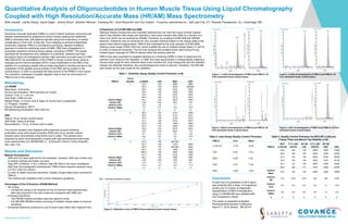
- 1. Presented at APA 2019 Quantitative Analysis of Oligonucleotides in Human Muscle Tissue Using Liquid Chromatography Coupled with High Resolution/Accurate Mass (HR/AM) Mass Spectrometry Nidhi Jaiswal1, Jianbo Zhang2, Sarah Kriger1, Jeremy Elison1, Brandon Wilcock1, Yuanfeng Wu1, Scott Reuschel1 and Troy Voelker1; 1Covance Laboratories Inc., Salt Lake City, UT; 2Sarepta Therapeutics, Inc., Cambridge, MA Introduction Duchenne muscular dystrophy (DMD) is a rare X-linked recessive neuromuscular disease characterized by progressive severe muscle wasting and weakness. DMD is ultimately fatal, with patients typically dying from respiratory or cardiac complications in their mid- to late-20s. Exon skipping by phosphorodiamidate morpholino oligomer (PMO) is considered a promising, disease-modifying approach to treat the underlying cause of DMD. PMO was conjugated to a proprietary peptide to enhance tissue uptake, providing a PPMO. This poster describes the development and validation of a sensitive, selective and high- throughput liquid chromatography-tandem high resolution-accurate mass (LC/HR- AM) method for the quantitation of the PPMO in human muscle tissue using an analogue as the internal standard (ISTD). A key modification to the PMO is the addition of a proprietary peptide that provides specificity to binding and due to the metabolism of the peptide several entities of the PMO will be present in the muscle tissue, in order to quantitate the total amount of the PPMO in the muscle. The extraction undergoes a peptide digestion step to form an end product of PMO-A prior to the analysis. Methodology LC-HRAM Mass Spec: Q Exactive Source and Ionization: HESI (positive ion mode) Column: C18, 2.1 x 50 mm Flow Rate: 0.400 mL/min Mobile Phase: A: formic acid in water; B: formic acid in acetonitrile LC Program: Gradient Source Temperature: 350°C MS Monitoring Parameters: 600-1200 m/z SPE Aliquot: 30 μL human muscle tissue SPE Plate: Oasis HLB Plate Reconstitution: 175 µL of formic acid in water The muscle samples were digested with proteinase enzyme following purification using solid phase extraction (SPE) and 30 µL sample volume. Extracts were reconstituted using formic acid in water. The extracts were analyzed by liquid chromatography coupled with high-resolution/accurate-mass mass spectrometry (LC-HR/AM MS) i.e., Q Exactive (Thermo Fisher Scientific, San Jose, CA). Comparison of Full MS-SIM and tSIM Although desired compounds and unwanted interferences can have the same nominal masses (which may interfere with single unit resolution), their exact masses often differ by a fraction of a mass unit, which can be resolved by HR/AM. Therefore, by coupling Full MS-SIM with HR/AM detection, selectivity may be achieved for very complex matrices based on the charge state of the parent mass without fragmentation. PMO-A was evaluated by full scan analysis (Full MS-SIM) utilizing a scan range of 600-1200 m/z, which enabled the use of multiple charge states (11 and 9) in order to maximize sensitivity. The full scan analysis also enabled future data mining for any missed trypsin cleavage for PMO-A interest within the existing data file. PMO-A was also evaluated by targeted selected ion monitoring (tSIM) in order to determine the optimum scan mode for the validation. In tSIM, the mass spectrometer is independently trapping a narrow mass range for each selected mass in the inclusion list. Only compounds with the selected masses are detected; therefore, the overall background noise is reduced. Therefore, Full MS-SIM was chosen as the scan mode for the validation. Results and Discussion Method Development ▶ SPE and LLE were explored for the extraction. However, SPE was chosen due to cleaner extracts and better recovery. ▶ Oligo RP-2.0x50mm, C18 2.1x50mm, Max RP-30x2.0 mm were considered; after final chromatographic development, PMO-A were baseline resolved using a C18 2.1x50 mm column. ▶ In order to obtain improved sensitivity, multiple charge states were summed for PMO-A. ▶ The method was validated under current regulatory guidelines. Advantages of the Q Exactive (HRAM Method) ▶ No Tuning − Compound tuning is not required for the Q Exactive mass spectrometer; data was acquired in full scan mode and compared with tSIM, prm. ▶ Increased Sensitivity − The HR/AM method provided improved signal to noise. − Full MS-SIM HR/AM enables summing of multiple charge states to improve sensitivity. ▶ Enhanced selectivity achieved by use of exact mass rather than fragment ions. PMO-A Curve Number LLOQQC LQC MQC HQC 1 44.6 148 2280 4680 58.9 155 2240 &&4820 54.6 146 2310 4450 53.8 155 2300 4450 53.6 156 2250 4550 56.9 162 2210 4610 Intrarun Mean 53.7 154 2270 4590 Intrarun SD 4.92 5.82 38.3 143 Intrarun %CV 9.2 3.8 1.7 3.1 Intrarun %Bias 7.4 2.7 13.5 14.8 n 6 6 6 6 2 55.0 157 2110 4390 56.8 154 2110 4310 47.6 149 2140 4410 51.3 156 2010 4570 53.8 152 1960 4600 52.2 149 2060 4780 Intrarun Mean 52.8 153 2070 4510 Intrarun SD 3.21 3.43 68.9 173 Intrarun %CV 6.1 2.2 3.3 3.8 Intrarun %Bias 5.6 2.0 3.5 12.8 n 6 6 6 6 3 61.8 166 &&1550 60.4 148 2100 4580 60.9 155 &&1590 4820 60.0 147 1860 4420 &63.3 136 2060 4460 57.2 140 2120 4000 Intrarun Mean 60.6 149 1880 4460 Intrarun SD 2.04 10.8 258 299 Intrarun %CV 3.4 7.2 13.7 6.7 Intrarun %Bias 21.2 -0.7 -6.0 11.5 n 6 6 6 5 PMO-A %CV %Bias LLOQ QC 11.50 16.60 LQC 8.20 -0.70 MQC 10.80 0.50 HQC 5.00 11.80 DQC 13.80 -0.50 PMO-A MTX LLOQ QC F/T 4 -20C LQC/5C B/T WI LQC F/T 4 -20C HQC/5C B/T WI HQC DQC 48.6 146 150 4300 4500 21900 46.8 162 163 4780 3720 22300 53.2 145 139 4520 4230 18800 53.6 140 146 4330 4270 20100 53.7 147 159 4290 4460 14900 59.0 162 141 4120 4540 21200 Intrarun Mean 52.5 150 150 4390 4290 19900 Intrarun SD 4.31 9.35 9.67 230 305 2750 Intrarun %CV 8.2 6.2 6.4 5.2 7.1 13.8 Intrarun %Bias 5.0 0.0 0.0 9.8 7.3 -0.5 n 6 6 6 6 6 6 Conclusion A lower limit of quantitation of 50.0 ng/mL was achieved with a linear 1/x2 regression model over 2.0 orders of magnitude. Quantification of PMO-A therapeutics using LC-HR/AM MS was validated with GLP acceptance criteria. Table 1. Intra/Inter Assay Quality Control Precision (n=6) Table 2. Inter-Assay Quality Control Precision Table 3. Quality Control Precision for MTX Eff LLOQ and Freeze-Thaw and Bench-Top Stability (n=6) Figure 3. Blank chromatograms of PMO-A and PMO-A -IS from extracted human muscle tissue. Figure 2. ULOQ chromatograms of PMO-A and PMO-A -IS from extracted human muscle tissue. Figure 4. QC0 chromatograms of PMO-A and PMO-A -IS from extracted human muscle tissue. Figure 5. Calibration curve of PMO-A from extracted human muscle tissue. Figure 1. LLOQ chromatograms of PMO-A and PMO-A -IS from extracted human muscle tissue. &&: > meeting acceptance criteria The poster is presented at Applied Pharmaceutical Analysis Conference, Sept 9-11, 2019, Boston , MA 02116