More Related Content
Similar to Articulo medico hemorragia postparto
Similar to Articulo medico hemorragia postparto (20)
Articulo medico hemorragia postparto
- 1. Downloaded
from
http://journals.lww.com/jaapa
by
BhDMf5ePHKbH4TTImqenVHI/MjQoo0Y+dIpcg/LM8v3ZXZTOuzxkn4UO7kbJLHYD
on
04/06/2020
CME
JAAPA Journal of the American Academy of PAs www.JAAPA.com 29
cesarean).1
Early or primary postpartum hemorrhage, the
most common type, occurs within the first 24 hours of
delivery; secondary postpartum hemorrhage occurs after
the first 24 hours. In the United States, maternal mortality
has more than doubled over the past 30 years, and post-
partum hemorrhage accounts for 11% of these pregnancy-
related deaths.2
Other common causes of maternal deaths
include infection and complications due to cardiovascular
events. Racial disparities persist, as black women in the
United States have more than a threefold risk of dying due
to pregnancy complications compared with white women.2,3
Postpartum hemorrhage is the leading cause of maternal
mortality globally, causing almost 25% of all pregnancy-
related deaths.4
Women living in low-income countries are
particularly at risk for dying of a postpartum hemorrhage.4
CAUSES
The causes of postpartum hemorrhage can be classified by
the 4 Ts mnemonic: tone, trauma, tissue, and thrombin
(Table 1). Uterine atony is the most common cause of
postpartum hemorrhage, causing up to 80% of all cases.1
Uterine atony is caused by dysfunctional hypocontractility
of the myometrium during the immediate puerperium.
Uterine atony can develop in women with leiomyomata,
A
23-year-old woman is brought to the ED after
delivering a baby at home with a doula within the
past hour. She is pale and unable to answer questions.
Her nightgown is blood-soaked, and her vital signs include
a BP of 94/60 mm Hg and pulse of 110 beats/minute.
GENERAL FEATURES
Postpartum hemorrhage is defined as a blood loss of 1,000
mL or more or signs and symptoms of hypovolemia within
the first 24 hours after delivery and up to 12 weeks post-
partum, regardless of method of delivery (vaginal or
ABSTRACT
Postpartum hemorrhage is the leading cause of maternal
morbidity and mortality worldwide, and incidence in the
United States, although lower than in some resource-limited
countries, remains high. Women of color are at a dispro-
portionate risk of developing a life-threatening postpar-
tum hemorrhage. Risk assessment tools are available but
because they lack specificity and sensitivity, all pregnant
women are considered at risk. Early identification of and
intervention in a hemorrhage requires an interdisciplinary
team approach to care and can save the lives of thousands
of women each year.
Keywords: postpartum hemorrhage, pregnancy, complica-
tions in pregnancy, labor and delivery, uterine atony
Learning objectives
Understand the risk factors and common causes of post-
partum hemorrhage.
Describe the initial management of postpartum hemor-
rhage.
Elyse J. Watkins is an associate professor in the PA and DMSc pro-
grams at the University of Lynchburg in Lynchburg, Va., and a lecturer
in the PA program at Florida State University in Tallahassee, Fla. At
the time this article was written, Kelley Stem was a student in the PA
program at Florida State University. She now practices at North Florida
Women’s Care in Tallahassee. The authors have disclosed no potential
conflicts of interest, financial or otherwise.
DOI:10.1097/01.JAA.0000657164.11635.93
Copyright © 2020 American Academy of PAs
Postpartum
hemorrhage
Elyse J. Watkins, DHSc, PA-C, DFAAPA;
Kelley Stem
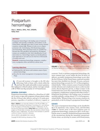
FIGURE 1. Uterine atony (A) is failure of the uterus to contract normally
(B) following delivery and is a common cause of postpartum hemorrhage.
B
A
Copyright © 2020 American Academy of Physician Assistants
- 2. CME
30 www.JAAPA.com Volume 33 • Number 4 • April 2020
multifetal gestations, polyhydramnios, and fetuses who
are large for gestational age (fetal macrosomia, defined as
a weight of 8 lb, 13 oz [4,000 g] or greater).5
Potential
pharmacologic causes of uterine atony include magnesium
sulfate (used for neuroprotection in patients with pre-
eclampsia with severe features and in patients with eclamp-
sia) and nifedipine (used for hypertension in pregnancy).
Chorioamnionitis, placental abruption, and a placenta that
implants into the lower uterine segment can cause uterine
atony and subsequent postpartum hemorrhage.1,6
Trauma from instrumentation to assist with delivery also
can cause postpartum hemorrhage.7
Patients who experi-
ence prolonged labor, particularly when uterine stimulants
such as IV oxytocin and vaginal prostaglandins are used,
can develop postpartum hemorrhage.8
Uterine rupture can
occur in patients undergoing a trial of labor after cesarean
delivery, and the risk is significantly increased if the patient
has had a low-vertical or high-vertical uterine incision with
previous cesarean deliveries.8
Placental anomalies also can place a patient at increased
risk for postpartum hemorrhage.7
These factors include
retained placental fragments as well as the spectrums of
placenta previa and placenta accreta.8
In placenta previa
spectrum, the placenta is attached to the uterine wall either
partially or completely covering the internal cervical os.
Placenta accreta spectrum is a condition in which the
placenta abnormally invades the uterine wall; this condi-
tion is divided into three categories: accreta, increta, and
percreta, depending on the depth of invasion into the
myometrium. Placenta percreta, the most invasive type, is
characterized by the placenta growing through the uterine
wall and potentially invading nearby organs.9
Coagulopathies may be another cause of postpartum
hemorrhage and can be either inherited or acquired.10
Von
Willebrand disease is one of the more common inherited
coagulopathies that can cause postpartum hemorrhage.11
Acquired coagulopathies include HELLP syndrome (hemo-
lysis, elevated liver enzymes, and low platelets) and dis-
seminated intravascular coagulopathy (DIC).12
Placental
abruption, amniotic fluid embolism, sepsis, fetal demise,
and HELLP syndrome can cause DIC.13
In a patient who presents with an acute disorder of
coagulation and postpartum hemorrhage, the two most
common causes are placental abruption and amniotic fluid
embolism.13
Patients with placental abruption will have
pelvic pain. Vaginal bleeding may not always be present
if bleeding is intrauterine, and if the patient is being mon-
itored with a tocodynamometer, uterine tachysystole (rapid
contractions) will be evident. Patients with an amniotic
fluid embolism develop rapid respiratory and hemodynamic
compromise and DIC. Morbidity and mortality from an
amniotic fluid embolism remain high.14
Other common primary causes include cervical and
vaginal lacerations and uterine inversion.8
Uterine inversion
is a medical emergency and requires prompt attention by
a trained healthcare provider. Uterine inversion occurs
when the fundus of the uterus is pulled into the uterine
cavity causing the uterus to be turned inside-out.15
The
inversion may only be palpable in the vaginal canal or it
can protrude through the introitus. A common secondary
cause is subinvolution of the uterus or placental site.1
Subinvolution occurs when the uterus does not return to
its normal size and can be caused by retained placental
fragments or endometritis.
RISK FACTORS
Risk factors for postpartum hemorrhage include being a
woman of color, a previous history of postpartum hemor-
Key points
Postpartum hemorrhage is the leading cause of maternal
morbidity and mortality, particularly in low-income
countries.
Women of color are at higher risk for postpartum
hemorrhage than white women.
Because risk assessment tools only identify 85% of
women with postpartum hemorrhage, consider all
pregnant women at risk.
TABLE 1. Risk factors for postpartum hemorrhage1
Medical or surgical history
• Previous postpartum hemorrhage
• Leiomyomata
• Previous cesarean delivery or other uterine instrumentation
Fetal issues
• Multifetal gestation
• Polyhydramnios
• Large-for-gestational-age fetus
• Fetal macrosomia (birthweight greater than 8 lb, 13 oz [4,000 g])
Maternal issues
• Hypertensive disorders of pregnancy
• Anemia
• Inherited coagulopathy such as von Willebrand disease
• Acquired coagulopathy such as HELLP syndrome
• Trial of labor after cesarean delivery
• Prolonged labor
• Induction and augmentation of labor
• Arrest of progress during the second stage of labor
• Prolonged third stage of labor
• Instrumentation during delivery (forceps)
Placental/uterine issues
• Placental abruption
• Placenta previa
• Retained placenta
• Chorioamnionitis
• Acute uterine inversion
• Subinvolution of the uterus
Copyright © 2020 American Academy of Physician Assistants
- 3. Postpartum hemorrhage
JAAPA Journal of the American Academy of PAs www.JAAPA.com 31
rhage, hematocrit less than 30%, retained placenta, arrest
of progress during the second stage of labor, a prolonged
third stage of labor (defined as more than 30 minutes for
the placenta to separate from the uterus), fetal macrosomia,
hypertensive disorders, and induction and augmentation of
labor.3,8
A general classification of risk factors may be orga-
nized according to the following classifications: medical or
surgical history, fetal issues, maternal issues, and placental/
uterine issues (Table 1). However, many women develop
postpartum hemorrhage without any known risk factors.
CLINICAL ASSESSMENT
Although risk assessment tools can help identify women
who may experience a postpartum hemorrhage, they may
only identify up to 85% of women with postpartum hem-
orrhage. As such, all pregnant women should be considered
at risk for postpartum hemorrhage.1
The initial assessment must focus on the patient’s hemo-
dynamic status; intervene immediately if the patient has
signs of hemodynamic compromise. When a postpartum
hemorrhage is suspected, emergency intervention with a
rapid response team to ensure coordinated care and to
prevent cardiovascular collapse is essential. In addition,
ascertain if the placenta has been delivered. If the placenta
has been delivered, examine it for missing fragments. If
the placenta is still intact, use controlled cord traction to
deliver it. Physical assessment of the patient may reveal a
boggy uterus. The fundus may be palpable above the level
of the umbilicus.
Visual estimation of blood loss and the weighing of
blood-soaked products have historically been used when
caring for women during labor and delivery. However,
visual estimation is associated with a significant underes-
timation of actual blood loss and should only be used when
other objective measures are not available.16
Calibrated
drapes have been developed to help objectively quantify
blood loss and are readily available at most US hospitals.
The American College of Obstetricians and Gynecologists
recently published recommendations on using tools to
accurately quantify blood loss and underscoring the impor-
tance of objective measurements to help reduce morbidity
and mortality.16
Heart rate and BP are the two most commonly used vital
signs to help diagnose a hemorrhage, but they lack specific-
ity.17
In addition, women who are experiencing a hemorrhage
may not develop tachycardia or hypotension until significant
blood loss (greater than 1,000 mL) has occurred. Signs of
a hemorrhage include heart rate greater than 110 beats/
minute, BP of 85/45 mm Hg or less, Spo2
less than 95%,
delayed capillary refill, decreased urine output, and pallor.
Often these changes will not be apparent until the patient
develops shock.17
The ratio of the heart rate over the systolic
BP is called the shock index and may be helpful in assessing
patients with significant bleeding events. A shock index
greater than 1 requires immediate management.6,17
TABLE 2. Laboratory testing in postpartum
hemorrhage
Test Clinical correlation
Blood urea nitrogen
• Elevated in renal failure
• Elevation after resuscitation could
indicate hemolysis
D-dimer Elevated in hemorrhage
Fibrinogen
• Low or normal if coagulopathy is
present
• Very low in amniotic fluid embolism
and placental abruption
Hemoglobin and
hematocrit
May not initially be low in acute
hemorrhage
Liver enzymes Elevated in HELLP syndrome
Lactate Elevated in septic shock
Serum calcium Can be low in hemorrhage
Serum magnesium Can be low in hemorrhage
Serum potassium Can be low in hemorrhage
TABLE 3. Pharmacologic agents used in the
prevention and treatment of postpartum hemorrhage
Drug Use and dosage Notes
Oxytocin
• Prevention and
treatment
• 10 to 40 units
IV or 10 units
intramyometrially
• Used in the third stage
of labor to help prevent
postpartum hemorrhage,
and used first-line in
treatment.
• Can be administered IV
or intramyometrially.
Tranexamic acid
• Treatment
• 1 g IV
Must be used early in the
treatment protocol.
Methylergonovine
• Treatment
• 0.2 mg
• Contraindicated in
patients with hyper-
tensive disorders or
cardiovascular disease.
• Can be given IM, IV, or
intramyometrially.
Carboprost
• Treatment
• 0.25 mg
• Can be given IM or
intramyometrially.
• Contraindicated in
patients with asthma.
Misoprostol
• Treatment
• 600 to 1,000
mcg
• Can be given orally,
sublingually, or rectally
as a suppository.
Copyright © 2020 American Academy of Physician Assistants
- 4. CME
32 www.JAAPA.com Volume 33 • Number 4 • April 2020
Other signs and symptoms associated with hypovolemia
include lightheadedness, palpitations, confusion, syncope,
fatigue, air hunger, and diaphoresis.
DIAGNOSIS
The diagnosis of postpartum hemorrhage is based on the
patient’s physical assessment and the clinician’s clinical
acumen, because many of the objective measures indepen-
dently lack specificity and sensitivity. A baseline complete
blood cell count, coagulation studies (including fibrinogen),
and blood type and antibody screen, if not already known,
should be ordered. Hemoglobin and hematocrit levels are
not generally useful in the initial diagnosis unless a previous
hemoglobin or hematocrit is available for comparison.
Order a metabolic panel to assess for electrolyte abnor-
malities and renal compromise; also order d-dimer, fibrin-
ogen, liver enzymes, and serum lactate levels (Table 2).
Identifying the probable cause of hemorrhage is impera-
tive; thoroughly inspect and palpate the patient’s perineum,
vaginal vault, and uterine cavity. Ultrasonography is a quick
and effective tool that can be used to assess the pelvis for
retained placenta, hematomas, or peritoneal blood.
TREATMENT
Early diagnosis and intervention are essential in reducing
mortality from postpartum hemorrhage and a coordinated
team effort must be used. Simultaneously, clinicians must
manage the patient’s hypovolemia and shock and identify
the cause of the hemorrhage. If the patient has a massive
hemorrhage, notify the rapid response team and use life
support measures.
Two essential initial interventions for postpartum hem-
orrhage are oxytocin and uterine massage. Bimanual
compression of the uterus also can be performed. Ensure
that the patient has an indwelling urinary catheter to
monitor urinary output, because anuria is associated with
massive hemorrhage. Implement resuscitation measures
including elevating the patient’s legs, administering oxygen,
and infusing 0.9% sodium chloride solution or Ringer
lactate via a 14-gauge catheter.
Early administration of the antifibrinolytic tranexamic
acid has been shown to reduce maternal mortality from
postpartum hemorrhage.1,18
When hemorrhage occurs,
tranexamic acid should be given within 3 hours of delivery.
Rapid transfusion of 2 to 4 units of packed red blood cells
is recommended.1
Type-specific is preferred, but type O
Rh-negative blood may be used. Monitor for a coagu-
lopathy; 4 units of fresh frozen plasma are initially used
to help correct a coagulation defect. If the patient’s fibrin-
ogen levels are significantly decreased, administer cryo-
precipitate. If significant thrombocytopenia persists,
administer platelet concentrates. The usual ratio of packed
red blood cells to fresh frozen plasma to platelets is 1:1:1.
Further management is undertaken depending on the
cause of the hemorrhage.19
Uterine atony, the most common
cause of postpartum hemorrhage, is managed as described
above, with the addition of ergonovine, carboprost, and
misoprostol (Table 3).1
Carboprost is contraindicated in
patients with a history of asthma, and hypertension is a
contraindication for methylergonovine.1
Other interventions
for uterine atony include intrauterine tamponade with a
uterine balloon or gauze, B-Lynch suturing of the uterus,
arterial ligation, and uterine artery embolization.19
Defin-
itive surgical management with hysterectomy may be
necessary. The management of other causes of postpartum
hemorrhage is beyond the scope of this article.
CONCLUSION
Identifying patients at risk of developing postpartum hem-
orrhage is advised, but all pregnant women should be
considered at risk, as many without known risk factors will
develop postpartum hemorrhage. Early identification and
intervention are critical to help reduce morbidity and mor-
tality. The use of oxytocin, uterine massage, and controlled
umbilical cord traction are three crucial components of the
active management of the third stage of labor and may help
reduce the incidence of postpartum hemorrhage. Using a
team-based approach to the care of a laboring woman and
implementing hospital-specific protocols will help reduce
themortalityassociatedwithpostpartumhemorrhage. JAAPA
Earn Category I CME Credit by reading both CME articles in this
issue, reviewing the post-test, then taking the online test at http://
cme.aapa.org. Successful completion is defined as a cumulative
score of at least 70% correct. This material has been reviewed and is
approved for 1 hour of clinical Category I (Preapproved) CME credit
by the AAPA. The term of approval is for 1 year from the publication
date of April 2020.
REFERENCES
1. American College of Obstetricians and Gynecologists. Practice
Bulletin No. 183: postpartum hemorrhage. Obstet Gynecol.
2017;130(4):e168-e186.
2. Centers for Disease Control and Prevention. Pregnancy mortality
surveillance system. www.cdc.gov/reproductivehealth/maternal
infanthealth/pregnancy-mortality-surveillance-system.htm. Accessed
December 16, 2019.
3. Petersen EE, Davis NL, Goodman D, et al. Vital signs: pregnancy-
related deaths, United States, 2011-2015, and strategies for
prevention, 13 states, 2013-2017. MMWR Morb Mortal Wkly
Rep. 2019;68(18):423-429.
4. World Health Organization. WHO recommendations on preven-
tion and treatment of postpartum haemorrhage and the WOMAN
Trial. www.who.int/reproductivehealth/topics/maternal_perinatal/
pph-woman-trial/en. Accessed December 19, 2019.
5. Wetta LA, Szychowski JM, Seals S, et al. Risk factors for uterine
atony/postpartum hemorrhage requiring treatment after vaginal
delivery. Am J Obstet Gynecol. 2013;209:51.e1,51 -56.
6. Yiadom MYAB, Carusi D. Postpartum hemorrhage in emergency
medicine. https://emedicine.medscape.com/article/796785-over-
view. Accessed December 19, 2019.
7. Sheiner E, Sarid L, Levy A, et al. Obstetric risk factors and
outcome of pregnancies complicated with early postpartum
hemorrhage: a population-based study. J Matern Fetal Neonatal
Med. 2005;18(3):149-154.
8. Oyelese Y, Ananth CV. Postpartum hemorrhage: epidemiology, risk
factors, and causes. Clin Obstet Gynecol. 2010;53(1):147-156.
Copyright © 2020 American Academy of Physician Assistants
- 5. Postpartum hemorrhage
JAAPA Journal of the American Academy of PAs www.JAAPA.com 33
9. Wu S, Kocherginsky M, Hibbard JU. Abnormal placentation:
twenty-year analysis. Am J Obstet Gynecol. 2005;192:1458 -1461.
10. Gillissen A, van den Akker T, Caram-Deelder C, et al., TeM-
pOH-1 Study Group. Coagulation parameters during the course
of severe postpartum hemorrhage: a nationwide retrospective
cohort study. Blood Adv. 2018;2(19):2433-2442.
11. Govorov I, Loefgren S, Chaireti R, et al. Postpartum hemorrhage
in women with von Willebrand disease—a retrospective
observational study. PLoS ONE. 2016;11(10):e0164683.
12. James AH, Grotegut C, Ahmadzia H, et al. Management of
coagulopathy in postpartum hemorrhage. Semin Thromb Hemost.
2016;42(07):724-731.
13. Thachil J, Toh C-H. Disseminated intravascular coagulation in
obstetric disorders and its acute haematological management.
Blood Reviews. 2009;23:167-176.
14. Fong A, Chau C , Pan D, Ogunyemi D. Morbidities associated
with a disproportionately high risk of amniotic fluid embolism:
a contemporary population-based study. Am J Obstet Gynecol.
2013; 208(1):S328 -S329.
15. Poon SS, Chean CS, Barclay P, Soltan A. Acute complete uterine
inversion after controlled cord traction of placenta following vagi-
nal delivery: a case report. Clin Case Repr. 2016;4(7):699-702.
16. American College of Obstetricians and Gynecologists. ACOG
Committee Opinion No. 794. Quantitative blood loss in
obstetrical hemorrhage. www.acog.org/Clinical-Guidance-and-
Publications/Committee-Opinions/Committee-on-Obstetric-
Practice/Quantitative-Blood-Loss-in-Obstetric-Hemorrhage.
Accessed January 15, 2020.
17. Nathan HL, El Ayadi A, Hezelgrave NL, et al. Shock index: an
effective predictor of outcome in postpartum haemorrhage?
BJOG. 2015;122:268 -275.
18. WOMAN Trial Collaborators. Effect of early tranexamic acid
administration on mortality, hysterectomy, and other morbidities
in women with post-partum haemorrhage (WOMAN): an
international, randomised, double-blind, placebo-controlled trial.
Lancet. 2017;389:2105 -2116.
19. Chandraharan E, Krishna A. Diagnosis and management of
postpartum haemorrhage. BMJ. 2017;358:j3875.
Copyright © 2020 American Academy of Physician Assistants
![CME
30 www.JAAPA.com Volume 33 • Number 4 • April 2020
multifetal gestations, polyhydramnios, and fetuses who
are large for gestational age (fetal macrosomia, defined as
a weight of 8 lb, 13 oz [4,000 g] or greater).5
Potential
pharmacologic causes of uterine atony include magnesium
sulfate (used for neuroprotection in patients with pre-
eclampsia with severe features and in patients with eclamp-
sia) and nifedipine (used for hypertension in pregnancy).
Chorioamnionitis, placental abruption, and a placenta that
implants into the lower uterine segment can cause uterine
atony and subsequent postpartum hemorrhage.1,6
Trauma from instrumentation to assist with delivery also
can cause postpartum hemorrhage.7
Patients who experi-
ence prolonged labor, particularly when uterine stimulants
such as IV oxytocin and vaginal prostaglandins are used,
can develop postpartum hemorrhage.8
Uterine rupture can
occur in patients undergoing a trial of labor after cesarean
delivery, and the risk is significantly increased if the patient
has had a low-vertical or high-vertical uterine incision with
previous cesarean deliveries.8
Placental anomalies also can place a patient at increased
risk for postpartum hemorrhage.7
These factors include
retained placental fragments as well as the spectrums of
placenta previa and placenta accreta.8
In placenta previa
spectrum, the placenta is attached to the uterine wall either
partially or completely covering the internal cervical os.
Placenta accreta spectrum is a condition in which the
placenta abnormally invades the uterine wall; this condi-
tion is divided into three categories: accreta, increta, and
percreta, depending on the depth of invasion into the
myometrium. Placenta percreta, the most invasive type, is
characterized by the placenta growing through the uterine
wall and potentially invading nearby organs.9
Coagulopathies may be another cause of postpartum
hemorrhage and can be either inherited or acquired.10
Von
Willebrand disease is one of the more common inherited
coagulopathies that can cause postpartum hemorrhage.11
Acquired coagulopathies include HELLP syndrome (hemo-
lysis, elevated liver enzymes, and low platelets) and dis-
seminated intravascular coagulopathy (DIC).12
Placental
abruption, amniotic fluid embolism, sepsis, fetal demise,
and HELLP syndrome can cause DIC.13
In a patient who presents with an acute disorder of
coagulation and postpartum hemorrhage, the two most
common causes are placental abruption and amniotic fluid
embolism.13
Patients with placental abruption will have
pelvic pain. Vaginal bleeding may not always be present
if bleeding is intrauterine, and if the patient is being mon-
itored with a tocodynamometer, uterine tachysystole (rapid
contractions) will be evident. Patients with an amniotic
fluid embolism develop rapid respiratory and hemodynamic
compromise and DIC. Morbidity and mortality from an
amniotic fluid embolism remain high.14
Other common primary causes include cervical and
vaginal lacerations and uterine inversion.8
Uterine inversion
is a medical emergency and requires prompt attention by
a trained healthcare provider. Uterine inversion occurs
when the fundus of the uterus is pulled into the uterine
cavity causing the uterus to be turned inside-out.15
The
inversion may only be palpable in the vaginal canal or it
can protrude through the introitus. A common secondary
cause is subinvolution of the uterus or placental site.1
Subinvolution occurs when the uterus does not return to
its normal size and can be caused by retained placental
fragments or endometritis.
RISK FACTORS
Risk factors for postpartum hemorrhage include being a
woman of color, a previous history of postpartum hemor-
Key points
Postpartum hemorrhage is the leading cause of maternal
morbidity and mortality, particularly in low-income
countries.
Women of color are at higher risk for postpartum
hemorrhage than white women.
Because risk assessment tools only identify 85% of
women with postpartum hemorrhage, consider all
pregnant women at risk.
TABLE 1. Risk factors for postpartum hemorrhage1
Medical or surgical history
• Previous postpartum hemorrhage
• Leiomyomata
• Previous cesarean delivery or other uterine instrumentation
Fetal issues
• Multifetal gestation
• Polyhydramnios
• Large-for-gestational-age fetus
• Fetal macrosomia (birthweight greater than 8 lb, 13 oz [4,000 g])
Maternal issues
• Hypertensive disorders of pregnancy
• Anemia
• Inherited coagulopathy such as von Willebrand disease
• Acquired coagulopathy such as HELLP syndrome
• Trial of labor after cesarean delivery
• Prolonged labor
• Induction and augmentation of labor
• Arrest of progress during the second stage of labor
• Prolonged third stage of labor
• Instrumentation during delivery (forceps)
Placental/uterine issues
• Placental abruption
• Placenta previa
• Retained placenta
• Chorioamnionitis
• Acute uterine inversion
• Subinvolution of the uterus
Copyright © 2020 American Academy of Physician Assistants](data:image/gif;base64,R0lGODlhAQABAIAAAAAAAP///yH5BAEAAAAALAAAAAABAAEAAAIBRAA7)