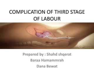
1. COMPLICATION OF THIRD STAGE
OF LABOUR
Prepared by : Shahd shqerat
Baraa Hamammrah
Dana Bewat
2. OUTLINE
• INTODUCTION.
• Complication of the third stage of labor
• Rupture of the uterus ( What it is?, Cases,
signs, Management).
• Inversion of the uterus ( Types , causes ,
Management).
• Retained placenta ( risk factor , Management)
• Shock (causes , clinical signs , Management).
3. INTRODUCTION
The third stage of labour start with the
complete delivery of the fetus and ends
with completed delivery of the placenta
and its membrane
Length of the third stage it self 5 – 15
minutes and may last up to 1 hour
4. Complication of the third stage of labor
• PPH (post partum hemorrege )
• Rupture of the uterus
• Inversion of the uterus
• Retained placenta
• Shock
5. RUPTURE OF THE UTERUS
• Rupture of the uterus is one of the most serious complications in
midwifery and obstetrics. It is often fatal for the fetus and may also be
responsible for the death of the mother. however, of the nine maternal
deaths from haemorrhage, only one was associated with uterine
rupture (Norman 2011). uterine rupture remains a significant problem
worldwide. With effective antenatal and intrapartum care, some cases
of uterine rupture may be avoided.
• Rupture of the uterus is defined as being complete or incomplete:
1. complete rupture involves a tear in the wall of the uterus with or
without expulsion of the fetus.
2. incomplete rupture involves tearing of the uterine wall but not the
perimetrium.
6.
7. CAUSES
1. previous history of caesarean section.
2. high parity.
3. use of oxytocin, particularly where the woman is of high
parity.
4. use of prostaglandins to induce labour, in the presence of
an existing scar.
5. trauma, as a result of a blast injury or an accident.
6. extension of severe cervical laceration upwards into the
lower uterine segment - the result of trauma during an
assisted birth.
8. Signs of rupture of the uterus
• The degree and speed of the woman's collapse and shock
depend on the extent of the rupture and the blood loss.
1. Abdominal pain or pain over previous caesarean section
scar.
2. Abnormalities of the fetal heart rate and pattern.
3. Vaginal bleeding.
4. Maternal tachycardia.
5. Poor progress in labour.
9. Management
• An immediate caesarean section is per formed in the hope
of procuring a live baby. Following the birth of the baby and
placenta, the extent of the rupture can be assessed. Choice
between the options to perform a hysterectomy or to
repair the rupture depends on the extent of the trauma
and the woman's condition. Further dinical assessment will
include evaluation of the need for blood replacement and
management of any shock.
10. Uterine inversion
• Uterine inversion means that the uterus has
turned inside out
• It is a serious complication of the third stage
of labour because if the inner surface of the
fundus appears at the vaginal outlet, it will
cause death of the mother
• Occurs 1 in 100,000
deliveries
11.
12. Types:-
I. Partial inversion:- the funds dose not pass
through the cervix
II. Complete inversion:- the fundus extruded into
the vagina
13. Causes:-
All causes are connected with applying force to the uterine
fundus when it is relaxed.
1. Exerting controlled cord traction when the uterus is
relaxed especially if the placenta is centrally sited in the
funds.
2. Attempting force to expel the placenta by using fundal
pressure when the uterus
3. Combining fundal pressure and cord traction to deliver the
placenta is atonic
4. Spontaneous occurrence, it is more likely to follow a
delivery when a multiparous mother has pushed
vigorously since she has very week muscle tone and
ligaments
14. Recognition:-
1. Sudden onset of shock
2. Sever pain due to dragged "compression and traction"
ovaries into the inverte fundus
3. Bleeding may or may not be present depending on
the degree of placent adherence to the uterine wall
4. On palpation, a concave shape will be felt at the
fundus if partially inverted
*If complete inversion occurs, none of the uterine parts
will be palpated "No Uterus felt in the abdomen
" Vaginal examination will reinforce "reveal" the inversion
15. Midwifery Care:-
• The best chance of replacing the uterus occurs
immediately following the investigation "Which is
a doctor procedure" by applying pressure to the
part nearest the cervix, working upwards to the
fundus on the principle of Last Out, First In
• No attempt must be made to remove the
placenta until the uterus becomes in the right
way out, otherwise haemorrhage can not
controlled
16. • If replacement of a totally inverted uterus is not
possible, it should be gently placed inside the
vagina to relieve traction on the ovaries and the
fallopian tubes
•
17. • Raising the foot of the bed will also help to
relieve the tension and alleviate shock
• Severe shock is an immediate consequence ,
so resuscitative measures must be initiated
prior to operative intervention
• Urgent cross-matching is done
18. • Give sedation for pain relief "Pethidine, Morphine...“
• The uterus may be replaced manually under general
anaesthesia when maternal condition is stable
• When the uterus returns to its normal position
methergine with active management must be started to
have a good contracted uterus before the hand is
withdrawn
• Then syntocinon drip will continue for the next 24 hours
to maintain uterine contraction In extreme cases ,
hysterectomy may be considered to save the mother's life
19. Retained placenta
• 10% of PPH cases.
• Retained placental tissue is most likely to
occur with a placenta that has an accessory
lobe, deliveries that are extremely preterm, or
variants of placenta accreta.
• Retained or adherent placental tissue prevents
adequate contraction of the uterus allowing
for increased blood loss.
20.
21. Risk factors for retained products of
conception include the following:
1. Prior uterine surgery or procedures .
2. Premature delivery.
3. Difficult or prolonged placental delivery .
4. Multilobed placenta.
5. Placental accreta
22. Management:
1. Prevent with syntocinon at anterior shoulder
delivery.
2. Active management of 3rd stage of labour.
3. Manual removal of placenta or the retained
pieces.
4. Explore for fragments (careful examination of
the placenta and membranes.)
5. Ultrasound
23. Shock
• Shock is the collapse caused by failure of the
circulatory system to meet the body's need for
oxygen, nutrients and removal of waste
substances
• Shock is a serious complication; if unrecognised
or inadequately treated, a chronic condition
develops and even death.
• There are two varieties of shock that can occur
during pregnancy, or more commonly after
delivery, which are haemorrhagic shock "hypo
volaemic shock" and non-haemorrhagic shock
24. The main causes of shock
1. Haemorrhage "ante partum or post partum“,
the common cause
2. Uterine causes include inversion or rupture
3. Acid aspiration syndrome
4. Pulmonary embolism
5. Amniotic fluid embolism
25. Clinical signs:-
• Low blood pressure, increased pulse rate;
"Not Reliable Alone"
• Cold skin, clammy, moised and pallor
• Air hunger "In Severe Cases"
• Diminished urinary out put "Oligouria or
anuria"
26. Management
• Urgent resuscitative measures are necessary before the condition
becomes irreversible
• The principles of under lying treatment whatever the cause of shock
are:-
1. Maintenance of an airway
2. Administration of IV fluids to increase blood volume until cross-
matching of blood is available
3. Raising the foot of the bed
4. A sedative may be given to keep the mother quiet and undisturbed
5. Administration of oxygen especially if dyspnoea is present
6. Avoid over heating the body, the pale, cold skin is evidence that the
body's defence mechanism is at work. The superficial arterioles and
capillaries have contracted in order to direct the blood to the
essential "Vital organs" of the body such as heart, brain and kidneys
29. Thrombophilia
Are a group of blood coagulation disorders, characterised by
an increase in the coagulability of the blood and the tendency
for thrombi or clots to form. They are multifactorial disorders in
which both environmental factors (age, smoking, overweight,
etc.) and genetic predisposition may play a rol .Evidence
supports an association between certain types of thrombophilia
and some specific problems in relation to the infertile couple.
These coagulation disorders
.
This condition lead to recurrent
pregnancy loss growth restriction, late miscarriages, stillbirth
and preeclampsia
30. Pregnancy and thrombophilia
Pregnancy is a hypercoagulability status because it contains which
includes hypercoagulability, venous stasis, and endothelial injury thus
promoting thrombosis
Hypercoagulability: pregnancy causes alterations in coagulation proteins
Factors I, II, VII, VIII, IX, and X increase in pregnancy. Resistance to the
anticoagulant protein C is increased and the protein S level decreases.
PAI-1 (Plasminogen Activator Inhibitor type 1) levels increase five-fold
which reduces fibrinolytic activity
.
Venous stasis is also present in pregnancy due to venous dilatation as a
result of progesterone effects that case relaxation of smooth muscle. Also
the pressure from the gravid uterus resist the venous retain
.
Endothelial injury may occur antepartum or during delivery
.
31. state Classification
Inherited
:
The most common inherited
disorders in pregnancy that cause
thrombophilia are
:
🔻Mutations in factor V gene
(factor V Leiden)
🔻Prothrombin (PT) G20210A
gene mutation
🔻Anti-thrombin deficiency
Deficiencies in protein C and
protein S
🔻
Acquired
The most common cause
of acquired thrombophilia
is Anti-phospholipid
syndrome which is
autoimmune disorder in
which the body produces
antibodies that attack
phospholipids and damage
the endothelium.TE, new
VTE in pregnancy, or
history of VTE in women
not previously
screened.10A gene
mutation (PGM)
Protein S deficiency
Protein C deficiency
32. Risk factors for thrombophilia
Transient
factors
Obstetric
factors
Pre-existing
factors
Systemic infection
Paraplegia or
immobility
Recent surgical
procedure
Ovarian
hyperstimulation
syndrome
Travel >4 Hours
Multiple pregnancy
Pre_eclampisa
Caesarean section
or forceps delivery
Prolonged labour
Postpartum
haemorrhage
Previous
thrombophilia
Family history of
VTE (e.g.
deficiency of
protein C or S,
antithrombin
deficiency,
prothrombin gene
variant, Factor V
Leiden)
cardiac disease
Age >35 years
Obesity (BMI >30
kg/m²)
Parity >3
Smoking
33. Intravenous drug
user
Varicose veins
.
Symptoms of Thrombophilia
Thrombophilia doesn’t have any symptoms unless
you have a blood clot. However, some symptoms
include:
tenderness,
or leg:
Arm
📌
warmth, swelling, pain
: shortness of breath
Heart
📌
light-headedness, sweating,
discomfort in the upper body,
chest pain and pressure
: shortness of
Lung
📌
breath, sweating, fever,
coughing up blood, rapid
heartbeat, chest pain
.
: trouble speaking, vision problems,
Brain
📌
dizziness, weakness in the face or limbs, sudden
severe headache
34. .
pain in the calf muscle,
Symptoms of DVT (
📌
like chest
PE symptoms
📌
warm, swelling) and
pain, shortness of breath and cough
.
36. The first step in the
diagnosis of thrombophilia
is usually a blood test to
check the level of
coagulation. This is usually
followed by a more specific
test that includes genetic
and/or coagulation testing
for the following factors
:
Standard antibody
Protein Z antibody
Anti-annexin V antibody
Factor V Leiden
Factor VIII
Factor XIII
ABO genotype
Homocysteine
Factor XII C46T polymorphism
These tests are usually done only when there is an
indication for them, such as incidents of
thrombosis, family history, recurrent miscarriages,
testing is performed remote (at least
Laboratory
etc.
)
six weeks
37. Treatment
Treatment of thrombophilia is based on the
individual age, family history and overall health
.
The major treatment for thrombophilia is
Anticoagulant medication includes aspirin, heparin
and LMWH (clexane). There is a risk for bleeding
associate with the use of anticoagulant therapy
.
Other lifestyle modification can also be considered
in the treatment of thrombophilia include: maintain
healthy body weight, stop smoking and exercises
regularly.
:
up, the patient should have
-
In terms of pregnancy follow
Doppler ultrasound
38. To check that blood flow in the umbilical artery is
adequate and that the baby receives sufficient nutrients
and oxygen. It also serves to see fetal growth and
development
.
Heart rate monitoring
Checks the baby's heart rate and its variation as the baby
moves. It is used to ensure that the baby receives enough
oxygen
Blood analysis
To verify that anticoagulation is within the expected
therapeutic range
Prevention of thrombophilia
Thrombophilia testing for individuals who are at
high risk for developing thrombophilia such as:
positive family history with thrombophilia,
immobilization, taking oral contraceptives is important
to prevent thrombus formation. Also these individuals
may take prophylactic Anticoagulants.
Maintain a healthy body weight, stop smoking,
exercise regularly and avoid long periods of inactivity or
bed rest
.
Individuals who have been diagnosed with
thrombophilia must be compliance with the
anticoagulant therapy (if it recommends) to avoid
thrombus formation
40. Made by: Ruba Ekhlayel 22010875
Lamia Sayara 22115024
Kefaya Omar 22012094
Doctor : Ibtesam dwekat
41. PPH:is one of the most common
obstetric emergencies, in UK
hemorrhage was the third most
common cause of death. Its
defined as:
*primary PPH: loss of ≥ 500 ml
blood from genital tract within 24
hours of delivery.
*secondary PPH : loss of ≥ 500 ml
blood from genital tract after 24
hours till 12 week post-delivery.
42. Its can be classify to:
A. Minor PPH if blood loss is
between 500 & 1000 ml, the loss of
this amount are relatively
common, and usually tolerated
well by the women.
B.Massive PPH loss over 1000 ml
this required the application of
PPH protocols.
43. Risk factors of PPH
PPH can be predicted and
preventive measures can be
undertaken if risk factors are
present :
.Maternal risk factors:
1.raised maternal age.
2.grand multiparty .
3.primiparity.
4. previous cesarean.
5.obesity.
6.uterine fibroid.
7. antepartum hemorrhage.
8.previous PPH.
Antepartum:
45. Aetiology of PPH:
The causes of PPH can be remembered as
five" T“ :
1.Tone.
2.Tissue.
3.Truma .
4.Thrombin.
5. Traction: uterine inversion.
1.Tone:
Uterine atony or a failure of uterus to
contract after delivery of placenta. it’s the
most common cause of PPH.atony occur due
to the:
*uterine over distention: polyhydramnios,
multiple gestation, fetal macrosomia.
*Rapid or prolonged labor.
*Oxytocin use
*High parity
*Chorioamnionitis
46. 2.Tissue :
retained part of placenta &/or
membranes.
3.Truma :
almost all types of delivery can
cause some degree of genital tract
trauma in form of perineal &
vaginal tears, but this occur mainly
after forceps delivery, cervix may
be torn if delivery occurred before
the cervix is fully dilated.
4.Thrombin:
Its mean clotting disorder which
occur in women with underlying
disorder like Von Willebrand
disease or platelet disorders .
5. Traction: uterine inversion.
47.
48. Diagnosis:
Early recognition of blood loss &
rapid action is vital in the
management of PPH
A appreciation of risk factors
*accurate estimation of blood loss
& recognition of the maternal signs
of cardiovascular compromise are
vital, these include tachycardia,
low BP, pallor, slow capillary refill.
49. Treatment:
In practice, diagnosis and
management of PPH occur
simultaneously.
Team work (senior
obstetrician,anaestheatist,senior
midwife)
1-Oxygen by mask initially.
2-Two large bore cannula(16-gauge
intravenous lines).
3-Rapid fluid resuscitation.
4-Full blood count and clotting
studies.
50. 5-cross-match units of blood &
transfuse blood as soon as possible
or give O negative until the blood
of the same group available.
6-Foley catheter
7-May need fresh frozen plasma,
platelets, cryoprecipitate.
51. Treat the cause:
Uterine atony:
which is the most common cause
of postpartum hemorrhage.
Because hemostasis associated
with
placental separation depends on
myometrium
contraction, atony is treated
initially by: 1. Bimanual uterine
compression and massage
2.uterotonics drugs that promote
uterine contraction, include
oxytocin, ergot alkaloids, and
prostaglandins
52. uterotonics drugs:
• 1. Oxytocin stimulates the upper
segment of the myometrium to contract
rhythmically, Oxytocin is an effective first
line treatment for postpartum
hemorrhage. 10 international units (IU)
should be injected intramuscularly, or 20
IU in 1 L of saline may be infused at a rate
of 250 mL per hour.
2.Methylergonovine (Methergine) and
ergometrine are ergot alkaloids that cause
generalized smooth muscle contraction in
which the upper and lower segments of
the uterus contract tetanically . A typical
dose of methylergonovine, 0.2 mg
administered IM, may be repeated as
required at intervals of two to four hours.
Because ergot alkaloid agents raise blood
pressure, they are contraindicated in
women with preeclampsia or
hypertension. Other adverse effects
include nausea and vomiting
53. 3. Prostaglandins enhance uterine
contractility and cause vasoconstriction.
The prostaglandin most commonly used is
15-methyl prostaglandin F2a, or
carboprost (Hemabate). Carboprost can
be administered intramyometrially or IM
in a dose of 0.25 mg; this dose can be
repeated every 15 minutes for a total
dose of 2 mg. carboprost should be used
with caution in patients with asthma or
hypertension. Side effects include nausea,
vomiting, diarrhea, hypertension,
headache, flushing, and pyrexia
4. Misoprostol is another prostaglandin
that increases uterine tone and decreases
postpartum bleeding. Misoprostol is
effective in the treatment of PPH but side
effects may limit its use. It can be
administered sublingually, orally, vaginally,
and rectally. Doses range from 200 to
1,000 mcg.
54. Secondary PPH
Loss of ≥ 500 ml of blood from genital
tract between 24 hours & 12 weeks
post-delivery. Its rare cause of massive
bleeding. Causes of PPH Main causes:
. Infection mainly (endometritis).
. Retained product of conception.
. Blood disorder.
Choriocarcinoma.
Clinical feature:
Bleeding usually slight to moderate,
but it may be life threatening,
fever subinvoluted uterus may be
tender
,anaemia,pallor.
Diagnosis:
1.CBC & blood film.
3. ULS to exclude retained pieces
55.
56. Treatment:
According to the cause this
involved:
1- Correction of anemia
2- Antibiotic
3- Medical or surgical way
to evacuated the uterus.
4- If the cause choriocarcinoma
chemotherapy may be used.
58. Long term complications:
1.Infections: puerperal infections )
2. Sheehan’s syndrome (necrosis of
anterior pituitary)
3. Chronic anemia
4. Chronic renal failure
5. infertility due to : a. Sheehan's
syndrome b. asherman syndrome
c.infection and tubal blockage
:
Uterine inversion
Its rare complication of the third
stage ,incidence 1:2000 - 1:6000.the
uterine fundus descend through the
uterine cavity, or rarely beyond the
intoitus,can be occurred after C/S or
vaginal delivery.
59. Causes
1. mismanagement of the third
stage – e.g. premature or excessive
cord traction during active
management of the third stage, a
combination of fundal pressure
and cord traction to deliver the
placenta , or use of fundal pressure
when the uterus is atonic during
placental delivery
2.abnormally adherent placenta .
3. spontaneous inversion of
unknown etiology 4.short umbilical
cord 5.sudden emptying of a
distended uterus.
60. Diagnosis:
The prolapsed uterus stretching the
cervix causes vagal stimulation, so the
women will have sign of
cardiovascular collapse & shock,
although bleeding is commonly
present, the symptoms will be out of
proportion to estimated blood loss.
The inverted uterus may be obvious at
the intoitus.lack of palpable uterus in
the abdomen, feeling of a "dimple" in
the uterine fundus on examination
61. Management:
• The inverted uterus usually
appears as a bluish-gray mass
protruding from the vagina
• Vasovagal effects producing vital
sign changes disproportionate to
the amount of bleeding may be an
additional clue.
• The placenta often is still
attached, and it should be left in
place until after reduction. Every
attempt should be made to replace
the uterus quickly
62. :
Retained placenta
Failure of placental delivery within 60
minutes after delivery of the fetus,
complicates 2% of births .
Risk Factors
• Previous retained placenta .
• Previous injury or surgery to the uterus.
• Preterm delivery.
• Multiparty.
• Induced labor.
Causes:
1. Constriction ring-reforming cervix
2. Full bladder
3. Uterine abnormality
4. Morbid adherence of the placenta:
Placenta Accreta
Placenta Increta
Placenta Percreta
63. Management:
Third stage
If the placenta is undelivered after
30 minutes consider:
Emptying bladder
Breastfeeding or nipple stimulation
Change of position – encourage an
upright position
4.using of uterotonics drug by
intravenous ,IM,intra umbilical
venous route
64. 5.if after20 minute of uterotonics
drug placenta not delivered
transferred the patient to the
theater for manual removal of
placenta under anesthesia.
6.if failed remove it by curettage.
7. if placenta was morbid adherent
hysterectomy may be done.