

Recommended
More Related Content
What's hot
What's hot (20)
Similar to Mammography
Similar to Mammography (20)
Recently uploaded
Recently uploaded (20)
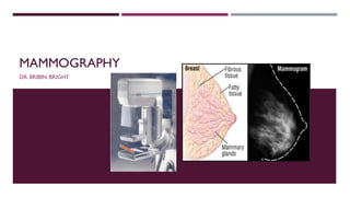
Mammography
- 2. MAMMOGRAPHY - INDICATIONS Screening of asymptomatic women. Screening of high risk women. Follow up of patients after mastectomy of same and opposite breast / same breast with implant Investigations of benign breast diseases with eczematous skin,nipple discharge , skin thickening . Investigation of a breast lump. Investigation of occult primary with secondaries . Male breast evaluation .
- 3. TYPE OF MAMMOGRAPHIC EXAMINATION Diagnostic mammography Is performed on patients with symptoms or elevated risk factors. Two or three views of each breast may be required. Screening mammography Is performed on asymptomatic women with the use of a two view protocol, usually medial lateral oblique and cranio caudal , to detect on unsuspected cancer
- 4. MAMMOGRAPHY - BASICS Uses low-energy x-rays (40kVp) for dectecting breast pathologies. Higher kV - Poor Contrast Lower kV - Good Contrast Tissues within breast have very small difference in attenuation property - Lacks contrast <40kVp - Good photoelectric effect - brings out contrast between the tissues within the breasts. Better contrast Better detailing of structures
- 5. INSTRUMENTATIONS HIGH FREQUENCY X-RAY GENERATOR X-RAYTUBE ANODE CATHODE BEAM PORT K-EDGE FILTERS COMPRESSION PADDLE SPOT COMPRESSION PARALLEL LINEAR GRID IMAGE RECEPTOR AEC (AUTOMATIC EXPOSURE CONTROL)
- 6. X-RAY TUBE Rotating Anode type - Glass or Metal ceramic Advantage -Decreases off focus radiation -IncreasesTube lighting Cathode - Dual focalspots 0.3mm (Imaging) 0.1 mm (Magnification) ( Focal spot - Penumbra - Resolution) Helps identifiying microcalcification
- 7. ANODE (TARGET) Usually-Tungsten (xray /Floro/ CT/ cathlab) MOLYBDENUM -17.5 & 19.6 keV RHODIUM -20.2 & 22.7 keV CHARACTERISTIC RADIATION Characteristic x-ray Characteristic x-rays are produced after ionization of a k- shell electron.When an outer-shell electron fills the vacancy in the k shell, an x-ray is emitted. In mammography,17.5- 19.5kev characteristic x-ray is produced with Mo and 23kev is produced with Rh
- 9. ANODE ANGLE = 16° to 0 to -9° EFFECTIVE ANODE ANGLE - Angle of anode relative to the horizontal tube mount XRAYTUBE TILT -To reduce unwanted radiation - focus only to the breast area
- 10. SOURCETO IMAGE DISTANCE (SID) - lesser than usual Radiography -60/65 cm (Chest = 18) - short SID - Heel Effect utilized At Cathode → chestwall – Thicker tissues SIDE At Anode → Nipple region – LessThickness SIDE Non-uniformity of X-ray Intensity Along the cathode side - More Intensity Along the Anode side - Less Intensity
- 11. PARTS OF MAMMOGRAPHY EQUIPMENT Beam port - 1 mm Beryllium Since metal/ Glass is used in XRAYTUBE metal will absorb the low energy photons from xray tube K-edge filters - Cuts unwanted Low energy photons (RADIATION) / High energy photons (SCATTER – Reduces contrast) (0.03mm Mo / 0.025mm Rh)
- 12. PARTS OF MAMMOGRAPHY EQUIPMENT To decrease Breast thickness, spreads out the tissue - Spreads out superimposed anatomy. Scatter Radiation - contrast Radiation dose to the breast tissue Geometric Blurring (closer to image receptor ) Decreases the kinetic blur . Makes breast thickness uniform in film density. Differentiates the easily compressible cysts and fibro- glandular tissue from the more rigid carcinomas Separates the super imposed breast lesions . Compression paddle
- 13. PARTS OF MAMMOGRAPHY EQUIPMENT GRIDS Stationary grids or grids placed in between the screen and the film are no longer used as the thin grid lines compromised on the quality of the image . Covered tiny details such as microcalcifications Hence oscillating grids are used o Grid ratio of 4 :1 or 5 : 1 The grid lines are eliminated by the motion of the grid . Grids improve the image quality and cause a significant reduction
- 14. PARTS OF MAMMOGRAPHY EQUIPMENT Spot.compression - for imaging particular region Parallel Linear grid - (4:1 to 5:1) low grade ratio grids used to increase the Contrast Image Receptor Screen film CR Cassette Flat panel detector - (Digital Mammography) AEC -Automatic Exposure Control KeV selected → MAS automatically chosen.
- 15. FLAT PANEL DETECTORS - INDIRECT Indirect Amorphous silicon technology Xray scintillator (Gradoliniun Sulphate / Csl) Produces light flashes when Xray beam falls over it. Silicon Photodiode array (Photo detector) Made of Amorphous silicon Converts light photons from the scintillator into electrical signal TFT-electronic switch - Reads electrical signal - Gives positional information -This data is sent to AEC (Analogue to Digital Converter) AEC Converts it into digital signal and are finally sent to Computer for processing Disadvantage - LIGHT SCATTER Scattering of light within the phosphors leading to unsharpness
- 16. FLAT PANEL DETECTORS - DIRECT Crystals of amorphous selenium directly converts Xrays into electrical signal that is stored in a capacitor (ready to be read)
- 17. MAGNIFICATION TECHNIQUE To increase the visibility of finer details like microcalcification Increase O ID - object to Image Distance Breast is kept over a raised platform No need for grid as there is presence of air gap
- 18. SCREEN FILM MAMMOGRAPHY Old method The image is created directly on a film-Non modifiable Less sensitive for women with dense breasts Screen film cassette used Single emulsion film Single screen Terbium activated Gadolinium oxysulphide Screen emits green light Film will be sensitive to greenlight (ORTHOCHROMATIC FILM)
- 19. COMPUTER RADIOGRAPHY (CR) MAMMOGRAPHY A/K/A Filmless radiography Electronic radiography Digital storage phosphor imaging Xray images are acquired in digital format Photostimulable phosphor plates used Imaging plate has high sensitivity to xrays COMPONENTS storage phosphor cassette Storage phosphor reader Bar code scanner Workstation Photostimuliable Phosphor (PSP) Cassette PSP Receptor materials . BaFBr : Eu2+ . BaF (Br1): Eu 2+ .Ba SrFBr:Eu 2+
- 20. DIGITAL MAMMOGRAPHY Also called full-field digital mammography (FFDM) -in which the x-ray film is replaced by electronics that convert x-rays into mammographic pictures of the breast. Display Monitor - HIGH RESOLUTION (5 MP) Direct Digital Mammography - Flat panel detector with amorphous selenium array Indirect Digital Mammography -Flat panel scintillator with amorphous silicon diode array
- 21. DIGITAL MAMMOGRAPHY ADVANTAGE Better contrast,more details.Better delineation of parenchyma & subcutaneous tissue Better noise level reduction Tolerance to over/under exposure (Avoids Repetition? Post-processing techniques for better diagnosis Radiation dose is decreased- Mean glandular dose in mammography Screen Film (2-3m bGy) Digital (20-30% reduction) Image can be seen quickly and stored digitally(less storage space )
- 22. LIMITATIONS OF 2D MAMMOGRAPHY 20% of cancers will be missed 10% recalled for additional workup 75-80% of biopsies result in benign lesions sensitivity decreases with increased breast density 2D mammograms,take images only from the front and side, this may create images with overlapping breast tissue
- 23. ADVANCES IN DIGITAL MAMMOGRAPHY Computer -Aided Diagnosis - uses software to detect area of clinical significance and highlight them for better output image Digital BreastTomosynthesis (DBT) XrayTube rotates /moves in a 50-55° arc around the breast, capturing several (11-25) low dose projections of the breast at different angles. Then reconstructed to 3D Projection images (1 mm slices) 3D MAMMOGRAPHY
- 24. DIGITAL BREAST TOMOSYNTHESIS - DBT (3D MAMMOGRAPHY ) Disadvantages of DBT • Twice the radiation compared to 2D mammo • More storage requirements • Takes longer time to read • Cannot be read on demand Advantages of DBT • Increased cancer detection rate • Lesions are better defined • Precise location of lesions • Less false positive recalls 3D MAMMOGRAPHY
- 25. HOW MAMMOGRAM IS PERFORMED ?
- 26. HOW MAMMOGRAM IS PERFORMED ? CRANIO CAUDAL (CCVIEW) demonstrate maximum tissue on both medial and lateral aspects of the breast with the retromammaryspace and some pectoral muscle The following points were analyzed on CC view: nipple should be in profile nipple should point straight and should not be pointing lateral or medial PND (pectoral nipple distance) must be within 1 cm of the same measurement of the MLO view.
- 27. HOW MAMMOGRAM IS PERFORMED ? MEDIOLATERAL OBLIQUE(MLOVIEW) demonstrate axilla,axillary tail,and inframammary fold with all the breast tissue The following points were analyzed on MLO view: Breast should be pulled out with nipple in profile The pectoralis muscle margin should be well visualized The lower edge of pectoralis muscle should be at the level of pectoralis–nipple line (PNL) or below PND must be within 1 cm of the same measurement of the MLO view. When MLO image of both breasts are viewed as mirror images,pectoralis muscle should meet in the midline and form a “V”.
- 28. OTHER VIEWS Latero-medial (LO) - from the outside towards the center Medio-lateral (ML) - from the center towards the outside Spot compression - compression on only a small area,to get more detail Cleavage view - both breast compressed,to see tissue near the center of the chest Magnification -to see borders of structures and calcifications
- 30. REPORTING A MAMMOGRAM Breast Imaging Reporting and Data System (BI-RADS) RISKASSESSMENT AND QUALITYASSURANCETOOL developed byAmerican college of radiology (ACR) that provides a widely accepted lexicon and reporting schema for imaging of the breast. All mammographic, ultrasound and breast MRI findings and reports should closely adhere to the BI-RADS lexicon and assessment categories.
- 33. THANKYOU