SlideShare a Scribd company logo
1 of 90
ANATOMY
• Modified sweat gland.
• 15-20 lobes
• Functional unit of breast is TDLU, which
consists of a lobule and its extra lobular
terminal duct.
• Each lobule consists of intralobular
segment of terminal ducts, ductules,
loose intra lobular stromal fibrous
tissue.
• TDLUs–site of most breast pathology
and ANDIs
Mammography and Tomosynthesis
• Dr Shamim Hyder Kalenchira
Lymphatic drainage
• Axillary or lateral pathway
– fed by Sappey’s Plexus, as well as by ducts satellite
lymphatics and by most of the parenchymal
lymphatics
– this pathway runs around the inferior edge of
the pectoralis major and reach the pectoral group
of axillary nodes
• internal mammary pathway
– originates from both the lateral and medial halves
of the breast and passes through the pectoralis
major; connections may lead across the median
plane and hence to the contralateral breast
• retromammary pathway
– comes from the posterior portion of the breast
Lymphatic drainage
• Lymph of the breast drains as:
– About 75% into axillary nodes(anterior group)
– 20% ---- internal mammary nodes
– 5% ---- posterior intercostal nodes
• Internal mammary nodes drain the lymph not
only from the inner half of the breast,but from
the outer half as well there is a tendency for the
lateral part of the breast to drain towards the
axilla and the medial part to the internal
mammary chain
• Subareolar plexus of Sappey – plexus of lymph
vessel present deep to the areola
MAMMOGRAPHY
Mammography equipment
• Generator
• Xray tube- fine focus of 0.2-0.5 mm with an additional 0.1mm
focus for magnification.
• Target- Molybdenum and rhodium.
• Beryllium window-minimises absorption of radiation within
the tube.
• Molybdenum filter- by transmitting only characteristic
radiation, absorbs unwanted radiation and forms a
monoenergetic beam.
• Compression device: 1-4mm thick plastic plate.
TARGET
• The xray tubes are designed with tungsten, molybdenum ,
rhodium targets.
• Tungsten target is operated under 30kVp with a 0.5
Aluminium filter- brehmsstrahlung and 12keV L-shell
characteristic xrays.
• Molybdenum target with Mo/Rh filter is more suitable for
mammography.
• Rhodium target filtered with Rh filter gives a similar spectrum
as molybdenum.
• Since the atomic number is higher, the brehmsstrahlung
radiation is also higher than molybdenum.
FILAMENT
• Positioned within a focussing cup with two focal spot sizes.
• Filament types are either double wound/flat ribbon/ circular
filament.
• Focal spot size is very critical in mammography where high
spatial resolution is required.
• Small focal spot is used with small anode angle, which permits
the use of high mA factors.
• Usually the cathode is positioned towards the chest wall ,
which makes the patient positioning east and takes care of
the heel effect.
FILTERS
• Thin beryllium window or borosilicate glass window is used to
reduce inherent filtration, since it offers low attenuation.
• For a tungsten target x ray tube , Mo or Rh filter is
recommended.
• In case of rhodium target x ray tube , rhodium filter of 0.025
mm is used which gives high quality x rays with higher
penetration.
• Combination is suitable for thicker and dense breast imaging.
GRIDS
• Stationary grids or grids placed in between the screen and the
film are no longer used as the thin grid lines compromised on
the quality of the image.
• Covered tiny details such as micro calcifications.
• Hence oscillating grids are used.
• Grid ratio of 4:1 or 5:1.
• The grid lines are eliminated by the motion of the grid.
• Grids improve the image quality.
HEEL EFFECT
• Shape of the breast requires higher intensity of radiation near
the chest wall, to create uniform exposure to the screen film.
• The cathode is positioned towards the chest wall and the
anode is towards the nipple.
• Permits easy positioning of the patient.
• Increases the intensity of radiation near the chest wall, where
greater penetration is needed.
• The anode is often grounded with zero potential and the
cathode is given higher potential.
Compression peddle
• Decreases the thickness of the breast, thus reduces the
scattered radiation- improves the contrast.
• Decreases the kinetic blur.
• Reduces the geometric unsharpness by homogenously
bringing the object close to the film.
• Makes the breast thickness uniform in film density.
• Differentiates the easily compressible cysts and fibroglandular
tissue from the more rigid carcinomas.
• Separates the super imposed breast lesions.
• Reduces radiation dose to the breast tissue.
• The compression device is
parallel to the receptor surface.
• Radiolucent plate that is flat
and parallel to the support
table.
• Should match the cassette size.
Automatic exposure control
• AEC system employs phototimers to measure the x ray
intensity and quality.
• Kept closer to the image receptor, to minimize the object to
image distance- improves spatial resolution.
Two types:
- Ionization chamber type.
- Solid state diode type.
SCREEN FILM SYSTEM
• The x ray film should have high resolution and small grain size
and used along with single intensifying screen
• The emulsion surface of the film must face the screen.
• Prevents loss of resolution due to light diffusion in the screen.
• Gadolinium oxysulifde activated with terbium is used
as screen phospor.
DOUBLE EMULSION TECHNIQUE
RADIOLOGICAL ANATOMY OF THE BREAST
• Schematically, the radiological
examination may show the
following normal anatomical
structures:
– Skin
– Nipple and areola
– Fatty tissue
– The breast tissue proper, or
corpus mammae
– Blood vessels
Mammography
• Overall breast density reflects ratio between glandular elements
(higher density) and fat (lower density); usually symmetric between
breasts.
• Fatty involution typically begins in lower-outer quadrant; progresses
with age to upper-outer quadrant
• American College of Radiology Breast Imaging and Reporting and
Database System (BI-RADS) density categories
A: Almost entirely fat
B: Scattered fibroglandular densities
C: Heterogeneously dense
D: Extremely dense
NIPPLE-AREOLA COMPLEX AND SKIN
• Normal skin thickness is usually < 2 mm
• Dermal papillae often visible as grooves in nipple
• Nipple usually everted
• Should be seen in profile on at least one of two standard
mammographic views
• New nipple inversion requires diligent search for subareolar
pathology, esp. if unilateral
Can be caused by periductal mastitis, esp. bilateral
Imaging Approaches
• Screening mammography
– CC and MLO views of each breast
– Additional views performed as
necessary to include all glandular
tissue
• Diagnostic mammography
– Examination is tailored to clinical
question,
– performed under direct supervision
of radiologist
– Any number of views are used to
answer the question
– • Is there a real abnormality?
– • If finding is real, what is the
morphology and level of suspicion
for cancer?
– Final assessment may require
additional modalities,
e.g., ultrasound or (rarely) MR
– Report should conclude with a Bl-
RADS final assessment and a
management recommendation
– Rare use of BI-RADS 0 if MR needed
due to inconclusive mammogram
and US, finding too vague to biopsy
Conventional Vs Digital Mammography
• Researches have proven that digital is superior to film for
women under the age of 50 and for women with dense
breasts.
• 25% less radiation to the patient
• Ease of storing, sharing and manipulating electronic images
Mammography
In digital mammography screen-film system has been
replaced by Detectors to give the digital images
Conventional
DIGITAL MAMMOGRAPHY
• The stages in digital imaging :
• Image capture by digital detector
• Conversion of latent image into digital data set
• Processing of image data
• Display of processed image
• Transmission and archival of data set
IMAGE ACQUISITION DEVICES
• CHARGE COUPLE DEVICE TECHNOLOGY
• Digital detectors consist of a
phosphor, which is optically
coupled to the CCD.
• ADVANTAGE- The slit
collimation of the X-ray beam
results in a significant
reduction in scatter.
• This eliminates the need for a
grid, leading to potential dose
savings .
SELENIUM-BASED DETECTORS
• Selenium based detectors consist of a thin layer of amorphous
selenium deposited on the imaging plate.
A uniform positive charge is built up on the surface of the selenium.
• When X-ray photons strike the surface of the selenium, electrons
are freed.
• This leads to a partial discharge of the uniform surface charge
pattern.
• The degree of discharge reflects the local radiation exposure,
leadingto a latent image in the form of a charge distribution
pattern.
• This charge pattern is then “read” and the resultant electrical signal
digitised.
ADVANTAGES OF DIGITAL
MAMMOGRAPHY
1. Acquired in less than a minute as compared to
conventional mammography .
2. Spot view digital systems are available to help
guide breast biopsy .
3. Superior contrast resolution especially in dense
breast tissues
4. Offers the possibility of three dimensional breast
tomography using relatively low doses .
MAMMOGRAPHY POSITIONING
Standard views:
45* Medio lateral Oblique (MLO view)/ Lundgren’s view
Craniocaudal view (CC view)
RCC RMLO
LCC LMLO
Left
Craniocaudal
Right
Craniocaudal
Right
Mediolateral
Oblique
Left
Mediolateral Oblique
Cranio caudal view
• The casette is placed under the breast at the level of the
inframammary fold.
• The breast is then pulled until the inframammary fold is taut.
• Compression is applied and x ray beam is directed vertically
from above.
• Postero medial aspect should also be included.
Mediolateral oblique view
• Best view to image all of the breast tissue and the pectoral
muscle.
• The C arm of the mammographic unit is rotated to 45 degree
so that the cassette is parallel to the pectoral muscle.
• The film holder is kept high up in the axillary fossa and the
patients arm is abducted at the elbow by 80 degrees.
• The x ray beam enters the breast from the medial side-
compression is applied to the pectoralis major muscle.
LAT
Left Axillary Tail
LCV
Left Cleavage
LFB
Left From Below
LLMO
Left Lateromedial
Oblique
LML
Let Mediolateral
LRL
Left Roll Lateral
LRM
Left Roll Medial
LLM
Left Lateromedial
MAMMOGRAPHIC SIGNS
• The detection and identification of elementary mammographic signs form
the basis for correctly interpreting breast pathologies and describing them
accurately in the mammographic report.
• The specific features are the basis for classifying the lesions as either
benign or malignant. These features define the positive predictive value
(PPV)
Mammographic signs can be described in terms of:
– Opacity (mass)
– Architectural distortion
– Calcifications
– Radiolucency
– Asymmetry
– Focal asymmetry
– Skin thickening and retraction
– Oedema and trabecular thickening
– Asymmetrically dilated ducts
OPACITY
• Definition
– A space-occupying lesion visible in two projections, implying the
concept of mass and volume.
– An alteration visible in just one projection can be described as a
density or focal asymmetry.
• Shape: oval (may include 2 or 3 lobulations), round or
irregular
• Margins: circumscribed, obscured, microlobulated,
indistinct, spiculated
• Density: high, equal, low or fat-containing.
• The margin of a lesion can be:
• Circumscribed (historically well-defined).
This is a benign finding.
• Obscured or partially obscured, when the margin
is hidden by superimposed fibroglandular tissue.
• Microlobulated. This implies a suspicious finding.
• Indistinct (historically ill-defined).
This is also a suspicious finding.
• Spiculated with radiating lines from the mass is a
very suspicious finding.
• The density of a mass is related to the
expected attenuation of an equal volume of
fibroglandular tissue.
High density is associated with malignancy.
It is extremely rare for breast cancer to be low
density.
ARCHITECTURAL DISTORTION
• The term architectural distortion is used, when the
normal architecture is distorted with no definite mass
visible.
This includes thin straight lines or spiculations radiating
from a point, and focal retraction, distortion or
straightening at the edges of the parenchyma..
• Architectural distortion can also be seen as an
associated feature.
For instance if there is a mass that causes architectural
distortion, the likelihood of malignancy is greater than
in the case of a mass without distortion.
• Classification
– Marginal
– Intra-parenchymal distortion.
Notice the distortion of the normal breast architecture on oblique view
(yellow circle) and magnification view.
CALCIFICATIONS
• Definition
– Calcifications are amorphous, radiopaque, acellular entities produced by
calcium deposits.
– They reflect the secretory or degenerative processes of the breast.
Malignant calcifications may be caused either by cellular secretion or by
calcification of necrotic cancer cells.
– Calcifications are among the most frequent mammographic signs and can
also be amongst the most difficult to define and interpret.
• In relation to their site of origin, calcifications are classified as
– Intraductal
– Lobular
• Evaluation
– Shape
– Distribution
– Site
– Size
– Number
• Lobular calcifications: This results in uniform, homogeneous and sharply outlined
calcifications, that are often punctate or round.
When the acini become very large, as in cystic hyperplasia, 'milk of calcium' may fill these
cavities.
• Intraductal calcifications: form a complete cast of the ductal lumen.
This explains why they often have a fine linear or branching form and distribution.
Intraductal calcifications are suspicious of malignancy and are classified as BI-RADS 4 or 5.
DUCTAL CALCIFICATIONS
LOBAR CALCIFICATIONS
.
Suspicious morphology
• Amorphous (BI-RADS 4B)
So small and/or hazy in appearance that a more specific particle
shape cannot be determined.
• Coarse heterogeneous (BI-RADS 4B)
Irregular, conspicuous calcifications that are generally between 0,5
mm and 1 mm and tend to coalesce but are smaller than dystrophic
calcifications.
• Fine pleomorphic (BI-RADS 4C)
Usually more conspicuous than amorphous forms and are seen to
have discrete shapes, without fine linear and linear branching
forms, usually < 0,5 mm.
• Fine linear or fine-linear branching (BI-RADS 4C)
Thin, linear irregular calcifications, may be discontinuous,
occasionally branching forms can be seen, usually < 0,5 mm.
Distribution of calcifications
• The arrangement of calcifications, the distribution, is at least as
important as morphology.
These descriptors are arranged according to the risk of malignancy:
• Diffuse: distributed randomly throughout the breast.
• Regional: occupying a large portion of breast tissue > 2 cm greatest
dimension
• Grouped (historically cluster): few calcifications occupying a small
portion of breast tissue: lower limit 5 calcifications within 1 cm and
upper limit a larger number of calcifications within 2 cm.
• Linear: arranged in a line, which suggests deposits in a duct.
• Segmental: suggests deposits in a duct or ducts and their branches.
• Site: Before starting to analyse calcifications, it is
advisable to ascertain that calcifications are truly
intra-mammary, to rule out skin or
pseudocalcifications due to artifacts.
• Size: Many different dimensions are reported, but
calcifications associated with malignancies are
generally between 0.5 and 2 mm, though often even
smaller.
• Number: There is no consensus . The number of
calcifications must be evaluated together with their
morphology and distribution.
RADIOLUCENCY
• A sharp-edged radiolucent mass may be seen/ with a
radiopaque rim that is generally complete or only partly
broken. Varying in size and sometimes occupying the whole
breast.
• These may be round, oval or more rarely lobulated.
• They are usually caused by lipomas/ pseudolipomas and foci
of fat necrosis.
ASYMMETRIC BREAST TISSUE
• Significant areas of fibroglandular tissue on one side that
appear different compared with the contralateral breast,
because they are either of greater extent or of higher density.
If there are no other abnormalities, this is simply a normal
variant of breast structure.
FOCAL ASYMMETRY
• Areas of glandular tissue without the characteristics of a true mass,
but recognizable with similar appearance in the craniocaudal ,
lateral and oblique projection.
• These may simply be normal local asymmetries but further
investigation is justified, with targeted magnification views and if
necessary ultrasound.
SKIN THICKENING AND RETRACTION
• Skin thickening and retraction can be diffuse or focal. Skin
thickening or even ulceration can occur as a result of tumor
direct invasion or through thickening and retraction of
Coopers ligaments.
• Skin changes can result from a contiguous malignancy,
infection or inflammation, primary skin disorders, lymphatic
or vascular obstruction. and systemic diseases affecting the
skin.
• Post-surgical scarring and fat necrosis can also cause skin
thickening and retraction.
• A high density, stellate
opacity with radiating
spicules into the
surrounding parenchyma is
seen in the inner subareolar
location with thickened and
retracted overlying areolar
skin.
OEDEMA
• Diffuse oedema causes generalized thickening of the
trabecular stromal net. There may also be thickening of the
skin.
• This finding is typical of benign processes such as mastitis,
post-radiotherapy changes and congestive heart failure
secondary to kidney failure.
• Thickened skin also invariably accompanies inflammatory
carcinoma in which a focal opacity may be visible
mammographically.
• Post-therapy follow-up may be advisable in these cases to
permit a differential diagnosis.
ASYMMETRICALLY DILATED DUCTS
• Linear, ribbon-like, tubular or branching, single or multiple
opacities, parallel and radiating in the retroareolar space.
• This appearance is typical of ectactic ducts and, less
commonly, of intraductal papilloma or carcinoma.
BI-RADS - Breast Imaging Reporting and Data
System
• BI-RADS 0: Screening mammogram with finding for which
additional evaluation is needed
– Also known as recall or call-back
• BI-RADS 1: Negative mammogram
• Bl-RADS 2: Mammogram with a benign finding
• Bl-RADS 3: Mammogram with a probably benign
finding, for which short-interval follow-up suggested
• BI-RADS 4: Mammogram with a suspicious
abnormality for which biopsy should be considered
• BI-RADS 5: Mammogram with a finding highly
suggestive of malignancy, for which appropriate
action should be taken
• BI-RADS 6: Mammogram with a known
biopsy-proven malignancy, for which appropriate
action should be taken
Breast Ductography
• Helps define the cause of unilateral, single-
pore, spontaneous nipple discharge.
• Allows diagnosis of the underlying condition,
definition of the extent of disease,
identification of central and peripheral lesions,
and guidance of surgical excision.
• It involves cannulation of the discharging duct,
injecting contrast material within and
obtaining 90 degree and craniocaudal views.
Normal ductogram
In this patient, contrast outlines (single arrows) a duct that is wider than normal
(dilated), and the contrast column is disrupted by the presence of a tumor (double
arrow). About half of the patients presenting with spontaneous nipple discharge are
found to have papillomas, which are benign, non-cancerous tumors. The tiny, blunt-
tipped needle used to inject the contrast is seen at the edge of the film (wide arrow).
Breast tomosynthesis
• Modification of a digital mammography unit to enable
the acquisition of a three-dimensional (3D) volume of
thin-section data.
• Images are reconstructed in conventional orientations
by using reconstruction algorithms similar to those
used in computed tomography (CT).
• Reduces or eliminates tissue overlap.
In breast tomosynthesis, a moving x-ray source
and a digital detector are used.
• The X-ray tube makes an arc, during which is acquired a series
of images, each of which is delivered a dose equal to a
fraction of that provided in a standard mammogram. During
the acquisition any detector element receive in time
sequence related information on each object volume
element. The set of digital projections thus contains a
complete structural information on all the object layers in the
form of raw data
• Acquisitions may be performed either with the
step-and-shoot method or with a continuous
exposure method.
• The image data are sent from the acquisition
workstation to the reading workstation. Images
are reconstructed by using a mathematical
algorithm similar to those used in CT
reconstructions, to generate a set of thin image
sections parallel to the breast platform
• Tomosynthesis, therefore, does not provide direct projection
images, but reconstructed images of any individual layers
through several available algorithms, more or less efficient,
each aimed to removing from reconstructed slice the upper
and lower layers "structured noise".
• DBT allows the detection of a greater number of expansive
lesions and a better morphological analysis of masses and
architectural distortions, thanks to the contrast of findings
greater than the background, given by the more shade
structures belonging to the upper and lower layers, and then
to the smaller amount of noise. It is thus exceeded one of the
limits of two-dimensional imaging, which is the masking of
lesions caused by superimposition of normal structures.
• A further advantage of DBT is given by the lack of need for
operator training (the breast is positioned just like a
conventional mammography in MLO and / or CC projection)
and for the radiologist (as he continues to perform diagnosis
from images with mammograms features).
Comparison of screening mammography with breast tomosynthesis in a 57-year-old
woman. (a) Digital mammogram shows a mass (arrows) in the lower outer part of the
left breast. The mass is not clearly visible because of surrounding dense tissue. (b)
Breast tomosynthesis image provides clearer depiction of the mass (arrows), which is
well circumscribed.
Invasive ductal carcinoma in a 45-year-old woman with a lump in the left breast for 6
months. (a) Digital mammogram clearly shows an oval-shaped relatively well-
circumscribed primary mass. (b) Breast tomosynthesis image provides better
depiction of the microlobulated, spiculated border of the mass (arrows), a finding
suggestive of malignancy.
Computer-Aided Detection
• CAD programs are commercially available systems that use
computer software to assist the mammographer in detecting or
identifying potentially suspicious abnormalities on a mammogram.
• The CAD program identifies potential abnormalities on the images
and marks areas on the study that the computer considers to be
suspicious.
• The radiologist reviews the flagged areas to assure that nothing
was missed.
• DM readily provides images that can be used with CAD. Film screen
mammography has to be digitized in order for CAD to be
performed.
Bibliography
• Bi-RADS for Mammography and Ultrasound 2013 Updated version
Harmien Zonderland and Robin Smithuis
• Diagnostic Radiology: Musculoskeletal and Breast Imaging by Berry
Manorama, Chowdhury Veena, Mukhopadhyay Sima, and Suri Sudha

More Related Content

What's hot

Recent advances in Mammography
Recent advances in MammographyRecent advances in Mammography
Recent advances in MammographyDr.Suhas Basavaiah
 
Beam modification devices
Beam modification devicesBeam modification devices
Beam modification devicesVibhay Pareek
 
Isodose curves RADIATION ONCOLOGY
Isodose curves RADIATION ONCOLOGYIsodose curves RADIATION ONCOLOGY
Isodose curves RADIATION ONCOLOGYPaul George
 
Mammography physics and technique
Mammography  physics and techniqueMammography  physics and technique
Mammography physics and techniqueArchana Koshy
 
Beam Modification in Radiotherapy
Beam Modification in RadiotherapyBeam Modification in Radiotherapy
Beam Modification in RadiotherapySantam Chakraborty
 
Icru 50,62,83 volume deliniation
Icru 50,62,83 volume deliniationIcru 50,62,83 volume deliniation
Icru 50,62,83 volume deliniationalthaf jouhar
 
Fractionation in Radiotherapy
Fractionation in RadiotherapyFractionation in Radiotherapy
Fractionation in Radiotherapyameneh haghbin
 
Radiotherapy lymphoma
Radiotherapy lymphoma Radiotherapy lymphoma
Radiotherapy lymphoma vrinda singla
 
Breast ultrasound
Breast ultrasoundBreast ultrasound
Breast ultrasoundairwave12
 
Intensity Modulated Radiation Therapy (IMRT)
Intensity Modulated Radiation Therapy (IMRT)Intensity Modulated Radiation Therapy (IMRT)
Intensity Modulated Radiation Therapy (IMRT)Dilshad KP
 
Interventional radiology part 1
Interventional radiology part 1Interventional radiology part 1
Interventional radiology part 1Chandni Wadhwani
 
Clinacal applications of PET/CT vs PET/MRI
Clinacal applications of PET/CT vs PET/MRIClinacal applications of PET/CT vs PET/MRI
Clinacal applications of PET/CT vs PET/MRIWalid Rezk
 
Digital breast tomosynthesis
Digital breast tomosynthesisDigital breast tomosynthesis
Digital breast tomosynthesisYashawant Yadav
 

What's hot (20)

Recent advances in Mammography
Recent advances in MammographyRecent advances in Mammography
Recent advances in Mammography
 
Ct abdomen
Ct abdomenCt abdomen
Ct abdomen
 
Beam modification devices
Beam modification devicesBeam modification devices
Beam modification devices
 
Isodose curves RADIATION ONCOLOGY
Isodose curves RADIATION ONCOLOGYIsodose curves RADIATION ONCOLOGY
Isodose curves RADIATION ONCOLOGY
 
BASIC MRI SEQUENCES
BASIC MRI SEQUENCESBASIC MRI SEQUENCES
BASIC MRI SEQUENCES
 
Mammography physics and technique
Mammography  physics and techniqueMammography  physics and technique
Mammography physics and technique
 
Beam Modification in Radiotherapy
Beam Modification in RadiotherapyBeam Modification in Radiotherapy
Beam Modification in Radiotherapy
 
Icru 50,62,83 volume deliniation
Icru 50,62,83 volume deliniationIcru 50,62,83 volume deliniation
Icru 50,62,83 volume deliniation
 
5 rs radiotherapy
5 rs radiotherapy5 rs radiotherapy
5 rs radiotherapy
 
MRI carcinoma Uterus and Cervix
MRI carcinoma Uterus and  CervixMRI carcinoma Uterus and  Cervix
MRI carcinoma Uterus and Cervix
 
Fractionation in Radiotherapy
Fractionation in RadiotherapyFractionation in Radiotherapy
Fractionation in Radiotherapy
 
Mammography
MammographyMammography
Mammography
 
MAMMOGRAM
MAMMOGRAM MAMMOGRAM
MAMMOGRAM
 
Radiotherapy lymphoma
Radiotherapy lymphoma Radiotherapy lymphoma
Radiotherapy lymphoma
 
Breast ultrasound
Breast ultrasoundBreast ultrasound
Breast ultrasound
 
Intensity Modulated Radiation Therapy (IMRT)
Intensity Modulated Radiation Therapy (IMRT)Intensity Modulated Radiation Therapy (IMRT)
Intensity Modulated Radiation Therapy (IMRT)
 
Interventional radiology part 1
Interventional radiology part 1Interventional radiology part 1
Interventional radiology part 1
 
Clinacal applications of PET/CT vs PET/MRI
Clinacal applications of PET/CT vs PET/MRIClinacal applications of PET/CT vs PET/MRI
Clinacal applications of PET/CT vs PET/MRI
 
Digital breast tomosynthesis
Digital breast tomosynthesisDigital breast tomosynthesis
Digital breast tomosynthesis
 
Mammography
MammographyMammography
Mammography
 

Similar to Breast Anatomy and Mammography Techniques

Similar to Breast Anatomy and Mammography Techniques (20)

FINAL MAMMO.pptx
FINAL MAMMO.pptxFINAL MAMMO.pptx
FINAL MAMMO.pptx
 
MAMMOGRAPHY.pptx
MAMMOGRAPHY.pptxMAMMOGRAPHY.pptx
MAMMOGRAPHY.pptx
 
mammography
mammography mammography
mammography
 
Mammographic equipment and its advancement
Mammographic equipment and its advancementMammographic equipment and its advancement
Mammographic equipment and its advancement
 
Mammography -A ppt bt J K PATIL, Prof,dept of radiology
Mammography -A ppt bt J K PATIL, Prof,dept of radiologyMammography -A ppt bt J K PATIL, Prof,dept of radiology
Mammography -A ppt bt J K PATIL, Prof,dept of radiology
 
Mammography-Physics-and-Technique-PDF.pdf
Mammography-Physics-and-Technique-PDF.pdfMammography-Physics-and-Technique-PDF.pdf
Mammography-Physics-and-Technique-PDF.pdf
 
Mammography
Mammography Mammography
Mammography
 
Mammography
MammographyMammography
Mammography
 
Radiotherapy in carcinoma breast
Radiotherapy in carcinoma breastRadiotherapy in carcinoma breast
Radiotherapy in carcinoma breast
 
Mammography.pptx
Mammography.pptxMammography.pptx
Mammography.pptx
 
Breast ultrasound techniques
Breast ultrasound techniquesBreast ultrasound techniques
Breast ultrasound techniques
 
Sonographic evaluation of breast Dr. Muhammad Bin Zulfiqar
Sonographic evaluation of breast Dr. Muhammad Bin ZulfiqarSonographic evaluation of breast Dr. Muhammad Bin Zulfiqar
Sonographic evaluation of breast Dr. Muhammad Bin Zulfiqar
 
Radiotherapy Breast Cancer
Radiotherapy Breast CancerRadiotherapy Breast Cancer
Radiotherapy Breast Cancer
 
Mammography
Mammography Mammography
Mammography
 
Imaging breast mammogram
Imaging breast mammogramImaging breast mammogram
Imaging breast mammogram
 
Imaging in breast cancer
Imaging in breast cancerImaging in breast cancer
Imaging in breast cancer
 
Breast cancer
Breast cancerBreast cancer
Breast cancer
 
Mammography (Radiology).pdf
Mammography (Radiology).pdfMammography (Radiology).pdf
Mammography (Radiology).pdf
 
breast-ultrasound.pptx
breast-ultrasound.pptxbreast-ultrasound.pptx
breast-ultrasound.pptx
 
Breast cancer awatif
Breast cancer awatifBreast cancer awatif
Breast cancer awatif
 

Recently uploaded

Call Girls Service Chennai Jiya 7001305949 Independent Escort Service Chennai
Call Girls Service Chennai Jiya 7001305949 Independent Escort Service ChennaiCall Girls Service Chennai Jiya 7001305949 Independent Escort Service Chennai
Call Girls Service Chennai Jiya 7001305949 Independent Escort Service ChennaiNehru place Escorts
 
Aspirin presentation slides by Dr. Rewas Ali
Aspirin presentation slides by Dr. Rewas AliAspirin presentation slides by Dr. Rewas Ali
Aspirin presentation slides by Dr. Rewas AliRewAs ALI
 
Housewife Call Girls Hoskote | 7001305949 At Low Cost Cash Payment Booking
Housewife Call Girls Hoskote | 7001305949 At Low Cost Cash Payment BookingHousewife Call Girls Hoskote | 7001305949 At Low Cost Cash Payment Booking
Housewife Call Girls Hoskote | 7001305949 At Low Cost Cash Payment Bookingnarwatsonia7
 
Ahmedabad Call Girls CG Road 🔝9907093804 Short 1500 💋 Night 6000
Ahmedabad Call Girls CG Road 🔝9907093804  Short 1500  💋 Night 6000Ahmedabad Call Girls CG Road 🔝9907093804  Short 1500  💋 Night 6000
Ahmedabad Call Girls CG Road 🔝9907093804 Short 1500 💋 Night 6000aliya bhat
 
Kesar Bagh Call Girl Price 9548273370 , Lucknow Call Girls Service
Kesar Bagh Call Girl Price 9548273370 , Lucknow Call Girls ServiceKesar Bagh Call Girl Price 9548273370 , Lucknow Call Girls Service
Kesar Bagh Call Girl Price 9548273370 , Lucknow Call Girls Servicemakika9823
 
Low Rate Call Girls Ambattur Anika 8250192130 Independent Escort Service Amba...
Low Rate Call Girls Ambattur Anika 8250192130 Independent Escort Service Amba...Low Rate Call Girls Ambattur Anika 8250192130 Independent Escort Service Amba...
Low Rate Call Girls Ambattur Anika 8250192130 Independent Escort Service Amba...narwatsonia7
 
Call Girl Bangalore Nandini 7001305949 Independent Escort Service Bangalore
Call Girl Bangalore Nandini 7001305949 Independent Escort Service BangaloreCall Girl Bangalore Nandini 7001305949 Independent Escort Service Bangalore
Call Girl Bangalore Nandini 7001305949 Independent Escort Service Bangalorenarwatsonia7
 
Call Girls Colaba Mumbai ❤️ 9920874524 👈 Cash on Delivery
Call Girls Colaba Mumbai ❤️ 9920874524 👈 Cash on DeliveryCall Girls Colaba Mumbai ❤️ 9920874524 👈 Cash on Delivery
Call Girls Colaba Mumbai ❤️ 9920874524 👈 Cash on Deliverynehamumbai
 
Call Girl Service Bidadi - For 7001305949 Cheap & Best with original Photos
Call Girl Service Bidadi - For 7001305949 Cheap & Best with original PhotosCall Girl Service Bidadi - For 7001305949 Cheap & Best with original Photos
Call Girl Service Bidadi - For 7001305949 Cheap & Best with original Photosnarwatsonia7
 
Russian Call Girls in Pune Tanvi 9907093804 Short 1500 Night 6000 Best call g...
Russian Call Girls in Pune Tanvi 9907093804 Short 1500 Night 6000 Best call g...Russian Call Girls in Pune Tanvi 9907093804 Short 1500 Night 6000 Best call g...
Russian Call Girls in Pune Tanvi 9907093804 Short 1500 Night 6000 Best call g...Miss joya
 
Call Girls Chennai Megha 9907093804 Independent Call Girls Service Chennai
Call Girls Chennai Megha 9907093804 Independent Call Girls Service ChennaiCall Girls Chennai Megha 9907093804 Independent Call Girls Service Chennai
Call Girls Chennai Megha 9907093804 Independent Call Girls Service ChennaiNehru place Escorts
 
Hi,Fi Call Girl In Mysore Road - 7001305949 | 24x7 Service Available Near Me
Hi,Fi Call Girl In Mysore Road - 7001305949 | 24x7 Service Available Near MeHi,Fi Call Girl In Mysore Road - 7001305949 | 24x7 Service Available Near Me
Hi,Fi Call Girl In Mysore Road - 7001305949 | 24x7 Service Available Near Menarwatsonia7
 
Call Girls Service Jaipur Grishma WhatsApp ❤8445551418 VIP Call Girls Jaipur
Call Girls Service Jaipur Grishma WhatsApp ❤8445551418 VIP Call Girls JaipurCall Girls Service Jaipur Grishma WhatsApp ❤8445551418 VIP Call Girls Jaipur
Call Girls Service Jaipur Grishma WhatsApp ❤8445551418 VIP Call Girls Jaipurparulsinha
 
Call Girls Service Noida Maya 9711199012 Independent Escort Service Noida
Call Girls Service Noida Maya 9711199012 Independent Escort Service NoidaCall Girls Service Noida Maya 9711199012 Independent Escort Service Noida
Call Girls Service Noida Maya 9711199012 Independent Escort Service NoidaPooja Gupta
 
Russian Call Girls in Pune Riya 9907093804 Short 1500 Night 6000 Best call gi...
Russian Call Girls in Pune Riya 9907093804 Short 1500 Night 6000 Best call gi...Russian Call Girls in Pune Riya 9907093804 Short 1500 Night 6000 Best call gi...
Russian Call Girls in Pune Riya 9907093804 Short 1500 Night 6000 Best call gi...Miss joya
 
Call Girls Service Pune Vaishnavi 9907093804 Short 1500 Night 6000 Best call ...
Call Girls Service Pune Vaishnavi 9907093804 Short 1500 Night 6000 Best call ...Call Girls Service Pune Vaishnavi 9907093804 Short 1500 Night 6000 Best call ...
Call Girls Service Pune Vaishnavi 9907093804 Short 1500 Night 6000 Best call ...Miss joya
 
Russian Call Girls Chennai Madhuri 9907093804 Independent Call Girls Service ...
Russian Call Girls Chennai Madhuri 9907093804 Independent Call Girls Service ...Russian Call Girls Chennai Madhuri 9907093804 Independent Call Girls Service ...
Russian Call Girls Chennai Madhuri 9907093804 Independent Call Girls Service ...Nehru place Escorts
 
Russian Call Girls in Bangalore Manisha 7001305949 Independent Escort Service...
Russian Call Girls in Bangalore Manisha 7001305949 Independent Escort Service...Russian Call Girls in Bangalore Manisha 7001305949 Independent Escort Service...
Russian Call Girls in Bangalore Manisha 7001305949 Independent Escort Service...narwatsonia7
 
Call Girls In Andheri East Call 9920874524 Book Hot And Sexy Girls
Call Girls In Andheri East Call 9920874524 Book Hot And Sexy GirlsCall Girls In Andheri East Call 9920874524 Book Hot And Sexy Girls
Call Girls In Andheri East Call 9920874524 Book Hot And Sexy Girlsnehamumbai
 

Recently uploaded (20)

Call Girls Service Chennai Jiya 7001305949 Independent Escort Service Chennai
Call Girls Service Chennai Jiya 7001305949 Independent Escort Service ChennaiCall Girls Service Chennai Jiya 7001305949 Independent Escort Service Chennai
Call Girls Service Chennai Jiya 7001305949 Independent Escort Service Chennai
 
Aspirin presentation slides by Dr. Rewas Ali
Aspirin presentation slides by Dr. Rewas AliAspirin presentation slides by Dr. Rewas Ali
Aspirin presentation slides by Dr. Rewas Ali
 
Housewife Call Girls Hoskote | 7001305949 At Low Cost Cash Payment Booking
Housewife Call Girls Hoskote | 7001305949 At Low Cost Cash Payment BookingHousewife Call Girls Hoskote | 7001305949 At Low Cost Cash Payment Booking
Housewife Call Girls Hoskote | 7001305949 At Low Cost Cash Payment Booking
 
Ahmedabad Call Girls CG Road 🔝9907093804 Short 1500 💋 Night 6000
Ahmedabad Call Girls CG Road 🔝9907093804  Short 1500  💋 Night 6000Ahmedabad Call Girls CG Road 🔝9907093804  Short 1500  💋 Night 6000
Ahmedabad Call Girls CG Road 🔝9907093804 Short 1500 💋 Night 6000
 
Kesar Bagh Call Girl Price 9548273370 , Lucknow Call Girls Service
Kesar Bagh Call Girl Price 9548273370 , Lucknow Call Girls ServiceKesar Bagh Call Girl Price 9548273370 , Lucknow Call Girls Service
Kesar Bagh Call Girl Price 9548273370 , Lucknow Call Girls Service
 
Low Rate Call Girls Ambattur Anika 8250192130 Independent Escort Service Amba...
Low Rate Call Girls Ambattur Anika 8250192130 Independent Escort Service Amba...Low Rate Call Girls Ambattur Anika 8250192130 Independent Escort Service Amba...
Low Rate Call Girls Ambattur Anika 8250192130 Independent Escort Service Amba...
 
Call Girl Bangalore Nandini 7001305949 Independent Escort Service Bangalore
Call Girl Bangalore Nandini 7001305949 Independent Escort Service BangaloreCall Girl Bangalore Nandini 7001305949 Independent Escort Service Bangalore
Call Girl Bangalore Nandini 7001305949 Independent Escort Service Bangalore
 
Call Girls Colaba Mumbai ❤️ 9920874524 👈 Cash on Delivery
Call Girls Colaba Mumbai ❤️ 9920874524 👈 Cash on DeliveryCall Girls Colaba Mumbai ❤️ 9920874524 👈 Cash on Delivery
Call Girls Colaba Mumbai ❤️ 9920874524 👈 Cash on Delivery
 
Call Girl Service Bidadi - For 7001305949 Cheap & Best with original Photos
Call Girl Service Bidadi - For 7001305949 Cheap & Best with original PhotosCall Girl Service Bidadi - For 7001305949 Cheap & Best with original Photos
Call Girl Service Bidadi - For 7001305949 Cheap & Best with original Photos
 
Russian Call Girls in Pune Tanvi 9907093804 Short 1500 Night 6000 Best call g...
Russian Call Girls in Pune Tanvi 9907093804 Short 1500 Night 6000 Best call g...Russian Call Girls in Pune Tanvi 9907093804 Short 1500 Night 6000 Best call g...
Russian Call Girls in Pune Tanvi 9907093804 Short 1500 Night 6000 Best call g...
 
Call Girls Chennai Megha 9907093804 Independent Call Girls Service Chennai
Call Girls Chennai Megha 9907093804 Independent Call Girls Service ChennaiCall Girls Chennai Megha 9907093804 Independent Call Girls Service Chennai
Call Girls Chennai Megha 9907093804 Independent Call Girls Service Chennai
 
Hi,Fi Call Girl In Mysore Road - 7001305949 | 24x7 Service Available Near Me
Hi,Fi Call Girl In Mysore Road - 7001305949 | 24x7 Service Available Near MeHi,Fi Call Girl In Mysore Road - 7001305949 | 24x7 Service Available Near Me
Hi,Fi Call Girl In Mysore Road - 7001305949 | 24x7 Service Available Near Me
 
Call Girls Service Jaipur Grishma WhatsApp ❤8445551418 VIP Call Girls Jaipur
Call Girls Service Jaipur Grishma WhatsApp ❤8445551418 VIP Call Girls JaipurCall Girls Service Jaipur Grishma WhatsApp ❤8445551418 VIP Call Girls Jaipur
Call Girls Service Jaipur Grishma WhatsApp ❤8445551418 VIP Call Girls Jaipur
 
Call Girls Service Noida Maya 9711199012 Independent Escort Service Noida
Call Girls Service Noida Maya 9711199012 Independent Escort Service NoidaCall Girls Service Noida Maya 9711199012 Independent Escort Service Noida
Call Girls Service Noida Maya 9711199012 Independent Escort Service Noida
 
Russian Call Girls in Pune Riya 9907093804 Short 1500 Night 6000 Best call gi...
Russian Call Girls in Pune Riya 9907093804 Short 1500 Night 6000 Best call gi...Russian Call Girls in Pune Riya 9907093804 Short 1500 Night 6000 Best call gi...
Russian Call Girls in Pune Riya 9907093804 Short 1500 Night 6000 Best call gi...
 
Call Girls Service Pune Vaishnavi 9907093804 Short 1500 Night 6000 Best call ...
Call Girls Service Pune Vaishnavi 9907093804 Short 1500 Night 6000 Best call ...Call Girls Service Pune Vaishnavi 9907093804 Short 1500 Night 6000 Best call ...
Call Girls Service Pune Vaishnavi 9907093804 Short 1500 Night 6000 Best call ...
 
Russian Call Girls Chennai Madhuri 9907093804 Independent Call Girls Service ...
Russian Call Girls Chennai Madhuri 9907093804 Independent Call Girls Service ...Russian Call Girls Chennai Madhuri 9907093804 Independent Call Girls Service ...
Russian Call Girls Chennai Madhuri 9907093804 Independent Call Girls Service ...
 
Russian Call Girls in Bangalore Manisha 7001305949 Independent Escort Service...
Russian Call Girls in Bangalore Manisha 7001305949 Independent Escort Service...Russian Call Girls in Bangalore Manisha 7001305949 Independent Escort Service...
Russian Call Girls in Bangalore Manisha 7001305949 Independent Escort Service...
 
Call Girls In Andheri East Call 9920874524 Book Hot And Sexy Girls
Call Girls In Andheri East Call 9920874524 Book Hot And Sexy GirlsCall Girls In Andheri East Call 9920874524 Book Hot And Sexy Girls
Call Girls In Andheri East Call 9920874524 Book Hot And Sexy Girls
 
Escort Service Call Girls In Sarita Vihar,, 99530°56974 Delhi NCR
Escort Service Call Girls In Sarita Vihar,, 99530°56974 Delhi NCREscort Service Call Girls In Sarita Vihar,, 99530°56974 Delhi NCR
Escort Service Call Girls In Sarita Vihar,, 99530°56974 Delhi NCR
 

Breast Anatomy and Mammography Techniques

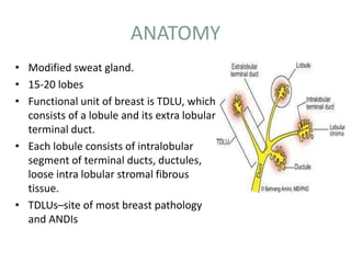
  • 1. ANATOMY • Modified sweat gland. • 15-20 lobes • Functional unit of breast is TDLU, which consists of a lobule and its extra lobular terminal duct. • Each lobule consists of intralobular segment of terminal ducts, ductules, loose intra lobular stromal fibrous tissue. • TDLUs–site of most breast pathology and ANDIs
  • 2. Mammography and Tomosynthesis • Dr Shamim Hyder Kalenchira
  • 3.
  • 4. Lymphatic drainage • Axillary or lateral pathway – fed by Sappey’s Plexus, as well as by ducts satellite lymphatics and by most of the parenchymal lymphatics – this pathway runs around the inferior edge of the pectoralis major and reach the pectoral group of axillary nodes • internal mammary pathway – originates from both the lateral and medial halves of the breast and passes through the pectoralis major; connections may lead across the median plane and hence to the contralateral breast • retromammary pathway – comes from the posterior portion of the breast
  • 5. Lymphatic drainage • Lymph of the breast drains as: – About 75% into axillary nodes(anterior group) – 20% ---- internal mammary nodes – 5% ---- posterior intercostal nodes • Internal mammary nodes drain the lymph not only from the inner half of the breast,but from the outer half as well there is a tendency for the lateral part of the breast to drain towards the axilla and the medial part to the internal mammary chain • Subareolar plexus of Sappey – plexus of lymph vessel present deep to the areola
  • 6.
  • 8. Mammography equipment • Generator • Xray tube- fine focus of 0.2-0.5 mm with an additional 0.1mm focus for magnification. • Target- Molybdenum and rhodium. • Beryllium window-minimises absorption of radiation within the tube. • Molybdenum filter- by transmitting only characteristic radiation, absorbs unwanted radiation and forms a monoenergetic beam. • Compression device: 1-4mm thick plastic plate.
  • 9.
  • 10. TARGET • The xray tubes are designed with tungsten, molybdenum , rhodium targets. • Tungsten target is operated under 30kVp with a 0.5 Aluminium filter- brehmsstrahlung and 12keV L-shell characteristic xrays. • Molybdenum target with Mo/Rh filter is more suitable for mammography. • Rhodium target filtered with Rh filter gives a similar spectrum as molybdenum. • Since the atomic number is higher, the brehmsstrahlung radiation is also higher than molybdenum.
  • 11. FILAMENT • Positioned within a focussing cup with two focal spot sizes. • Filament types are either double wound/flat ribbon/ circular filament. • Focal spot size is very critical in mammography where high spatial resolution is required. • Small focal spot is used with small anode angle, which permits the use of high mA factors. • Usually the cathode is positioned towards the chest wall , which makes the patient positioning east and takes care of the heel effect.
  • 12. FILTERS • Thin beryllium window or borosilicate glass window is used to reduce inherent filtration, since it offers low attenuation. • For a tungsten target x ray tube , Mo or Rh filter is recommended. • In case of rhodium target x ray tube , rhodium filter of 0.025 mm is used which gives high quality x rays with higher penetration. • Combination is suitable for thicker and dense breast imaging.
  • 13. GRIDS • Stationary grids or grids placed in between the screen and the film are no longer used as the thin grid lines compromised on the quality of the image. • Covered tiny details such as micro calcifications. • Hence oscillating grids are used. • Grid ratio of 4:1 or 5:1. • The grid lines are eliminated by the motion of the grid. • Grids improve the image quality.
  • 14.
  • 15. HEEL EFFECT • Shape of the breast requires higher intensity of radiation near the chest wall, to create uniform exposure to the screen film. • The cathode is positioned towards the chest wall and the anode is towards the nipple. • Permits easy positioning of the patient. • Increases the intensity of radiation near the chest wall, where greater penetration is needed. • The anode is often grounded with zero potential and the cathode is given higher potential.
  • 16.
  • 17. Compression peddle • Decreases the thickness of the breast, thus reduces the scattered radiation- improves the contrast. • Decreases the kinetic blur. • Reduces the geometric unsharpness by homogenously bringing the object close to the film. • Makes the breast thickness uniform in film density. • Differentiates the easily compressible cysts and fibroglandular tissue from the more rigid carcinomas. • Separates the super imposed breast lesions. • Reduces radiation dose to the breast tissue.
  • 18. • The compression device is parallel to the receptor surface. • Radiolucent plate that is flat and parallel to the support table. • Should match the cassette size.
  • 19. Automatic exposure control • AEC system employs phototimers to measure the x ray intensity and quality. • Kept closer to the image receptor, to minimize the object to image distance- improves spatial resolution. Two types: - Ionization chamber type. - Solid state diode type.
  • 20. SCREEN FILM SYSTEM • The x ray film should have high resolution and small grain size and used along with single intensifying screen • The emulsion surface of the film must face the screen. • Prevents loss of resolution due to light diffusion in the screen. • Gadolinium oxysulifde activated with terbium is used as screen phospor.
  • 21.
  • 23. RADIOLOGICAL ANATOMY OF THE BREAST • Schematically, the radiological examination may show the following normal anatomical structures: – Skin – Nipple and areola – Fatty tissue – The breast tissue proper, or corpus mammae – Blood vessels
  • 24.
  • 25. Mammography • Overall breast density reflects ratio between glandular elements (higher density) and fat (lower density); usually symmetric between breasts. • Fatty involution typically begins in lower-outer quadrant; progresses with age to upper-outer quadrant • American College of Radiology Breast Imaging and Reporting and Database System (BI-RADS) density categories A: Almost entirely fat B: Scattered fibroglandular densities C: Heterogeneously dense D: Extremely dense
  • 26.
  • 27. NIPPLE-AREOLA COMPLEX AND SKIN • Normal skin thickness is usually < 2 mm • Dermal papillae often visible as grooves in nipple • Nipple usually everted • Should be seen in profile on at least one of two standard mammographic views • New nipple inversion requires diligent search for subareolar pathology, esp. if unilateral Can be caused by periductal mastitis, esp. bilateral
  • 28. Imaging Approaches • Screening mammography – CC and MLO views of each breast – Additional views performed as necessary to include all glandular tissue • Diagnostic mammography – Examination is tailored to clinical question, – performed under direct supervision of radiologist – Any number of views are used to answer the question – • Is there a real abnormality? – • If finding is real, what is the morphology and level of suspicion for cancer? – Final assessment may require additional modalities, e.g., ultrasound or (rarely) MR – Report should conclude with a Bl- RADS final assessment and a management recommendation – Rare use of BI-RADS 0 if MR needed due to inconclusive mammogram and US, finding too vague to biopsy
  • 29. Conventional Vs Digital Mammography • Researches have proven that digital is superior to film for women under the age of 50 and for women with dense breasts. • 25% less radiation to the patient • Ease of storing, sharing and manipulating electronic images
  • 30. Mammography In digital mammography screen-film system has been replaced by Detectors to give the digital images Conventional
  • 31. DIGITAL MAMMOGRAPHY • The stages in digital imaging : • Image capture by digital detector • Conversion of latent image into digital data set • Processing of image data • Display of processed image • Transmission and archival of data set
  • 32.
  • 33. IMAGE ACQUISITION DEVICES • CHARGE COUPLE DEVICE TECHNOLOGY • Digital detectors consist of a phosphor, which is optically coupled to the CCD. • ADVANTAGE- The slit collimation of the X-ray beam results in a significant reduction in scatter. • This eliminates the need for a grid, leading to potential dose savings .
  • 34. SELENIUM-BASED DETECTORS • Selenium based detectors consist of a thin layer of amorphous selenium deposited on the imaging plate. A uniform positive charge is built up on the surface of the selenium. • When X-ray photons strike the surface of the selenium, electrons are freed. • This leads to a partial discharge of the uniform surface charge pattern. • The degree of discharge reflects the local radiation exposure, leadingto a latent image in the form of a charge distribution pattern. • This charge pattern is then “read” and the resultant electrical signal digitised.
  • 35. ADVANTAGES OF DIGITAL MAMMOGRAPHY 1. Acquired in less than a minute as compared to conventional mammography . 2. Spot view digital systems are available to help guide breast biopsy . 3. Superior contrast resolution especially in dense breast tissues 4. Offers the possibility of three dimensional breast tomography using relatively low doses .
  • 37. Standard views: 45* Medio lateral Oblique (MLO view)/ Lundgren’s view Craniocaudal view (CC view) RCC RMLO LCC LMLO Left Craniocaudal Right Craniocaudal Right Mediolateral Oblique Left Mediolateral Oblique
  • 38. Cranio caudal view • The casette is placed under the breast at the level of the inframammary fold. • The breast is then pulled until the inframammary fold is taut. • Compression is applied and x ray beam is directed vertically from above. • Postero medial aspect should also be included.
  • 39.
  • 40. Mediolateral oblique view • Best view to image all of the breast tissue and the pectoral muscle. • The C arm of the mammographic unit is rotated to 45 degree so that the cassette is parallel to the pectoral muscle. • The film holder is kept high up in the axillary fossa and the patients arm is abducted at the elbow by 80 degrees. • The x ray beam enters the breast from the medial side- compression is applied to the pectoralis major muscle.
  • 41.
  • 42. LAT Left Axillary Tail LCV Left Cleavage LFB Left From Below LLMO Left Lateromedial Oblique LML Let Mediolateral LRL Left Roll Lateral LRM Left Roll Medial LLM Left Lateromedial
  • 43. MAMMOGRAPHIC SIGNS • The detection and identification of elementary mammographic signs form the basis for correctly interpreting breast pathologies and describing them accurately in the mammographic report. • The specific features are the basis for classifying the lesions as either benign or malignant. These features define the positive predictive value (PPV)
  • 44. Mammographic signs can be described in terms of: – Opacity (mass) – Architectural distortion – Calcifications – Radiolucency – Asymmetry – Focal asymmetry – Skin thickening and retraction – Oedema and trabecular thickening – Asymmetrically dilated ducts
  • 45. OPACITY • Definition – A space-occupying lesion visible in two projections, implying the concept of mass and volume. – An alteration visible in just one projection can be described as a density or focal asymmetry. • Shape: oval (may include 2 or 3 lobulations), round or irregular • Margins: circumscribed, obscured, microlobulated, indistinct, spiculated • Density: high, equal, low or fat-containing.
  • 46.
  • 47. • The margin of a lesion can be: • Circumscribed (historically well-defined). This is a benign finding. • Obscured or partially obscured, when the margin is hidden by superimposed fibroglandular tissue. • Microlobulated. This implies a suspicious finding. • Indistinct (historically ill-defined). This is also a suspicious finding. • Spiculated with radiating lines from the mass is a very suspicious finding.
  • 48.
  • 49. • The density of a mass is related to the expected attenuation of an equal volume of fibroglandular tissue. High density is associated with malignancy. It is extremely rare for breast cancer to be low density.
  • 50.
  • 51. ARCHITECTURAL DISTORTION • The term architectural distortion is used, when the normal architecture is distorted with no definite mass visible. This includes thin straight lines or spiculations radiating from a point, and focal retraction, distortion or straightening at the edges of the parenchyma.. • Architectural distortion can also be seen as an associated feature. For instance if there is a mass that causes architectural distortion, the likelihood of malignancy is greater than in the case of a mass without distortion. • Classification – Marginal – Intra-parenchymal distortion.
  • 52. Notice the distortion of the normal breast architecture on oblique view (yellow circle) and magnification view.
  • 53. CALCIFICATIONS • Definition – Calcifications are amorphous, radiopaque, acellular entities produced by calcium deposits. – They reflect the secretory or degenerative processes of the breast. Malignant calcifications may be caused either by cellular secretion or by calcification of necrotic cancer cells. – Calcifications are among the most frequent mammographic signs and can also be amongst the most difficult to define and interpret. • In relation to their site of origin, calcifications are classified as – Intraductal – Lobular • Evaluation – Shape – Distribution – Site – Size – Number
  • 54. • Lobular calcifications: This results in uniform, homogeneous and sharply outlined calcifications, that are often punctate or round. When the acini become very large, as in cystic hyperplasia, 'milk of calcium' may fill these cavities. • Intraductal calcifications: form a complete cast of the ductal lumen. This explains why they often have a fine linear or branching form and distribution. Intraductal calcifications are suspicious of malignancy and are classified as BI-RADS 4 or 5.
  • 57. .
  • 58.
  • 59. Suspicious morphology • Amorphous (BI-RADS 4B) So small and/or hazy in appearance that a more specific particle shape cannot be determined. • Coarse heterogeneous (BI-RADS 4B) Irregular, conspicuous calcifications that are generally between 0,5 mm and 1 mm and tend to coalesce but are smaller than dystrophic calcifications. • Fine pleomorphic (BI-RADS 4C) Usually more conspicuous than amorphous forms and are seen to have discrete shapes, without fine linear and linear branching forms, usually < 0,5 mm. • Fine linear or fine-linear branching (BI-RADS 4C) Thin, linear irregular calcifications, may be discontinuous, occasionally branching forms can be seen, usually < 0,5 mm.
  • 60.
  • 61. Distribution of calcifications • The arrangement of calcifications, the distribution, is at least as important as morphology. These descriptors are arranged according to the risk of malignancy: • Diffuse: distributed randomly throughout the breast. • Regional: occupying a large portion of breast tissue > 2 cm greatest dimension • Grouped (historically cluster): few calcifications occupying a small portion of breast tissue: lower limit 5 calcifications within 1 cm and upper limit a larger number of calcifications within 2 cm. • Linear: arranged in a line, which suggests deposits in a duct. • Segmental: suggests deposits in a duct or ducts and their branches.
  • 62.
  • 63. • Site: Before starting to analyse calcifications, it is advisable to ascertain that calcifications are truly intra-mammary, to rule out skin or pseudocalcifications due to artifacts. • Size: Many different dimensions are reported, but calcifications associated with malignancies are generally between 0.5 and 2 mm, though often even smaller. • Number: There is no consensus . The number of calcifications must be evaluated together with their morphology and distribution.
  • 64. RADIOLUCENCY • A sharp-edged radiolucent mass may be seen/ with a radiopaque rim that is generally complete or only partly broken. Varying in size and sometimes occupying the whole breast. • These may be round, oval or more rarely lobulated. • They are usually caused by lipomas/ pseudolipomas and foci of fat necrosis.
  • 65.
  • 66. ASYMMETRIC BREAST TISSUE • Significant areas of fibroglandular tissue on one side that appear different compared with the contralateral breast, because they are either of greater extent or of higher density. If there are no other abnormalities, this is simply a normal variant of breast structure.
  • 67. FOCAL ASYMMETRY • Areas of glandular tissue without the characteristics of a true mass, but recognizable with similar appearance in the craniocaudal , lateral and oblique projection. • These may simply be normal local asymmetries but further investigation is justified, with targeted magnification views and if necessary ultrasound.
  • 68. SKIN THICKENING AND RETRACTION • Skin thickening and retraction can be diffuse or focal. Skin thickening or even ulceration can occur as a result of tumor direct invasion or through thickening and retraction of Coopers ligaments. • Skin changes can result from a contiguous malignancy, infection or inflammation, primary skin disorders, lymphatic or vascular obstruction. and systemic diseases affecting the skin. • Post-surgical scarring and fat necrosis can also cause skin thickening and retraction.
  • 69. • A high density, stellate opacity with radiating spicules into the surrounding parenchyma is seen in the inner subareolar location with thickened and retracted overlying areolar skin.
  • 70. OEDEMA • Diffuse oedema causes generalized thickening of the trabecular stromal net. There may also be thickening of the skin. • This finding is typical of benign processes such as mastitis, post-radiotherapy changes and congestive heart failure secondary to kidney failure. • Thickened skin also invariably accompanies inflammatory carcinoma in which a focal opacity may be visible mammographically. • Post-therapy follow-up may be advisable in these cases to permit a differential diagnosis.
  • 71. ASYMMETRICALLY DILATED DUCTS • Linear, ribbon-like, tubular or branching, single or multiple opacities, parallel and radiating in the retroareolar space. • This appearance is typical of ectactic ducts and, less commonly, of intraductal papilloma or carcinoma.
  • 72. BI-RADS - Breast Imaging Reporting and Data System • BI-RADS 0: Screening mammogram with finding for which additional evaluation is needed – Also known as recall or call-back • BI-RADS 1: Negative mammogram • Bl-RADS 2: Mammogram with a benign finding • Bl-RADS 3: Mammogram with a probably benign finding, for which short-interval follow-up suggested
  • 73. • BI-RADS 4: Mammogram with a suspicious abnormality for which biopsy should be considered • BI-RADS 5: Mammogram with a finding highly suggestive of malignancy, for which appropriate action should be taken • BI-RADS 6: Mammogram with a known biopsy-proven malignancy, for which appropriate action should be taken
  • 74.
  • 75.
  • 76. Breast Ductography • Helps define the cause of unilateral, single- pore, spontaneous nipple discharge. • Allows diagnosis of the underlying condition, definition of the extent of disease, identification of central and peripheral lesions, and guidance of surgical excision. • It involves cannulation of the discharging duct, injecting contrast material within and obtaining 90 degree and craniocaudal views.
  • 78. In this patient, contrast outlines (single arrows) a duct that is wider than normal (dilated), and the contrast column is disrupted by the presence of a tumor (double arrow). About half of the patients presenting with spontaneous nipple discharge are found to have papillomas, which are benign, non-cancerous tumors. The tiny, blunt- tipped needle used to inject the contrast is seen at the edge of the film (wide arrow).
  • 79. Breast tomosynthesis • Modification of a digital mammography unit to enable the acquisition of a three-dimensional (3D) volume of thin-section data. • Images are reconstructed in conventional orientations by using reconstruction algorithms similar to those used in computed tomography (CT). • Reduces or eliminates tissue overlap.
  • 80. In breast tomosynthesis, a moving x-ray source and a digital detector are used.
  • 81. • The X-ray tube makes an arc, during which is acquired a series of images, each of which is delivered a dose equal to a fraction of that provided in a standard mammogram. During the acquisition any detector element receive in time sequence related information on each object volume element. The set of digital projections thus contains a complete structural information on all the object layers in the form of raw data
  • 82. • Acquisitions may be performed either with the step-and-shoot method or with a continuous exposure method. • The image data are sent from the acquisition workstation to the reading workstation. Images are reconstructed by using a mathematical algorithm similar to those used in CT reconstructions, to generate a set of thin image sections parallel to the breast platform
  • 83.
  • 84. • Tomosynthesis, therefore, does not provide direct projection images, but reconstructed images of any individual layers through several available algorithms, more or less efficient, each aimed to removing from reconstructed slice the upper and lower layers "structured noise". • DBT allows the detection of a greater number of expansive lesions and a better morphological analysis of masses and architectural distortions, thanks to the contrast of findings greater than the background, given by the more shade structures belonging to the upper and lower layers, and then to the smaller amount of noise. It is thus exceeded one of the limits of two-dimensional imaging, which is the masking of lesions caused by superimposition of normal structures.
  • 85. • A further advantage of DBT is given by the lack of need for operator training (the breast is positioned just like a conventional mammography in MLO and / or CC projection) and for the radiologist (as he continues to perform diagnosis from images with mammograms features).
  • 86. Comparison of screening mammography with breast tomosynthesis in a 57-year-old woman. (a) Digital mammogram shows a mass (arrows) in the lower outer part of the left breast. The mass is not clearly visible because of surrounding dense tissue. (b) Breast tomosynthesis image provides clearer depiction of the mass (arrows), which is well circumscribed.
  • 87. Invasive ductal carcinoma in a 45-year-old woman with a lump in the left breast for 6 months. (a) Digital mammogram clearly shows an oval-shaped relatively well- circumscribed primary mass. (b) Breast tomosynthesis image provides better depiction of the microlobulated, spiculated border of the mass (arrows), a finding suggestive of malignancy.
  • 88. Computer-Aided Detection • CAD programs are commercially available systems that use computer software to assist the mammographer in detecting or identifying potentially suspicious abnormalities on a mammogram. • The CAD program identifies potential abnormalities on the images and marks areas on the study that the computer considers to be suspicious. • The radiologist reviews the flagged areas to assure that nothing was missed. • DM readily provides images that can be used with CAD. Film screen mammography has to be digitized in order for CAD to be performed.
  • 89.
  • 90. Bibliography • Bi-RADS for Mammography and Ultrasound 2013 Updated version Harmien Zonderland and Robin Smithuis • Diagnostic Radiology: Musculoskeletal and Breast Imaging by Berry Manorama, Chowdhury Veena, Mukhopadhyay Sima, and Suri Sudha