
Recommended
More Related Content
What's hot
What's hot (20)
Similar to Complications of Local anesthesia (part I) for B.D.S & M.D.S
Similar to Complications of Local anesthesia (part I) for B.D.S & M.D.S (20)
Recently uploaded
Recently uploaded (20)
Complications of Local anesthesia (part I) for B.D.S & M.D.S
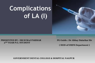
- 1. Complications of LA (I) GOVERNMENT DENTAL COLLEGE & HOSPITAL NAGPUR PRESENTED BY : DR SURAJ PARMAR 2ND YEAR P.G. STUDENT PG Guide : Dr Abhay Datarkar Sir ( HOD of OMFS Department )
- 2. • Local Complications • Systemic Complications
- 3. Local complication Needle breakage Prolonged anesthesia or paresthesia Facial nerve paralysis Trismus Soft tissue injury Hematoma Pain on injection Infection Edema Sloughing of tissues Post anesthetic intraoral lesions
- 4. Needle breakage • Extremely rare • Break at the hub ( most rigid portion of needle )
- 5. Causes • Weakening of dental needle by bending it before it insertion into patient’s mouth. • Sudden unexpected movement by the patient as needle penetrates muscle or contacts periosteum. • Defect in manufacture of needle.
- 6. Management • Remove needle if it is visible with help of a hemostat. • If not visible take radiographs of the region . • If needle is lost into the tissue spaces ,e.g. pterygomandibular space, infratemporal space, assure patient and review regularly. • 3D CT scanning recommended.
- 7. Prevention • Use larger gauge needles • 25 gauze needles are appropriate for IANB , PSA ASA, maxillary and mandibular block. • Use long needle for injection requiring penetration of significant (> 18 mm depth of tissues ) • Do not insert a needle into tissue to its hub • Do not redirect a needle once it is inserted in tissues. • Withdraw needle almost completely before redirecting it.
- 8. Persistent anesthesia or paresthesia • Clinical response : • Sensation of numbness • Swelling • Tingling • Itching
- 9. Causes • Trauma to any nerve • Injection of LA contaminated by alcohol or sterilizing solution near a nerve • Trauma to nerve sheath • Haemorrhage into or around the neural sheath
- 10. • According to Hass, paresthesia was reported most often after the administration of 4 % LA, either Prilocaine HCL and Articaine HCL.
- 11. Prevention • Strict adherence to injection protocol • Proper care and handling of dental cartridge
- 12. Management • Most case resolve within 8 weeks • Reassurance to the patient • Examine the patient in person • Reschedule the patient for examination every 2 months for as long as the sensory deficit persist. • Dental treatment may continue , but avoid readministering LA into region of the previously traumatized nerve. • Use alternate LAtechniques if possible.
- 13. Facial nerve paralysis • Usually occurs in inferior alveolar nerve block when anesthetic is introduced into the deep lobe of parotid gland.
- 14. Causes • Introduction of LA into capsule of parotid gland • Directing the needle posteriorly or inadvertently deflecting it in a posterior direction during an IANB • Over insertion during a Vazirani- Akinosi nerve block, may place the tip of needle within the body of parotid gland.
- 15. Problem • Loss of function to the muscle of facial expression : transitory • Unilateral paralysis and be unable to use of these muscles. • Primary problem : cosmetic • Secondary problem : unable to voluntarily close one eye • Cornea retain its innervation, thus if it is innervated, the corneal reflex is intact and tears lubricate the eye.
- 16. Prevention • By adhering to protocol with inferior alveolar and vazirani-akinosi nerve block.
- 17. Management • Reassure the patient. • Contact lenses should be removed untill muscular movements returns. • Eye patch should be applied to the affected eye untill muscle tone returns. • If resistance offered by patient advise patient to manually close lower eyelid periodically to keep cornea lubricated.
- 18. Trismus • Prolonged tetanic spasm of jaw muscles. • Normal opening of mouth is restricted.
- 19. Causes • Trauma to the muscle or blood vessels in the infratemporal fossa • Local anesthetic solution into which alcohol or cold sterilizing solution have diffused produce irritation of tissues leading potentially to trismus. • Injection of LAs either intramuscularly or supramuscularly leads to rapidly progressive necrosis of exposed muscle fibers. • Haemorrhage
- 20. • Low grade infection after injection. • Multiple needle penetrations . • Excessive volumes of local anesthetic solution deposited into a restricted area produce distention of tissues which may lead to post injection trismus.
- 21. Problem • Average inter-incisal opening in cases of trismus is 13.7 mm ( range 5-23 mm ). • In the acute phase of trismus , pain produced by hemorrhage leads to muscle spasm and limitation of movement. • Second or chronic phase usually develops if treatment is not begun.
- 22. • Chronic hypomobility is secondary to organization of hematoma with subsequent fibrosis and scar contracture. • Infection also may produce hypomobility through increased pain , increased tissue reaction , scarring.
- 23. Prevention • Use a sharp, sterile, disposable needle. • Properly care for and handle dental local anesthetic cartridges. • Use aseptic technique. • Practice atraumatic insertion and injection technique. • Avoid repeat injections and multiple insertions into same area . • Use minimum effective volume of local anesthetic
- 24. Management • Heat therapy • Warm saline rinses • Analgesics • Muscle relaxant to manage initial phase of muscle spasm • Physiotherapy
- 25. • Avoid further dental treatment in the involved region until symptoms resolve and patient is more comfortable. • Improvement within 48 to 72 hours. • If pain and dysfunction continue unabated beyond 48 hours, consider possibility of infection. • Antibiotics should be added to the treatment regimen and continued for 7 days. • Complete recovery from injection related trismus takes about 6 weeks ( range of 4-20 weeks )
- 26. • TMJ involvement is rare in first 4 to 6 weeks after injection. • Surgical intervention to correct chronic dysfunction may be indicated in some instances.
- 27. Soft tissue injury • Self-inflicted trauma to lips and tongue is frequently caused by patient inadvertently biting or chewing these tissues while still anesthetized.
- 28. Causes • Most frequently in younger children, in mentally or physically disabled children or adults and in older old patient . • However it can and dose occur in patients of all ages. • The primary reason is the fact that soft tissue anesthesia lasts significantly longer than does pulpal anesthesia.
- 29. Problem • It can lead to swelling and significant pain when the anesthetic effects resolve. • Ayounger child or a handicapped individual may have difficulty coping with the situation and may lead to behavioral problems.
- 30. Prevention • Acotton roll can be placed between the lip and the teeth if they are still anesthetized at the time of discharge. • The cotton roll is secured with dental floss wrapped around theteeth. • The patient and the guardian against eating, drinking hot fluids, and biting on the lips or tongue to test for anesthesia. • Aself-adherent warning sticker may be used on children.
- 31. Management • Analgesics for pain, as necessary. • Antibiotics, as necessary, in the unlikely situation that infections results . • Warm saline rinses to aid in decreasing any swelling that may be present. • Petroleum jelly or other lubricant to cover a lip lesion and minimise irritation.
- 32. Hematoma • Defind as effusion of blood into extravascular space can result from inadvertently nicking a blood vessel during the injection of LA. • Hematoma developing subsequent to nicking of an artery usually increases rapidly in size untill treatment is instituted, because of significantly greater blood pressure within artery. • Nicking vein may or may not result in hematoma. • Tissue density surrounding injured vessels is determining factor.
- 33. Causes • Damage blood vessel by needle during penetration • Hematoma after inferior alveolar nerve block are usually only visible intraorally, whereas PSA hematomas are visible extraorally.
- 34. Problem • Rarely produces significant problem aside from resulting bruise which may or may not be visible extraorally. • Possible complication of hematoma include trismus & pain. • Swelling and discoloration usually subside within 7 to 14 days.
- 35. Prevention • Knowledge of normal anatomy involved in proposed direction is important. • Modify the injection technique as dictated by patient’s anatomy. • Use a short needle for PSA nerve block to decrease the risk of hematoma. • Minimize number of needle penetration into tissue. • Never use a needle as a probe in tissues.
- 36. Management • Immediate : swelling become evident during or immediate after LAs , direct pressure should be applied to the site of bleeding. • IANB : pressure is applied to medial aspect of ramus • ASA : pressure is applied to skin directly over infraorbital foramen • Incisive or mental nerve block : pressure is placed directly over the mental foramen. On the skin or mucous membrane.
- 37. • PSA : hematoma progresses over a period of days inferiorly and anteriorly toward lower anterior region of cheek. • Digital pressure can be applied to soft tissues in the mucobuccal fold as far distally as can be tolerated by patient ( without eliciting a gag reflex ) • Applied pressure in medial and superior direction • Ice should be applied extraorally to increase pressure on the site and help constrict the vessel
- 38. • Advise patient about possible soreness and limitation of movement ( trismus ) • If soreness develop. Advise patient to take an analgesic • Heat may be applied to region beginning the next day. • Ice may be applied to th region immediately on recognition of developing hematoma. • With or without treatment , a hematoma will be present for 7 to 14 days. • Avoid additional dental therapy in the region untill symptoms and signs resolve.
- 39. Pain on injection • This increases patient's anxiety; and may lead to a sudden unexpected movement by the patient and increases the risk of needle breakage.
- 40. Causes • Careless injection technique. • Dull needles: Needles become dull due to multiple injections. • Rapid deposition of local anesthetic solution. • Needles with barbs: There is pain while withdrawal of the needle from the tissues. • Temperature: Extremes of temperature such as warm or hot or very • cold (refrigerated) local anesthetic solution.
- 41. Prevention • Adhere to proper technique of injection. • Use sharp needles. • Use topical ansthetic properly before injection. • Use sterile local anesthetic solutions. • Inject LAs slowly. • Be certain that temperature of solution is correct. A solution that is too hot or too cold may be more uncomfortable than one at room temperature.
- 42. Management • No management is necessory. • Step should taken recurrence of pain associated with injection of local anesthetic.
- 43. Burning on injection • Altered pH of the solution • a. Presence of vasoconstrictor: The pH of local anesthetic solution without the vasoconstrictor is approximately 5 to 5.5; and that with a vasoconstrictor is approximately 3 to 3.5. It is more acidic. • b. Old solution: The end result of oxidation of Na bisulfite is Na- bisulfate which is more acidic. • Non-isotonic local anesthetic solution. • Deposition of excessive amount of local anesthetic solutions
- 44. • Other causes: • i. Rapidity of injection: Especially in adherent tissues and confined areas, such as palatal mucoperiosteum. • ii. Contamination of local anesthetic solution, especially when cartridges are stored in alcohol or other cold sterilizing solutions; leads to diffusion of these solutions into the cartridge. • iii. High temperature of local anesthetic solution. Solutions warmed to body temperature are usually considered to be "too hot" by the patients.
- 45. Problem • When a burning sensation occurs as a result of rapid injection, contaminated solution or overly warm solution , there is a greater likelihood that tissue damaged, with subsequent development of other complications such as post anesthetic trismus, edema and possible paresthesia.
- 46. Prevention • Slow injection. Ideal rate is 1 ml/min; while recommended rate is 1.8 ml/min. • Cartridges should be stored in a suitable container at room temperature without alcohol or any other cold sterilizing solutions. • The excess of cold sterilising solution should be removed by dipping the cartridge in sterile water or normal saline. • Use recently manufactured cartridges, as far as possible, to circumvent • the problem of increased acidic medium of the solution, because of oxidation of vasoconstrictor
- 47. Management • Because most instances transiet and do not lead to prolonged tissue involvement, formal treatment is not usually indicated.
- 48. Infection • The incidence of injection-related infection has become less following introduction of pre-sterilised disposable needles and cartridges.
- 49. Causes • Contamination of the needles. Needles touching mucous membrane other than the area of insertion of the needle result in contamination of the needle. It is the major cause of post-injection infection. • Contamination of the local anesthetic solution. It is also rare, as the solutions are pre-sterilized. • Contamination of needles or solutions may cause a low-grade infection, if placed in the deeper tissues, which may lead to trismus. • Low-grade infection is not recognized immediately. The patient usually complains of pain and dysfunction in the immediate post- injection phase.
- 50. • Improper injection technique: It includes the following: • a. Improper handling of local anesthetic equipment (storage of the cartridges). • b. Improper preparation of the site. • c. Inadequate washing of operator's hands. • d. Needle passing through an area of infection. It may disseminate infection. • Local anesthetic solution deposited under pressure; as in intraligamentary injection. It is claimed to deposit bacteria, in healthy tissues and thus spreading the infections.
- 51. Prevention • Preparation of the site prior to needle penetration: Apply antiseptic, dry the area, and then apply topical anesthetic agent. • Careful handling of the needles. Avoid contamination of needles through contact with non-sterile surfaces. • Avoid multiple penetrations with the same needle. • Use pre-sterilized disposable needles. • Proper cleansing of operator's hands. • Avoid passing the needles through infected areas.
- 52. • Proper handling of dental cartridges: • i. Store cartridges aseptically, as far as possible, in a container covered with a lid all the time. Once the container is opened, cartridges should be stored dry in their original container or in another suitable sterile container that is kept covered at all times. • ii. Avoid contamination of plunger and the diaphragm prior to their use. The diaphragm-end of cartridge should be wiped with a sterile disposable sponge soaked with an antiseptic prior to its insertion into the syringe and fixing of the needle.
- 53. • iii. Use cartridges available in blister packing. • iv. Use cartridges only once (one patient). An attempt to use a portion for one patient and the remaining for another patient increases the possibility of cross-infection
- 54. Management • The management is symptomatic; and it consists of the following: • i. Analgesics • ii. Antibiotics • iii. Physiotherapy • iv. Heat therapy • v. Anti-inflammatory drugs • vi. Muscle relaxants, and • vii. Incision and drainage, if necessary.
- 55. Edema • Edema of tongue, pharynx and larynx may develop into potentially lifethreatening situations.
- 56. Causes • Trauma during injection • Infection • Allergy • Hematoma • Injection of irritating solutions such as cold-sterilising solutions. • Each factor should be considered with regard to its prevention and management
- 57. • Hereditary angioedema is a condition characterized by the sudden onset of brawny non pitting edema affecting the face , extremities and mucosal surface of intestines and respiratory tract , often without obvious precipitating factors. • Manipulation within the oral cavity, including LAs administration may precipitate an attack. • Lips, eyelid & tongue are often involved.
- 58. Problem • Airway obstruction • Pain & dysfunction of region
- 59. Prevention • Preoperative assessment: Complete medical evaluation of the patient, particularly history of allergy to any drug. • Careful handling of local anesthesia armamentarium. • Atraumatic anesthetic technique.
- 60. • Find out the cause. • In cases of traumatic injection and introduction of irritating solutions, the edema is minimal and resolves in a few days, and therapy is sometimes not required. • Analgesics for pain. • In case of infection, start suitable antibiotics. • If allergy: Administer antihistaminics orally and/or IM; sometimes it can be life-threatening. Consultation with an allergic specialist is mandatory.
- 61. • If breathing is compromised because of edema the following steps are taken: • i. Patient is placed in supine position. In case of tongue or oropharyngeal region, right or left lateral position is taken. • ii.Institute Basic Life Support (BLS); Airway, Breathing, and Circulation (ABC). • iii. Emergency Medical Services (EMS) are summoned. • iv. Administer O2.
- 62. • v. Administer epinephrine: 0.3 mg (adults), 0.15 mg (child) IM/IV every 5 minutes until respiratory distress resolves. • vi. Administer antihistaminics. • vii. Administer corticosteroids. • 5. Refer to oral and maxillofacial surgeon. • 6. Life-threatening situations may require cricothyroidotomy. Transfer the patient to a general hospital with an ICU facility
- 63. Sloughing of tissues • Various Forms • i. Epithelial desquamation or ulceration. • ii. Sterile abscess. • iii. Tissue necrosis or sloughing
- 64. Causes • 1. Predisposition: Commonly seen in hard palate, as in the region of .distribution of nasopalatine and greater palatine nerves, because the mucoperiosteum is firmly attached to the bone. • It occurs at the site of injection. Necrosis leads to painful ulceration and sloughing. • 2. Deposition of excessive volume of local anesthetic agent with a vasoconstrictor. • 3. Rapid deposition of the local anesthetic solution with undue pressure.
- 65. • 4. Application of topical local anesthetic agent for prolonged period (epithelial desquamation). • 5. Use of high concentration of vasoconstrictors (usually epinephrine), resulting in tissue ischemia and necrosis (Guinta, 1975). • Sterile abscess occurs secondary to prolonged ischemia, resulting from resulting from epinephrine.
- 66. Prevention • Avoid using excessive amounts of local anesthetic agent (minimal effective dose). • Avoid using vasoconstrictors of high concentration. • Avoid rapid deposition and with excessive pressure. • Excessive volume may have reaction secondary to excessive pressure. • Warn the patient against application of hot items; while the tissues are still anesthetised.
- 67. Management • Symptomatic: The management depends upon the extent of injury • consists of analgesics, topical anesthetics, and bland diet, etc. • It usually resolves in 1-2 weeks. • An established abscess may require incision and drainage.
- 68. Post anesthetic intraoral lesion • Patients' reporting of development of ulcerations around the site of injection a few days after intraoral injection of local anesthetic agent. • Patient complains of intense pain.
- 69. Causes • Recurrent Aphthous Stomatitis (RAS): It is a frequent manifestation, developing in gingival tissues (movable part, i.e. not attached to the bone) such as buccal vestibule. • Herpes Simplex/Herpes Labialis: It is related to reactivation of dormant Herpes Simplex Virus (HSV) particles by the trauma of injection. • It is usually seen in patients with history of recurrent herpes labialis, particularly, in the terminal area of distribution of trigeminal nerve (inferior alveolar nerve, or superior labial branch of infraorbital nerve), in a previously anesthetised nerve.
- 70. • HSV can develop intraorally, although it is commonly observed extraorally. • It is manifested as small bumps on tissues attached to underlying bone; such as soft tissues of the hard palate. • Trauma to the tissues by a needle, local anesthetic agent, cotton swab, or any other instrument, may activate the latent form of the disease process.
- 71. Prevention • Pre-anesthetic assessment: History of recurrent herpetic infections. • Delay surgical intervention in the active stage. • In susceptible patients, intraoral lesions cannot be prevented from developing. • Intraoral Herpes Simplex, may be prevented, or its manifestations may be minimized, if treated in its prodromal phase • (Prodrome: Mild burning or itching sensation at the site where the virus is present, e.g. lip.
- 72. Management • Explanation and assurance are integral parts of the management. • The management, otherwise is symptomatic and includes: • (i) Analgesics, • (ii) Topical anesthetics, e.g. viscous lidocaine, applied topically to affected painful areas, • (iii) Antiviral agents, (acyclovir) applied QID over the affected area. • It minimizes the acute phase.
- 73. •Thank you…..