
Recommended
More Related Content
What's hot
What's hot (20)
Similar to human body sense
Similar to human body sense (20)
Recently uploaded
Recently uploaded (20)
human body sense
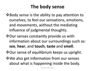
- 1. The body sense Body sense is the ability to pay attention to ourselves, to feel our sensations, emotions, and movements, without the mediating influence of judgmental thoughts. Our senses constantly provide us with information about our surroundings such as: see, hear, and touch, taste and smell. Our sense of equilibrium keeps us upright. We also get information from our senses about what is happening inside the body. by bashir awil ismail
- 2. Body sense are 1. Sensation of skin 2. Sensation of hearing 3. Sensation of taste 4. Sensation of smell 5. Sensation of vision by bashir awil ismail
- 3. SENSORY PATHWAY The impulses involved in sensations follow very precise pathways, which all have the following parts: 1.Receptors: detect changes (stimuli) and generate impulses. 2. Sensory neurons: transmit impulses from receptors to the central nervous system. These sensory neurons are found in both spinal nerves and cranial Nerves. 3. Sensory tracts: white matter in the spinal cord or brain that transmits the impulses to a specific part of the brain. 4. Sensory areas: most are in the cerebral cortex. by bashir awil ismail
- 4. CHARACTERISTICS OF SENSATIONS 1.Projection: the sensation seems to come from the area where the receptors were stimulated. 2. Intensity: some sensations are felt more distinctly and to a greater degree than are others. A weak stimulus such as dim light will affect a small number of receptors, but a stronger stimulus, such as bright sunlight, will stimulate many more receptors. 3. Contrast: simultaneous sensation on a current sensation, which may then be exaggerated/greater or diminished/smaller. If on a very hot day, you jump into a swimming pool, the water may feel quite cold at first. 4. Adaptation: becoming unaware of a continuing stimulus. The water in the swimming pool that seemed cold at first seems to warm up after a few minutes. 5. After-image: the sensation remains in the consciousness even after the stimulus has stopped. A familiar example is the bright after-image seen after watching a flashbulb go off. by bashir awil ismail
- 5. CUTANEOUS SENSES ( touch) The dermis of the skin and the subcutaneous tissue contain receptors for the sensations of touch, pressure, heat, cold, and pain. Cutaneous Senses include touch and everything else we feel through our skin: temperature, pressure, vibration, and pain. For example, when we run our fingers over a rough surface, receptors in the skin of our fingertips send information about the surface’s texture to our brain. by bashir awil ismail
- 6. by bashir awil ismail
- 7. Continue… Epidermis: is the thin, outer layer of the skin that is visible to the eye and works to provide protection to the body. It is responsible for the protection of the body. Dermis: is the second and thickest layer of the three major layers of skin, located between the epidermis and subcutaneous tissues. Hypodermis: is the innermost and thickest layer of skin. by bashir awil ismail
- 8. Skin nerves Meissner corpuscle: - stack of flattened disks in the dermis just below epidermis; they respond to touch, pressure, vibration. Pacinian corpuscle: - onion-like capsule located deep in the skin; they responsible for sensitivity to vibration and pressure. Nocieceptor: are sensory receptors that detect signals from damaged tissue and chemical released from the damaged tissues. Thermoreceptors: are specialized nerve cells that are able to detect differences in temperature, heat and cold. by bashir awil ismail
- 9. SENSE OF TASTE The receptors for taste are found in taste buds, most of which are in papillae on the tongue These chemoreceptors detect chemicals in solution in the mouth. The chemicals are foods and the solvent (if the mouth is very dry, taste is very indistinct). There are five general types of taste receptors: sweet, sour, salty, bitter, and savory. The sense of taste is important because it makes eating enjoyable. Some medications may interfere with the sense of taste, and this sense becomes less acute as we get older. by bashir awil ismail
- 10. Taste buds anatomy by bashir awil ismail
- 11. by bashir awil ismail
- 12. SENSE OF SMELL the sense of smell is the process of creating the perception of smell. It occurs when an odor binds to a receptor within the nose, transmitting a signal through the olfactory system. The receptors for smell (olfaction) are chemoreceptors that detect vaporized chemicals that have been sniffed into the upper nasal cavities Just as there are specific taste receptors, there are also specific scent receptors, and research indicates that humans have several hundred different receptors. by bashir awil ismail
- 13. Continue… When stimulated by vapor molecules, olfactory receptors generate impulses carried by the olfactory nerves (1st cranial) through the ethmoid bone to the olfactory bulbs. The pathway for these impulses ends in the olfactory areas of the temporal lobes. Vapors may stimulate many combinations of receptors, and it has been estimated that the human brain is capable of distinguishing among 10,000 different scents. by bashir awil ismail
- 14. Olfactory anatomy by bashir awil ismail
- 15. Continue… olfactory bulb: is a neural structure of the vertebrate forebrain involved in olfaction, the sense of smell. cribriform plate: is a sieve-like structure between the anterior cranial fossa and the nasal cavity. It is supports the olfactory bulb. olfactory nerve: is the first cranial nerve and conveys special sensory information related to smell. It is the shortest of the cranial nerves. Nasal cavity: is a large, air-filled space above and behind the nose in the middle of the face. olfactory tract: connects the olfactory bulb to the remainder of the cerebral cortex. by bashir awil ismail
- 16. HUNGER AND THIRST Hunger and thirst may be called visceral sensations, in that they are triggered by internal changes. Hunger is a sensation that seems to be far more complex than was first thought. The receptors for both senses are specialized cells in the hypothalamus. The receptors for thirst detect changes in the body water content, that occurs when your body needs water. by bashir awil ismail
- 17. Continue… The sensation of hunger is related to contractions of the stomach muscles. Hunger is a normal sensation that makes you want to eat. Your body tells your brain that your stomach is empty. Hunger controlled by a part of your brain called the hypothalamus. Fullness is a feeling of being satisfied. Your stomach tells your brain that it is full. by bashir awil ismail
- 18. Sensation of eyes • The human eyeball(bulbus oculi) is approximately globe shaped, with diameter of about 24mm. • Eyeball is made of two segments an anterior part and posterior part. • Orbital cavity is a bony cavity of the eyeball that keeps eyes from external forces. • eyelids: protect eyeball from foreign particles and open and close voluntarily as well as by reflex actions. by bashir awil ismail
- 19. THE EYE The eye or the organ of sight is situated in the orbital cavity of the skull. Each eyeball is similar to a camera and which produces images. It is well protected by bony walls of the orbit, also contains: muscles of eyeball, nerves, blood vessels, lacrimal gland by bashir awil ismail
- 20. Anatomy of the eyes Human eye ball it looks like globe shaped, with a diameter of about 24mm. Eye ball is made up of two segments, an anterior part and posterior part. Eyelids: protect eyeball from foreign particles coming in contact with its surface and cut of the light during sleep. Eyelids opens and closed voluntarily. As well as reflex action. Conjunctiva: is thin mucus membrane, which covers the exposed part of the eye. provides protection and lubrication of the eye by the production of mucus and tears. by bashir awil ismail
- 21. Continue… Lacrimal gland: secrete the watery component of tears and are located behind the outer part of each upper lid. It function is production of tears which moist and cleanse of eyes surface. Tear contains lysozyme that kills bacteria. Lacrimal gland by bashir awil ismail
- 22. Wall of the eye ball Wall of the eye ball is composed of three layers: 1. Outer layer (tunica externa or tunica fibrosa) Sclera Cornea 2. Middle layer ( TUNICA MEDIA OR TUNICA VASCULOSA). Choroid Ciliary body Iris 3. Inner layer ( tunica interna, or retina). It is delicate light sensitive membrane that forms the innermost layer of eyeball. Pigmented layer Sensitive or neuron layer by bashir awil ismail
- 23. Anatomy of the eye by bashir awil ismail
- 24. Continue… SCLERA: forms the outermost layer of the eyeball It provides protection to the delicate structure within the eye. CHOROID: is a thin pigmented membrane, dark brown in color which is situated in between sclera (externally) and retina (internally). CORNEA: is the transparent convex anterior portion of the outer layer of eyeball, which covers the iris and pupil. by bashir awil ismail
- 25. Continue… CILIARY BODY: is the continuation of choroid consisting of smooth muscle fibers, i.e., the ciliary- muscle IRIS: is the pigmented membrane surrounds the pupil. RETINA: Retina is the inner most layer of the eyeball. It is translates the light impulses into electronic impulse which travels down the optic pathways to the brain. Fovea: is a tiny pit located in the retina that provides the clearest vision. by bashir awil ismail
- 26. Continue… optic nerve: is located in the back of the eye. The function of the optic nerve is to transfer visual information from the retina to the vision centers of the brain via electrical impulses. by bashir awil ismail
- 27. Continue… Blind spot: a small area of the retina where the optic nerve enters the eye; this type of blind spot occurs normally in all eyes. by bashir awil ismail
- 28. Continue… Ciliary muscles: focusing muscles that change the shape of the lens. Suspensory ligaments: connectors that join ciliary muscle to the lens. by bashir awil ismail
- 29. Continue… vitreous humour: is a clear, colourless fluid that fills the space between the lens and the retina of the eye. 99% of it consists of water and the rest is a mixture of collagen, proteins, salts and sugars. by bashir awil ismail
- 30. Visual sense pathway Visual process in series of actions that take place during visual perception. the image of an object seen by the eyes comes cornea. Next comes the pupil, the opening that lets light enter the eye and ultimately reach the retina. The retina converts the image formed by the light rays into nerve impulses. the energy in visual spectrum is converted into electrical impulses by rods and cones of retina through some chemical reactions. the impulses from rods and cones reach the cerebral cortex through optic nerve and sensation of vision produced in cerebral cortex. by bashir awil ismail
- 31. The visual pathway contain six components: 1) Optic nerve 2) Optic chiasma 3) Optic tract 4) Lateral geniculate body 5) Optic radiation 6) Primary visual cortex by bashir awil ismail
- 32. Continue… 5. Optic nerve (CN II):is a paired cranial nerve that transmits visual information from the retina to the brain. 6. Optic chiasm:it is medial fibers of each optic nerve cross the midline and join the uncrossed lateral fibers of opposite side to form the optic tract. 7. Optic tracts: is formed by uncrossed fibers of optic nerve, on the same side and crossed fibers of optic nerve from opposite side. by bashir awil ismail
- 33. Continue… 8. lateral geniculate body: Part of the thalamus, that the majority of the fibers of optic tract terminate, and forms subcortical center for visual sensation. 9. Optic radiation: are axons from the neurons in the lateral geniculate nucleus to the primary visual cortex. It functions to transmit visual input coming from the retina, the optic nerve, and the optic tract. 10. Primary visual cortex: The primary visual cortex is the part of the neocortex that receives visual input from the retina. by bashir awil ismail
- 34. Continue… by bashir awil ismail
- 35. Hearing sense by bashir awil ismail
- 36. Anatomy of the ear: Ear consisting of three parties: 1. External ear 2. Middle ear 3. Internal ear by bashir awil ismail
- 37. by bashir awil ismail
- 38. Continue.. 1. External ear divided into three parties: pinna: the pinna consists of fibrocartilaginous plate covered by connective tissue and skin, that characterized folded and ridged. External auditory canal: it is starts from the concha and extends inside as a slightly curved canal, with length of about 55mm. It consists outer cartilaginous part and inner bony part. Tympanic membrane(eardrum): thin layer of tissue in the human ear that receives sound vibrations from the outer air and transmits them to the auditory ossicles. by bashir awil ismail
- 39. 2. Middle ear It is a small, narrowed irregular laterally compressed chamber, situated within the temporal bone. Middle ear contains the following parties: 1) Auditory ossicles 2) Auditory muscles 3) Eustachian tube. by bashir awil ismail
- 40. Auditory ossicles Auditory ossicles are the three bones, which are arranged in the form of a chain, extending across the middle ear from the tympanic membrane to oval window. Their functions is transmitting vibrations from eardrum to inner ear. Auditory ossicles bones are: malleus, incus, stapes. by bashir awil ismail
- 41. a by bashir awil ismail
- 42. Auditory muscles: There are two skeleton muscles attached to ossicles, that are: Tensor tympani: is a larger of the two muscles of tympanic cavity. Stapedius: is a smallest skeleton muscle in human body with length just over 1mm. by bashir awil ismail
- 43. Auditory muscles by bashir awil ismail
- 44. Eustachian tube( auditory tube) It is flattened canal extending from the anterior wall of middle ear to nasopharynx. It is upper part is surrounded by the bony wall and lower part is surrounded by fibrocartilaginous plate. It function is balanced pressure. by bashir awil ismail
- 45. Internal ear Internal ear is membranous structure, enclosed by a bony labyrinth in petrous part of temporal bone. It consist of sense organs of hearing and equilibrium. Sense organ of hearing is the cochlea, and sense organ for equilibrium is the vestibular apparatus. by bashir awil ismail
- 46. The end by bashir awil ismail