anatomy of pelvic hip
•Download as PPTX, PDF•
0 likes•238 views

pelvic hip

Recommended
More Related Content
What's hot
What's hot (20)
Similar to anatomy of pelvic hip
Similar to anatomy of pelvic hip (20)
Recently uploaded
Recently uploaded (20)
anatomy of pelvic hip
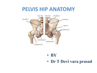
- 1. PELVIS HIP ANATOMY • BY • Dr T Devi vara prasad
- 2. Anatomy of the pelvis • The pelvis is a symmetrical bony ring interposed between the vertebrae of the sacral spine and the lower limbs, which are articulated through complex joints, the hips. It supports the spinal column and connects the upper body to the lower extremities. • It consists of three strong bones fused together: the ilium, ischium and pubis. These bones merge to form bilateral concave sockets named acetabulum • The ilium is the largest flat bone located on either side of the upper portion of the pelvis, including the iliac crest, which is the protruding tip bone of the pelvis that is easily felt with palpation. • The pubis is the smallest bone at the anterior side of the pelvis. Both ends of the pelvis are fused by the symphysis pubis consisting of a cartilaginous flexible tissue.
- 3. The sacrum The sacrum consists of five fused vertebrae (S1 to S5) adopting a triangular shape located in the posterior side of the pelvis. The upper, wider region of the sacrum articulates with the ilium (sacroiliac joint) on each side. In the lower portion it connects to the tail bone or coccyx formed by five small fused bones. The sacrum is particularly important in forming and stabilising the pelvic ring via the presence of numerous ligaments between bones and allows the connection of muscles of the pelvis and muscles of the hip joint (gluteus maximus, iliacus and piriformis).
- 4. The hip is a major ball-and-socket joint connecting the long bones of the lower limbs (femur) to the pelvis. This joint allows a wide range of movements of the lower limbs and is used when walking, running, climbing, lunging and bending. Because it bears the body weight, the hip joint is supported by large muscles, strong tendons and ligaments. The hip joint is protected by articular cartilage, which is a layer of elastic yet tough connective tissue surrounding both the femoral head and the acetabulum. The cartilage ad rotating inside the socket and importantly it absorbs shock impacts. Overuse of the cartilage is often leading to degeneration or osteoarthritis of the hip. Anatomy of the hip • The hip joint consists of the round head of the thighbone named femur, which inserts into the pelvis ring in a socked called acetabulum. The femoral facilitates the movement of the joint by reducing the friction of the femoral hehead continues distally with the femoral neck and the greater trochanter. The latter is the protruding portion of the femur bone that can be felt laterally at the higher thigh, below the pelvis.
- 5. Anatomy of the femur • The femur or thighbone is the longest and strongest bone in the human skeleton. It extends from the hip to the knee joint. The femoral head is the distal (upper) end of the femur that inserts into the acetabulum of the hip joint. As it descends towards the knee, it is separated from the longest tubular bone, or shaft, by the femoral neck.
- 6. Ligaments of the pelvis and hip • The bones of the pelvis are held together by a large number of ligaments and muscles. The stability and flexibility of thehip joint is provided by two structures: the ligaments, made of strong connective tissue, which connect bones to bones, and the tendons, which connect muscles to bones. • Iliofemoral ligament is the strongest ligament in the body. It is Y-shaped and extends from the lower front iliac spine of the coccyx to the femur trochanter (intertrochanteric line). This ligament prevents the extension of the femur in a standing position • Ischiofemoral ligament begins at the ischium, posteriorly to the acetabulum • Pubofemoral ligament originates lateral to the pubis adjacent to the iliofemoral ligament. • Ligamentum teres is smaller ligament that connects the higher extremity of the femoral head to the acetabulum. It contains an artery that supplies blood to the femoral head.
- 7. The labrum and capsule • The labrum is a cuff made of fibro-cartilaginous rim covering the edge of the acetabular cavity of the pelvis. It consists of two parts, one in contact to the femoral head and the other connecting with the joint capsule. The labrum can be subject to tear and injury. • The capsule of the hip is an essential bundle of strong ligaments that surround the hip to consolidate the joint elements and hold the femoral head in place during movement. The ligaments are the iliofemoral, pubofemoral and ischiofemoral. The capsule produces synovial fluid to keep the hip joint lubricated and facilitate the sliding of the joint components.
- 8. Bursas of the hip A bursa is a fluid-filled sac, functioning as a cushion to absorb shock and facilitate gliding of muscles and bones around the joint. The hip has two main bursas: The great trochanteric bursa is located on the great trochanter where the large ilio-psoas muscles of the hip joint are attached. This is quite a large bursa and is known for the related pathology, hip bursitis (trochanteric bursitis). The ilio-psoas bursa is located on the inner side of the hip. Also this bursa is subject to inflammation or bursitis, albeit less commonly.
- 9. Muscles of the pelvis The muscles of the pelvis and hip control the vast range of movement of the legs and torso. On the posterior side they are the glutei and on the anterior side the hip muscles extending into the thighs.