
Recommended
More Related Content
What's hot
What's hot (20)
Similar to Anatomy of esophagus
Similar to Anatomy of esophagus (20)
More from anwaradil4
Recently uploaded
Recently uploaded (20)
Anatomy of esophagus
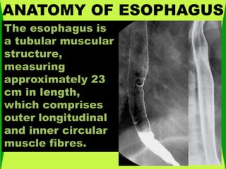
- 1. ANATOMY OF ESOPHAGUS The esophagus is a tubular muscular structure, measuring approximately 23 cm in length, which comprises outer longitudinal and inner circular muscle fibres.
- 2. ANATOMY OF ESOPHAGUS The proximal part of the esophagus consists of striated muscle fibres, whereas the distal part is composed of smooth muscle. The level of the transition zone between striated and smooth muscle is highly variable, with only the proximal 4 cm of the esophagus always composed of striated muscle.
- 3. ANATOMY OF ESOPHAGUS When the esophagus is partially collapsed, the normal longitudinal folds are seen as smooth, straight structures no more than 1 to 3 mm in width
- 4. ANATOMY OF ESOPHAGUS In some patients, fine transverse folds may also be observed in the esophagus because of transient contraction of the longitudinally oriented muscularis mucosae. These folds are almost always found to be associated with gastroesophageal reflux.12
- 5. ANATOMY OF ESOPHAGUS In contrast, focally spiculated transverse folds may be detected as a normal variant in upper thoracic esophagus at the junction of the striated and smooth muscle near the level of the aortic arch; these are thought to result from localized weakening of the amplitude of peristalsis in this region.
- 6. ANATOMY OF ESOPHAGUS Finally, in older patients, small nodules may be visible on the mucosal surface of the esophagus because of glycogenic acanthosis, a common degenerative condition of no clinical significance
- 7. ANATOMY OF ESOPHAGUS Extramural Esophageal Impressions Extramural esophageal impressions cause smooth and obtuse impressions on the esophagus similar to the extramural masses throughout the GI tract. Most causes are benign. External malignant masses may ultimately distort the mucosal contour but only after transmural metastatic invasion. Benign Malignant Postcricoid impression, Cricopharyngeal spasm, Cervical osteophytic disease, Retropharyngeal masses (e.g., goiter). Cardiomegaly, Vascular anomalies (aberrant vessels, aneurysms, vascular rings), Mediastinal adenopathy (e.g., tuberculosis) Mediastinal adenopathy (metastases)Lung cancerLung metastases
- 8. ANATOMY OF ESOPHAGUS Postcricoid Impression Also known as postcricoid defect of pharyngeal venous plexus, postcricoid impression is common and not pathological, and it is caused by redundant hypopharyngeal mucosa overlying the central pharyngeal venous plexus positioned approximately at the level of C6, just below the cricoid cartilage. It varies in appearance (sometimes smooth, sometimes web-like) and is recognized by its anterior location on the esophagus.
- 9. ANATOMY OF ESOPHAGUS Cricopharyngeal Spasm/hypertrophy This posterior esophageal impression is secondary to hypertrophy and spasm of the cricopharyngeus muscle, the lower portion of the inferior pharyngeal constrictor muscle located posteriorly at the level of C5-6 vertebral body.
- 10. ANATOMY OF ESOPHAGUS Vertebral Osteophytic Disease Anterior vertebral osteophytes or, rarely, anterior cervical disc herniation can cause discrete posterior esophageal indentations, which are usually smooth and sometimes marked
- 11. ANATOMY OF ESOPHAGUS Retropharyngeal Masses The cervical esophagus abuts the vertebral bodies, so pharyngeal disease that extends inferiorly and posterior to the esophagus will produce esophageal deviation. Such retropharyngeal masses include goiter, abscess, hematoma, lymphadenopathy, and parathyroid enlargement
- 12. ANATOMY OF ESOPHAGUS Extrinsic impressions on the esophagus. A. Normal impressions: 1.Aortic arch. 2.Left main bronchus. 3.Heart. 4.Esophageal hiatus.
- 13. ANATOMY OF ESOPHAGUS Abnormal extrinsic impression on the posterior wall of the esophagus caused by a large thoracic osteophyte.
- 14. ANATOMY OF ESOPHAGUS An aberrant right subclavian artery occurs in approximately 1 in 200 individuals and is due to the aberrant origin of the right subclavian artery, usually from a left-sided aortic arch, such that the vessel now has to reach the right axilla by crossing behind the esophagus at an oblique angle. Less commonly, it arises from a right aortic arch
- 15. ANATOMY OF ESOPHAGUS An aberrant left pulmonary artery is an anomaly resulting from the left lung being supplied from the right pulmonary artery, rather than the left pulmonary artery. Because of the location of its origin, this aberrant left pulmonary artery has to cross to the left side of the mediastinum and in doing so causes an anterior extrinsic compression of the thoracic esophagus at the level of the carina, giving the name to the so-called pulmonary sling