
Recommended
Recommended
More Related Content
What's hot
What's hot (12)
Similar to Adamantinoma 1
Similar to Adamantinoma 1 (20)
More from anwaradil4
Recently uploaded
Recently uploaded (20)
Adamantinoma 1
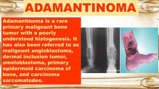
- 1. ADAMANTINOMA Adamantinoma is a rare primary malignant bone tumor with a poorly understood histogenesis. It has also been referred to as malignant angioblastoma, dermal inclusion tumor, ameloblastoma, primary epidermoid carcinoma of bone, and carcinoma sarcomatodes.
- 2. ADAMANTINOMA Approximately 165 cases have been reported in the literature, with all but 18 located in the tibia. Other bones affected are the jaw, ulna, humerus, femur, and fibula.
- 3. ADAMANTINOMA The mid-diaphysis of the long bones is the location in 75% of cases. The lesions are extremely rare in the spine. Most patients are between 10 and 40 years of age.
- 4. ADAMANTINOMA Radiographically, adamantinomas may be difficult to differentiate from other lesions because they can be situated in either the cortex or the medullary space. Cortical lesions may assume a lytic bone blister appearance, may appear as sawtooth loss of cortical bone with ragged margins, or may present as a multichambered, bubbly lesion.
- 5. ADAMANTINOMA The typical intramedullary lesion is usually one large, circumscribed area of radiolucency with a mottled increase in density scattered throughout the length of the lesion. Less commonly, a reticulated, honeycombed type of presentation occurs. Long- standing tumors produce marked cortical thickening and spool- shaped bulges of the outer cortex in an eggshell fashion.
- 6. ADAMANTINOMA Gradual expansion of bone with eventual cortical disruption and the development of a soft tissue mass occurs. Periosteal response is minimal and, if present, is usually of the laminated variety.
- 7. ADAMANTINOMA The most difficult and almost indistinguishable differential diagnosis is fibrous dysplasia, though the presence of periosteal response, moth-eaten destruction, no bowing or ground glass appearance, and younger age should allow correct diagnosis.