Presentation1.pptx, radiological imaging of bilaharziasis.
•Download as PPTX, PDF•
26 likes•1,935 views


Recommended
Recommended
More Related Content
What's hot
What's hot (20)
Viewers also liked
Viewers also liked (20)
Similar to Presentation1.pptx, radiological imaging of bilaharziasis.
Similar to Presentation1.pptx, radiological imaging of bilaharziasis. (20)
More from Abdellah Nazeer
More from Abdellah Nazeer (20)
Presentation1.pptx, radiological imaging of bilaharziasis.
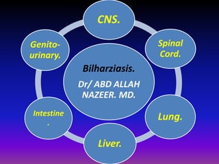
- 1. CNS. Bilharziasis. Dr/ ABD ALLAH NAZEER. MD. Spinal Cord. Lung. Liver. Genito-urinary. Intestine .
- 2. Schistosomiasis, also named bilharziasis (after the German physician Theodore Bilhartz, who worked in Egypt during the nineteenth century and was the first to discover the life cycle of the parasite), is one of the most prevalent infectious diseases in the world. It is endemic in more than 70 countries, mainly in the less developed countries, and is estimated to place approximately 20% of the endemic population at risk. The number of people affected is estimated to be 200 million. The schistosomes are a group of trematodes (flukes), some of which are pathogenic to humans. The principal parasites that affect humans are Schistosoma haematobium, Schistosoma mansoni, and Schistosoma japonicum; less prevalent parasites are Schistosoma intercalatum in Africa and Schistosoma mekongi in the Far East. presents the global distribution of schistosomal species. The most prevalent area for schistosomiasis is Africa, where S haematobium and S mansoni dominate.
- 4. Schema shows the two-stage life cycle of S haematobium.
- 5. Acute schistosomiasis: The symptoms of acute schistosomiasis are not directly caused by the parasites, but by your immune system (the body's defence against infection) reacting to the parasites. Symptoms include: a high temperature (fever) above 38ºC (100.4ºF) headache joint and muscle pain cough bloody diarrhea a dark red, blotchy, raised skin rash pain in the abdomen a general sense of feeling unwell In many cases, the symptoms usually get better by themselves within a few weeks. However, it is still important to seek treatment as the parasites will stay in your body and chronic schistosomiasis will develop. This does not always lead to symptoms, but the infection will remain and problems may develop at a later date.
- 6. Chronic schistosomiasis: If schistosomiasis is not treated, the parasites remain in your body and will go on to cause further symptoms. The immune system reacting to the eggs may damage your organs, but fails to kill the parasites. The symptoms of chronic schistosomiasis depend on where in the body the parasites have travelled to. If the parasites travel to the digestive system, they can cause the following symptoms: feeling tired all the time (fatigue), abdominal pain, bowel problems – such as mild or severe watery diarrhea that contains blood and mucus If the parasites travel to the urinary system, they can cause the following symptoms: symptoms of cystitis– such as pain when urinating frequent need to urinate, blood in your urine. If the parasites travel to the heart or lungs, they can cause the following symptoms: persistent cough in some cases, people cough up blood , wheezing, feeling breathless and very tired after physical activity . If the parasites travel to the central nervous system or brain, they can cause the following symptoms: seizures (fits), headache, back pain, urinary incontinence, weakness and numbness in your legs, dizziness, feeling sick The parasites can also sometimes travel to the female genitals, where they can cause the following symptoms: bleeding after sex, genital ulcers, irregular menstruation, pelvic pain.
- 7. Involvement of the central nervous system in Schistosoma mansoni and S. haematobium infection. involvement of the CNS in Schistosoma mansoni (SM) and S. haematobium (SH) infection was undertaken. Deposition of ova in the brain has been reported in SM infection and less of SH infection. In uncomplicated schistosomiasis, SH ova are more likely than SM ova to be deposited in the brain and may be carried there by the vertebral venous plexus. The deposition of SM ova in the brain and meninges is more frequent in hepatosplenic schistosomiasis, especially with corpulmonale, and the route to the brain may be through pulmonary arteriovenous shunts. Asymptomatic deposition of ova is common but epilepsy may occur. The formation of large granulomas and cerebral hemorrhage are rare complications. Adult SM and SH have been observed in cerebral vessels.
- 8. ETIOLOGY AND EPIDEMIOLOGY The term neuroschistosomiasis (NS) refers to the symptomatic or asymptomatic involvements of the central nervous system (CNS) by schistosomes. When associated with clinical symptoms, it is one of the most severe presentations of schistosomal infection. NS can be caused by Schistosama japonicum, S. mansoni, and S. haematobium. Considering the symptomatic form, the last two species are almost always associated with a myeloradicular syndrome and the first species, with cerebral disease. Symptomatic cerebral NS has been recorded in about 2-4% of individuals infected with S. japonicum (Watt et al. 1986). On the other hand, this form of presentation is very rare in association with the other two species.
- 9. Noncontrast axial CT of the brain showing edema areas in the right cerebellum, schistosomiasis (B) Axial T1-weighted MRI of the brain showing isointense signal in the right cerebellum, (C) Axial T2-weighted MRI of the brain showing hyperintense signal in the right cerebellum, (D) Axial T1-weighted MRI after gadolinium injection showing intensely enhancing nodules in the right cerebellum.
- 10. schistosomiasis with neurological Complications.
- 11. Schistosoma mansoni cerebral schistosomiasis. Brain magnetic resonance imaging shows a heterogeneously enhancing lesion with surrounding edema.
- 13. Cerebral schistosomiasis. (A) Axial T2-weighted MR image shows vasogenic edema and mass effect involving left frontal lobe. (B) Axial and (C) coronal contrast-enhanced T1-weighted MR image shows typical pattern of enhancement with multiple areas of central linear enhancement surrounded by enhancing punctate nodules.
- 14. Magnetic resonance images (A-D) and histologic features (E and F) in a patient with cerebral schistosomiasis. A, Coronal T1-weighted gradient echo image. B, T2-weighted gradient echo image. C, T1-weighted image after gadolinium injection, showing a nodular irregular lesion in the right parietal and insular cortex with mass effect, perilesional edema, and slight irregular contrast enhancement. D, T2-weighted image obtained 1 month after treatment, showing complete resolution of the lesion
- 15. Symptomatic spinal cord involvement is a rare but well-documented manifestation of schistosomiasis. A nonspecific intramedullary expansion in the caudal spinal cord is the most common finding in conventional and computed myelography. The literature concerning MR imaging of spinal cord schistosomiasis is scarce, and pathologically confirmed cases are mainly limited to isolated case reports. Schistosomiasis of the spinal cord should be considered in the differential diagnosis of lesions affecting the lower thoracic cord-conus medullaris-cauda equina in patients from endemic areas with history of exposure to Schistosomal infestation. MR imaging can reveal the true extent of the disease and can suggest the diagnosis through recognition of its signal intensity changes and enhancement forms. Accurate diagnosis allows early treatment and results in better prognosis of spinal cord schistosomiasis.
- 17. Schistosomiasis. A to D, MRI of lumbar spine revealed expansion of the medullary cone associated with spinal cord edema in the affected area associated with epidural enhancement adjacent to areas of medullary involvement. G and H, The roots of the cauda equine are thickened. E, F, and I, Magnetic resonance images after treatment reveal the lesions reduction.
- 18. Epidural bilharzioma. A, Axial T1-weighted MR image shows a homogeneous epidural mass with extension through the intervertebral foramina with a lobulated paraspinal mass. B, Axial T2-weighted MR image shows a mild heterogeneous hyperintensity and a contralateral displacement of spinal cord. C, Axial T1-weighted MR image after gadolinium injection shows a homogeneous enhancing of the lesion. D, Photomicrograph of frozen histopathologic section shows ovum of Schistosoma mansoni. Note characteristic broad lateral spine.
- 19. Sagital magnetic ressonance imaging in T1 phase (Figure 3a), no contrast T2 phase (Figure 3b), and contrast T2 phase (Figure 3c) in a spinal cord schistosomiasis patient.
- 20. Spinal cord schistosomiasis patient. It is observed a granular impregnation of gadolinium magnetic contrast in thoracic-lumbar spinal cord.
- 22. Masslike nodular enhancement form in spinal cord schistosomiasis. MR imaging of dorso-lumbar spine
- 23. Localization of spinal cord schistosomiasis. MR imaging of dorso-lumbar spine with moderate expansion of the distal cord and conus medullaris. Linear heterogenous contrast uptake at the enhanced study.
- 24. Radicular enhancement form in spinal cord schistosomiasis. Peripheral enhancement form of spinal cord schistosomiasis
- 25. Pulmonary involvement in schistosomiasis Although the lungs are not an end organ in the life cycle of schistosome infection in humans, pulmonary pathology exists. For many years, pulmonary pathology was described mainly as a late complication of the infection. It recently has been recognized that pulmonary involvement also may occur in the early, acute stages. Early-stage pulmonary involvement is unique to nonimmune patients, that is, populations never previously exposed to schistosomal infection, usually travelers from developed countries to endemic areas. Physicians who practice in Western countries who treat returning travelers are more likely to encounter pulmonary involvement in the early stages of infection rather than the later complications. Early and late pulmonary schistosomiasis are two different diseases with different clinical manifestations and different pathogenesis and pathology, not merely a difference in time of onset.
- 26. Early (acute) pulmonary schistosomiasis: Clinical manifestations. Early pulmonary manifestations occur usually 3 to 8 weeks after schistosome penetration. Patients with pulmonary schistosomiasis reported shortness of breath, wheezing, and dry cough, mainly while recumbent. Reports show that in some cases the pulmonary symptoms coincided with febrile illness (Katayama fever). Most patients, however, presented several weeks after the fever had subsided. Almost all the patients could recall having had febrile disease before the onset of pulmonary symptoms, but the pulmonary symptoms continued for weeks after the fever subsided. Pulmonary involvement can be divided into three types: 1. Symptomatic cases with radiologic findings (either chest radiograph or CT scan). The radiologic findings may be evident at presentation or, not uncommonly, may appear after antischistosomal treatment. In either case, cough may persist for several weeks despite the treatment.
- 27. 2. Symptomatic cases without radiologic findings. In some patients, the clinical course is similar to the previous one, but radiologic findings (either chest radiograph or CT scan) are absent. This result may be caused either by the small dimensions of the findings, which makes them invisible by conventional methods, or their transient nature. 3. Asymptomatic cases with radiologic findings. Cases in which there are pulmonary findings without a current history of pulmonary symptoms are rare. The incidence of these cases is unknown, because radiology is usually not performed for asymptomatic patients. The author was able to identify such cases since chest radiographs were performed as part of the evaluation of patients with suspected early schistosomiasis.
- 28. Pulmonary schistosomiasis. Chest radiography (pa) showing multiple small pulmonary nodules scattered over both lungs without obvious predilection.
- 29. Pulmonary schistosomiasis. Chest CT of the upper lungs showing blurred ground glass nodules scattered over both lungs
- 30. Chest radiograph that reveals several small round lesions in both lungs (arrows). (B) Chest radiograph that reveals diffuse, increased lung markings and prominent hilum with ill-defined nodules with pulmonary bilharziasis.
- 31. The progress of pulmonary hypertension secondary to S. mansoni.
- 32. pulmonary hypertension worsens, the cardiovascular changes become more marked, even in quite young patients. A In this 21-year -old Puerto Rican female, there is marked dilatation of the main pulmonary artery and the central pulmonary arteries, with rapid tapering of the peripheral branches.
- 33. The end stage of cardiopulmonary schistosomiasis. There is massive enlargement of the heart, and there are small bilateral pleural effusions, secondary to congestive cardiac failure. Pericardiocentesis removed 320 cc of straw-colored fluid. Thoracotomy showed multiple tiny nodules in the lungs and thick fibrous plaques. The pericardium was greatly thickened. Histology showed granulomas involving the lungs and pericardium and causing pulmonary arteriolitis.
- 34. CT scan through the bifurcation of the trachea that shows multiple lesions of different sizes in both lungs. (B) CT scan through a point just above the diaphragm that shows diffuse ground-glass areas with some nodularity. The findings are mainly at the lung periphery with pulmonary bilharziasis.
- 35. CT of chest showing peripheral nodular and patchy infiltrates in both lung fields with pulmonary bilharziasis.
- 36. Chest X-ray of a patient with Sch-PAH displaying the enlargement of the main pulmonary arteries.
- 37. Chest radiograph (A), magnetic resonance imaging (B), and pulmonary angiography (D) in patient with World Health Organization class IV pulmonary hypertension secondary to schistosomiasis from Brazil. C, Magnetic resonance imaging of the liver with typical Symmers’ fibrosis.
- 38. Hepatosplenic schistosomal disease and pulmonary hypertension and the pulmonary artery systolic pressure of 68 mmHg.
- 39. Chronic schistosomiasis with pulmonary hypertension. Chest radiograph (A and D)and CT demonstrate (B and C)marked dilatation of the main pulmonary artery.
- 40. Acute schistosomiasis in a 35 year-old man with fever, cough and dyspnea. Chest CT scans demonstrate bilateral diffuse patchy areas of ground glass opacities.
- 41. HEPATIC SCHISTOSOMIASIS. Schistosomiasis is caused by trematode blood flukes of the genus Schistosoma. Five species are responsible for human infections and numerous others only infect animals. The species that infect humans include Schistosoma haematobium, S. mansoni, S. japonicum, S. intercalaturn, and S. mekongi. Chronic infections with all Schistosoma species with the exception of S. haernatobiurn can cause significant morbidity and mortality as a result of granuloma formation in the intestine and liver. The resulting hepatic fibrosis can lead to portal hypertension that eventually can be complicated by splenomegally, esophageal varices, hematemesis, and death. S. haematobium primarily affects the urinary tract, resulting in chronic inflammation of the bladder, ureteral obstruction leading to hydronephrosis, stone formation, and urinary tract infections that can be complicated by gram-negative septicemia.% Although there have been reports of liver disease due to S. haematobium,3 these are extremely uncommon. Because the focus of this issue is on the liver, infection due to S. haematobium is not reviewed. S. mansoni and S. japonicum have been most intensely studied, and this article reflects available data on these two species unless otherwise indicated.
- 42. Hepatic schistosomiasis, or schistosomal hepatopathy, is the most common form of the chronic disease and usually results from heavy S. mansoni infection Clinical manifestations Clinical presentation of hepatic schistosomiasis markedly differs from that of cirrhosis. Although the symptoms and signs of portal hypertension and hypersplenism are dominant in schistosomiasis, the counter part of hepatocellular failure is absent. However, some patients with schistosomiasis progress to an end stage of the disease by exhibiting muscle wasting, hypoalbuminemia, ascites and coma. These observations led to the concept of compensated and decompensated schistosomiasis to differentiate patients with the sole manifestations of portal hypertension from those who, in addition, presented signs of hepatocellular failure.
- 43. Ultrasound of the patient showing periportal thickening (yellow arrows on the left side), thickening of the gallbladder wall (G = gallbladder; yellow arrow = thickened wall) and the enlarged spleen (S).
- 44. Non–contrast enhanced computed tomography (CT) liver showing (a) capsular calcifications (white arrowheads), an irregular hepatic contour, and extension of peri-portal fat deep into the liver (white arrows), and (b) septal calcifications (blackarrows) and junctional notches (white arrows).
- 45. (a) Ultrasonography of the liver showing diffuse peri-portal hyperechogenicity (white arrows). (b) Contrast enhanced computed tomography (CT) arterial phase image showing septal calcification (white arrow) in the right posterior segment and a hypervascular tumour in the right anterior segment (black arrowheads). (c) Contrast-enhanced CT porto-venous phase image showing washout of contrast in the tumour, suggestive of hepatocellular carcinoma (black arrowheads)
- 46. Hepatic schistosomiasis with portal hypertension and splenomegally.
- 47. CT from two years prior demonstrating fatty liver and periportal fibrosis. These images are similar in appearance to those of previous case reports demonstrating CT appearance of schistosomiasis. A, B. Arterial phase images show hypoattenuated round and linear branching lesions traveling adjacent to enhancing hepatic arterial branches (arrows). C, D. These lesions enhance during portal phase (arrows). E, F. The gallbladder wall is nodular and thickened and measures 4 mm.
- 50. MRI T2-weighted image of liver and spleen – a coronal section. Liver with periportal thickening (yellow arrow) around the portal vein (red arrow). There is a huge spleen (S). White arrow = stomach.
- 51. MRI T2-weighted image of liver and spleen – transversal section. There are four images showing different aspects of periportal fibrosis in the liver (the vein in the center appears dark – red arrow). Periportal thickening (yellow arrows). White arrows = stomach; S = spleen.
- 52. MRI T2-weighted image of liver and spleen – transversal section. The vessels are easily identified. Black arrow = splenic vein; yellow arrow = thickening around the gallbladder wall.G = gallbladder; S = spleen.
- 53. MRI T1-weighted image of liver and spleen with fat suppression – transversal section. S = spleen; G = gallbladder. The white areas around the vessels (fibrosis) and gallbladder (fibrosis) – yellow arrows - and also the blood inside the vessels, seen in previous images, became black/gray on the two upper figures. Note, in the two lower figures, that there is an enhancement of the gallbladder wall (yellow arrow) and of the periportal thickening (yellow arrow) after the intravenous infusion of contrast (gadolinium).
- 54. Liver fibrosis in hepatosplenic schistosomiasis (white areas around the vessels). There is a thrombus inside the portal vein (red arrow).
- 55. Angioresonance of the abdomen in hepatosplenic schistosomiasis with easy identification of the portal vessels. The red arrow is pointing to a huge collateral vessel (left gastric vein).
- 56. Intestinal schistosomiasis: Intestinal schistosomiasis is another well identified form of chronic schistosomal affection. Egg deposition and granuloma formation eventually leads to acute then chronic schistosomal colitis and is commonly associated with polyp formation. It frequently presents as abdominal pain, diarrhea, tenesmus and anal pain. Definite diagnosis of schistosomiasis disease depends on microscopy and egg identification. Marked progress regarding serologic diagnosis occurred with development of recent PCR techniques that can confirm schistosomal affection at any stage.
- 57. Schistosomal polyposis in the colon and rectum. A Multiple polyps throughout the rectosigmoid colon, which was displaced out of the pelvis by a large pericolic bilharzial (schistosomal) abscess
- 58. Colonic and rectal polyps are usually multiple and associated with a marked inflammatory reaction. A The hemicolectomy from an Egyptian patient with combined S. haematobium and S. mansoni infections.
- 59. Schistosomiasis mansoni can cause marked granulomatous thickening of the bowel wall and, eventually, strictures. A Transverse sections of a stenosed segment of bowel from an 18-year-old Egyptian male. Elsewhere in the colon there were multiple polyps.
- 60. The end results of schistosomiasis in the colon may be diffuse bowel wall thickening or local strictures. In these patients with diffuse loss of haustrations, there is narrowing and rigidity of the colon.
- 61. Schistosomal calcification of the intestine. A non-enhanced CT scan of a 46-year-old Egyptian showing dense calcification of the sigmoid colon.
- 62. Genitourinary schistosomiasis is produced by Schistosoma haematobium, a species of fluke that is endemic to Africa and the Middle East, and causes substantial morbidity and mortality in those regions. It also may be seen elsewhere, as a result of travel or immigration. S haematobium, one of the five fluke species that account for most human cases of schistosomiasis, is the only species that infects the genitourinary system, where it may lead to a wide spectrum of clinical symptoms and signs. In the early stages, it primarily involves the bladder and ureters; later, the kidneys and genital organs are involved. A definitive diagnosis of genitourinary schistosomiasis is based on findings of parasite ova at microscopic urinalysis. Clinical manifestations and radiologic imaging features also may be suggestive of the disease, even at an early stage: Hematuria, dysuria, and hemospermia, early clinical signs of an established S haematobium infection, appear within 3 months after infection. At imaging, fine ureteral calcifications that appear as a line or parallel lines on abdominopelvic radiographs and as a circular pattern on axial images from computed tomography (CT) are considered pathognomonic of early-stage schistosomiasis. Ureteritis, pyelitis, and cystitis cystica, conditions that are characterized by air bubble-like filling defects representing ova deposited in the ureter, kidney, and bladder, respectively, may be seen at intravenous urography, intravenous ureteropyelography, and CT urography. Coarse calcification, fibrosis, and strictures are signs of chronic or late-stage schistosomiasis. Such changes may be especially severe in the bladder, creating a predisposition to squamous cell carcinoma. Genital involvement, which occurs more often in men than in women, predominantly affects the prostate and seminal vesicles.
- 63. Pathologic changes in the urinary tract due to schistosomiasis are far more common in chronic infections than in acute ones. Such changes result from the deposition of eggs (not adult flukes) in and around vessels, which leads to chronic inflammatory lesions and induces an immune response with granuloma formation and associated fibrotic changes . The deposition of eggs in the bladder and ureter induces a chronic granulomatous reaction. The disease usually starts at the urinary bladder trigone and base, with the formation of submucosal granulomas leading to inflammatory patches and hematuria. Cystitis resembling that in tuberculosis results in “sandy patches” on the bladder wall that, in severe cases, may become a network of dense concentric calcifications . The degree of calcification is roughly correlated with the number of eggs deposited. Chronic irritation of the urothelium causes it to proliferate, producing budlike or polypoid structures. These structures differentiate into cystic deposits of cystitis cystica or intestinal columnar mucin-secreting glands, resulting in cystitis glandularis, which may develop into adenocarcinoma. Cystitis cystica, ureteritis cystica, and cystitis glandularis can be observed as polypoid filling defects on radiographs . Ureteral lesions due to S haematobium infection.
- 64. In long-standing infections, a cellular reaction to dead ova produces calcification and fibrosis, which are important contributors to squamous metaplasia and squamous cell carcinoma. Severe fibrosis classically involves the bladder and ureteral segments distal to the iliac vessels (hereafter referred to as “distal ureters”), diminishing their elasticity. Severely fibrotic ureters have a ragged outline and a beaded internal appearance, with irregular dilatation due to pseudotubercles in the submucosa. As the pseudotubercles heal, they may become fibrotic, a condition that may lead to ureteral stricture. Renal involvement in late-stage fibrosis, which usually results from vesicoureteral reflux, is manifested by renal calculi and hydronephrosis or pyonephrosis secondary to ureteral obstruction. Urethral involvement usually occurs in the form of fossa navicularis polyps, periurethral abscesses, and perineal and scrotal fistulas; strictures are uncommon . Schistosomal infection of the prostate gland and seminal vesicles is found at autopsy in 58% of male cadavers in geographic regions where S haematobium is endemic. The epididymis, spermatic cord, and testes are rarely affected. Prostatic involvement leads to initial enlargement of the prostate, followed by fibrosis and shrinkage with calcification of the gland. The seminal vesicles may become enlarged and calcified. Ejaculatory duct dilatation may result from distal fibrosis and obstruction. Genital schistosomiasis is not as common in women as in men, but lesions may be found in the vulva, vagina, and cervix, and more rarely in the ovaries, fallopian tubes, and uterus .
- 65. Excretory urogram demonstrates bilateral distal ureteral dilatation secondary to early-stage urinary tract infection by S haematobium. (7) Urinary tract schistosomiasis. (a) Excretory urogram shows substantial dilatation of the distal left ureter, likely a result of ureteral narrowing at the level of the pelvic bone. (b) Postmicturition radiograph shows persistent filling of the left ureter with a nondilated right ureter.
- 66. Urinary tract schistosomiasis. (a) Excretory urogram shows marked left hydroureteronephrosis. (b) Coronal three-dimensional urographic image obtained with computed tomography (CT) shows a long-segment stricture with proximal dilatation of the left ureter.
- 67. Urinary tract schistosomiasis. (a, b) Negative (a) and positive (b) anteroposterior radiographs show thin calcification of the bladder wall and the entire length of the left ureter (arrows in a). (c) Unenhanced axial pelvic CT image more clearly depicts the bladder wall calcification. (d) Sagittal reformatted CT image more clearly shows the distal left ureteral calcification (arrow).
- 68. (10a) Excretory urogram obtained in a 46-year-old woman shows multiple filling defects along the course of the distal left ureter (arrows). These findings represent ureteritis cystica due to schistosomiasis. (10b) Postmicturition radiograph shows multiple filling defects in the bladder, findings indicative of cystitis cystica due to schistosomiasis. The T-shaped object is an intrauterine contraceptive device. (11) Coronal three-dimensional CT urogram obtained in an 83- year-old man with hematuria shows multiple bilateral filling defects along the course of both ureters (arrows). These findings represent ureteritis cystica due to schistosomiasis.
- 69. Extensive bladder calcifications due to late-stage schistosomiasis. (a) Pelvic radiograph shows the classic appearance of the calcified bladder, which resembles a fetal head in the pelvis, with associated faint calcification of the distal right ureter (arrows). (b) Axial pelvic CT image shows thick calcification of an extensive region of the bladder wall and ringlike calcification of the wall of both ureters, with patent ureteral lumina. (c) Axial pelvic CT image obtained in another patient shows a completely calcified bladder wall and calcifications obstructing both ureteral lumina.
- 70. Squamous cell carcinoma of the bladder. (a) Axial T2-weighted magnetic resonance (MR) image shows a soft-tissue mass arising from the left lateral wall of the bladder with multiple small diverticula. (b) Axial T2-weighted MR image at the level of the bladder neck shows extension of the mass through the urinary bladder wall to the perivesical fat with infiltration of the left seminal vesicle.
- 71. Left hydroureteronephrosis due to schistosomiasis. (a) Coronal T2-weighted MR urogram shows marked left hydroureteronephrosis with a normal right kidney and right ureter. (b) Axial T2-weighted image depicts dilatation of the left ureter at the level of the bladder.
- 72. Calcification due to schistosomiasis of the seminal vesicles and vasa deferentia. (a) Unenhanced axial CT image shows calcification of both seminal vesicles. (b) Postmicturition radiograph from excretory urography shows calcification of the vasa deferentia and iliac vessels (arrows). (c) Coronal three-dimensional reformatted pelvic CT image shows calcification of both seminal vesicles (arrowheads) and the left vas deferens (arrow).
- 73. Transrectal US image of the prostate in a patient with genitourinary schistosomiasis shows a region of dense prostatic calcification that produces an anterior acoustic shadow.
- 74. Seminal vesiculitis due to schistosomiasis. (a) Transrectal US image of the seminal vesicles shows bilateral wall thickening (arrows). (b) High-resolution axial T2- weighted MR image of the pelvis shows thickened seminal vesicle walls (arrows).
- 75. Thank You.