Critical review on glass ionomer seal under composite resin of obturated root canals
•
1 like•497 views

This review article examines the need to seal the root canal orifice with glass ionomer cement beneath composite resin following endodontic treatment. The article summarizes various studies that have evaluated different materials for use as intracoronal seals, including glass ionomer cement, composite resin, mineral trioxide aggregate, and others. The results of these studies are conflicting, with some finding glass ionomer cement to be effective and others supporting alternative materials. Overall, the review was unable to definitively conclude whether an intracoronal seal is always needed or which material is best, finding that further high-quality research is still required to answer this question.

Recommended
Recommended
More Related Content
What's hot
What's hot (20)
Viewers also liked
Viewers also liked (9)
Similar to Critical review on glass ionomer seal under composite resin of obturated root canals
Similar to Critical review on glass ionomer seal under composite resin of obturated root canals (20)
Recently uploaded
Recently uploaded (20)
Critical review on glass ionomer seal under composite resin of obturated root canals
- 1. www.ijcmr.com International Journal of Contemporary Medical Research Volume 3 | Issue 5 | May 2016 | ICV: 50.43 | ISSN (Online): 2393-915X; (Print): 2454-7379 1406 Critical Review on Glass Ionomer Seal under Composite Resin of Obturated Root Canals Ranya F. Elemam1,2 , Ziad S. Abdul Majid3 ORIGINAL RESEARCH ABSTRACT Introduction: The root canal treated teeth need an adhesive seal for coronal leakage prevention. Glass ionomer sealant is the usual interface used between the coronal restoration and dental hard tissue however when composite resin material is used as a coronal restoration, some dental clinician prefer not to use it. The aim of this review is to determine the need to seal the orifice of an obturated root canal with glass ionomer under composite resin to prevent microleakage. Material and methods: Electronic searches were performed in the Pubmed and Scopus databases using relevant keywords. Textbook searching was also applied. Following selection, articles were fully reviewed to ensure that they met inclusion/ exclusion criteria. Results: The intracoronal sealing abilities of a wide variety of restorative materials have been investigated, assessed and compared within the dental literature. Conclusion: No definitive guidelines were found regarding the use of orifice sealing materials following endodontic treatment. This review was not able to answer the research question, and further investigation is required to achieve this goal. Keywords: Intra-orifice barriers, Composite resin, Glass ionomer, Micro leakage. INTRODUCTION Microbial infection via an inadequate coronal seal is one of the major factors associated with endodontic failure,1 and the literature suggests that coronal leakage is more likely to determine clinical success or failure than apical leakage.2 Placement of a material over the coronal gutta-percha to act as a barrier to coronal microleakage would be advantageous in reducing leakage and increasing the possibility of treatment success.3 The sealant material is placed into the canal orifice following removal of the coronal portion of gutta-percha and sealer. Many materials have been investigated for use as an intra-coronal seal to prevent microleakage, including Cavit, amalgam, intermediate restorative material (IRM), Super- EBA, composite resin, glass-ionomer cement (GIC), and mineral trioxide aggregate (MTA).1 Glass ionomer cement has been advocated for use as an intracanal barrier when microleakage or recurrent caries are likely because of its cariostatic and adhesive properties.4 Resin-modified glass ionomer material is one of the barrier materials used routinely to close the canal orifice after root canal obturation.5 It consists of glass ionomer and composite resin, having properties of both materials. Composite resin has excellent adhesive properties and is used commonly as a core in endodontically treated teeth.6 The aim of this review is to determine if there is a need, following endodontic treatment, to seal the root canal orifice with glass ionomer beneath composite resin to prevent microleakage. MATERIALAND METHODS Electronic searches were performed in the Pubmed and Scopus databases using the keywords: intraorifice barriers, composite resin, glass-ionomer, microleakage. Textbook searching was also applied for relevant information. Articles were first selected according to titles and abstracts, and they were then fully reviewed to ensure that they met the inclusion/exclusion criteria. Inclusion criteria Studies with all designs that used different materials and or techniques included. The study should refer to intracoronal orifice and micro leakage significance. Searches were limited to papers written in English and published between 2002 and 2014. The exclusion criteria All studies that failed to meet the inclusion criteria. If a study did not refer to the intraorifice barrier or explain its relation with microleakage, it was discarded. Studies that discussed a coronal barrier were also rejected. RESULTS Definition of an intraorifice sealing material and their importance The intra-orifice barrier is an effective treatment used in endodontically treated teeth by introducing an additional material into the canal orifice immediately after removal of the coronal portion of gutta-percha and sealer.7 Coronal leakage is a primary cause of endodontic failure.8 Sealing of the coronal part of the root canal is therefore indicated to reduce the chance of treatment deterioration.1 Sealing is of particular importance when the coronal restoration is lost or inadequately placed,9 or when there is delay in placing the final restoration.10 This is important for 1 Assistant Lecturer, Department of Restorative Dentistry and Periodontology, School of Dentistry, Libyan International Medical University (LIMU), Benghazi, Libya, 2 PhD Candidate, University of Porto, Porto, Portugal, 3 Orthodontic Resident, Division of Orthodontics and Dentofacial Orthopedics, Department of Developmental Dental Sciences, Faculty of Dentistry, Beirut Arab University, Beirut, Lebanon Corresponding author: Ranya F. Elemam, Department of Restorative Dentistry and Periodontology, School of Dentistry, Libyan International Medical University (LIMU), Benghazi, Libya. How to cite this article: Ranya F. Elemam, Ziad S. Abdul Majid. Critical review on glass ionomer seal under composite resin of obturated root canals. International Journal of Contemporary Medical Research 2016;3(5):1406-1408.
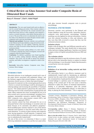
- 2. Elemam, et al. Glass Ionomer Seal under Composite Resin of Obturated Root Canals International Journal of Contemporary Medical Research ISSN (Online): 2393-915X; (Print): 2454-7379 | ICV: 50.43 | Volume 3 | Issue 5 | May 2016 1407 both anterior and posterior teeth.11 Cavit® , amalgam, intermediate restorative material (IRM® ), super-EBA, composite resin, glass ionomer cement and mineral trioxide aggregate (MTA) are commonly used materials.12,13 The use of colored materials is recommended so they can be easily identified in cases of retreatment or post restoration.11 Examples include the flowable composite resins PermaFlo® Pink or Purple (Ultradent), Flow-It® dark red (Pentron) or dark blue (DenMat). The use of a resin-modified glass-ionomer material over the gutta-percha followed by provision of a well-sealed temporary or permanent filling is suggested.11 The sealing abilities of a variety of intraorifice restorative materials and their capacity to prevent coronal micro leakage have been investigated, assessed and compared. MTA had the lowest rate of microleakage compared with composite resin or light-cured glass ionomer1 following completionofrootcanaltreatmentwithoutcoronalrestoration. Sealing with Cavit® gave better results than Vitremer® (glass- ionomer cement), and the flowable composite Flow-It® .14 Composite resin used alone or combined with Coltosol® showed a significant reduction in microleakage, whereas glass ionomer combined with Coltosol® resulted in less microleakage than the glass ionomer used alone.15 In an evaluation of the necessity to use an intraorifice seal in teeth with post space, a glass ionomer barrier over the gutta-percha coulSd reduce the risk of recontamination of the apical gutta-percha compared to those without glass ionomer but sealed with Vitrabond® .16 IRM® and Coltosol® were significantly better in preventing microleakage than chemically cured glass ionomer and dentinal adhesive.17 In a recent study the adhesive system CoroSeal® reduced coronal leakages more effectively than a flowable composite resin, fissure sealant or polycarboxylate cement18 Figure 1. DISCUSSION Conventional root filling materials such as gutta-percha and sealer do not provide adequate resistance to bacterial microleakage.21,22 Therefore, the coronal part of the root canal should be sealed to minimize the endodontic treatment failure rate.3 Previous research support the use of intra-orifice sealants, but there is little agreement on a standardized protocol or material to be used as a coronal barrier.23,24 Different studies have shown highly conflicting results regarding the sealing ability of different materials.1 The following criteria have been proposed by Wolcott et al. for an intracoronal barrier: (a) Easily placed by the specialist, (b) Bonds to tooth structure (retentiveness), (c) Effectively seals against microleakage, (d) Easily distinguishable from natural tooth structure and (e) Does not interfere with the final restoration of the access preparation. GIC is used commonly as an intraorifice barrier, and according to Mavec et al.,25 the literature supports the use of an intraorifice glass ionomer barrier to protect the root canal filling as a second line of defense for the temporary coronal seal. In their study, Parekh et al.3 found that microleakage was less beneath a seal of GIC plus composite resin as opposed to composite resin alone, and concluded that “LCGIC + Tetric N-Flow was found to be superior over other experimental materials as intraorifice barriers.” They suggested that the enhanced sealing ability of LCGIC may be attributed to: 1. Adhesion of LCGIC by development of an ion-exchange layer adjacent to dentin and 2. Shear bond strength of LCGIC which is higher than conventional GIC. Divya et al.26 also concluded that a GIC and composite combination can be recommended as coronal sealants, as did Deepali et al.27 who stated they had the “highest probability for achieving a maximal coronal seal.” Other studies have recommended other sealing agents: Slutzky- Goldberg et al.6 found GIC or MTA to be equivalent in their sealing abilities, and the results of Jiang et al.28 suggest that flowable composites can serve as ideal intra- orifice seals. Mineral trioxide aggregate and flowable composite was found to be preferred over glass ionomer as a coronal barrier by Sagar et al.,5 while El-Kady29 concluded that the use of a silorane based composite without the traditional glass ionomer base was best to decrease leakage to the root canal system. CONCLUSION The literature does not state clearly whether to use intra orifice sealant materials beneath final and temporary restorations. Although the routine is to place them under final restorations, no study has supported a single protocol. Consequently this review didn’t answer the research question, and a well- designed investigation is required to achieve this goal. REFERENCES 1. Yavari H, Samiei M, Eskandarinezhad M, Shahi S, Aghazadeh M, et al. An in vitro comparison of coronal microleakage of three orifice barriers filling materials. Iranian endodontic J. 2012;7:156-160. 2. Kurtzman GM. Improving Endodontic Success through Coronal Leakage Prevention. Inside Dentistry. 2005;1:2. 3. Parekh B, Irani R, Sathe S and Hegde V. Intraorifice sealing ability of different materials in endodontically treated teeth: An in vitro study. J Conserv Dent. 2014;17:234–237. 4. Malik G, Bogra P, Singh S, Samra R. Comparative evaluation of intracanal sealing ability of mineral trioxide aggregate and glass ionomer cement: An in vitro study. J Conserv Dent. 2013;16:540–545. 5. Sagar K, Sreenivasa Murthy B, Kumar M. Comparative Evaluation of Three Different Materials as Barriers to Coronal Microleakage in Root Filled Teeth: An in Vitro Figure-1: Placement of Sealing Materials.19,20
- 3. Elemam, et al. Glass Ionomer Seal under Composite Resin of Obturated Root Canals International Journal of Contemporary Medical Research Volume 3 | Issue 5 | May 2016 | ICV: 50.43 | ISSN (Online): 2393-915X; (Print): 2454-7379 1408 Study. 2012 Heal Talk. 6. Slutzky-Goldberg I, Slutzky H, Gorfil C, and Smidt A. Restoration of Endodontically Treated Teeth Review and Treatment Recommendations. Int J Dent. 2009;2009:150251. 7. Roghanizad N, Jones JJ. Evaluation of coronal microleakage after endodontic treatment. Journal of Endodontics.1996;22:471-3. 8. Saunders WP, Saunders EM. Coronal leakage as a cause of failure in root-canal therapy: a review. Endod Dent Traumatol. 1994;10:105-108. 9. Jenkins S, Kulild J, Williams K, Lyons W, Lee C. Sealing ability of three materials in the orifice of root canal systems obturated with gutta-percha. J Endod. 2005;32:225-227. 10. Varlan C, Dimitriu B,VarlanV, Bodnar D, Suciu. Current opinions concerning the restoration of endodontically treated teeth: basic principles. J Med Life. 2009;2:165- 172. 11. Qualtrough A, Morrow L, Brunton P. Principles of Operative Dentistry. Oxford-UK: Blackwell Munksgaard. 2005. 12. Chailertvanitkul P, Saunders WP, Saunders EM, MacKenzie D. An evaluation of microbial coronal leakage in the restored pulp chamber of root-canal treated multirooted teeth. Int Endod J. 1997;30:318-22. 13. Beckham BM, Anderson RW, Morris CF. An evaluation of three materials as barriers to coronal microleakage in endodontically treated teeth. J Endod. 1993;19:388-91. 14. Sauaia TS, Gomes BP, Pinheiro ET, ZaiaAA, Ferraz CC, et al. Microleakage evaluation of intraorifice sealing materials in endodontically treated teeth. Oral Surg Oral Med Oral Pathol Oral Radiol Endod. 2006;102: 242-246. 15. Damman D, Grazziotin-Soares R, Farina AP, Cecchin D. Coronal microleakage of restorations with or without cervical barrier in root-filled teeth. Microinfiltração coronária de restaurações com ou sem barreira cervical em dentes endodonticamente tratados. 2012;27:208- 212. 16. Mavec JC, McClanahan SB, Minah GE, Johnson JD, Blundell RE Jr. Effects of an intracanal glass ionomer barrier on coronal microleakage in teeth with post space. J Endod. 2006;32:120-122. 17. Zaia AA, Nakagawa R, De Quadros I, Gomes BP, Ferraz CC, et al. An in vitro evaluation of four materials as barriers to coronal microleakage in root-filled teeth. J Endod. 2002;35:729-734. 18. Bayram HM, Bayram E, Çelikten B, Bozkurt A. Fluid flow evaluation of coronal microleakage intraorifice barrier materials in endodontically treated teeth. Eur J Dent. 2013;7:359-62. 19. Wolcott JF, Hicks ML, Himel VT. Evaluation of pigmented intraorifice barriers in endodontically treated teeth. J Endod. 1999;25:589-592. 20. Bailon-Sanchez ME, Gonzalez-Castillo S, Gonzalez- Rodriguez MP, Poyatos-Martinez R, Ferrer-Luque CMN. Intraorifice sealing ability of different materials in endodontically treated teeth. Medicina oral, patologia oral y cirugia bucal. 2011;16:e105-9. 21. Kerby RE, Knobloch L. The relative shear bond strength of visible light-curing and chemical curing glass-ionomer cement to composite resin. Quintessence Int. 1992;23:641-4. 22. Diaz-Arnold AM, Wilcox LR. Restoration of endodontically treated anterior teeth: An evaluation of coronal microleakage of glass ionomer and composite resin materials. J Prosthet Dent. 1990;64:643-646. 23. Roghanizad N, Jones JJ. Evaluation of coronal microleakage after endodontic treatment. J Endod. 1996;22:471-473. 24. Galvan RR, Jr, West LA, Liewehr FR, Pashley DH. Coronal microleakage of five materials used to create an intracoronal seal in endodonticlly treated teeth. J Endod. 2000;28:59-61. 25. Mavec J, McClanahan S, Minah G, Johnson J, Blundell R. Effects of an Intracanal Glass Ionomer Barrier on Coronal Microleakage in Teeth with Post Space. JOE. 2006;32:120-122. 26. Divya K, Kala M, Bharati D. Comparative analysis of the sealing ability of various conventional restorative materials used in a double-seal technique as coronal sealants in root canal treatment -An in vitro. Endodontol. 2010;22:6-13. 27. Deepali S, Hegde M Coronal Microleakage of Four Restorative Materials Used in Endodontically Treated Teeth as A Coronal Barrier - An In Vitro Study. Endodontol. 2008;20:27-35. 28. Jiang Q, Zhang Q, and He J. An Evaluation of Intra- orifice Sealing Materials for Coronal Microleakage in Obturated Root Canals. Chin J Dent. 2009;12:1. 29. El-Kady A. In-Vitro Study Comparing the Coronal Sealing Ability of Silorane Versus Methacrylate-Based Composite Resins, with and Without Glass Ionomer Base, on the Endodontic Treatment Outcome. Egyptian Dent J. 2012;58:1-15. Source of Support: Nil; Conflict of Interest: None Submitted: 23-03-2016; Published online: 22-04-2016