13th publication - Dr Rahul VC Tiwari - Department of oral and Maxillofacial Surgery, SIBAR Institute of Dental Sciences, Takkellapadu,Guntur, Andhra Pradesh - 522509. IJCR JOURNALS
•
1 like•385 views

13th publication - Dr Rahul VC Tiwari - Department of oral and Maxillofacial Surgery, SIBAR Institute of Dental Sciences, Takkellapadu,Guntur, Andhra Pradesh - 522509. IJCR JOURNALS

Recommended
More Related Content
What's hot
What's hot (19)
Similar to 13th publication - Dr Rahul VC Tiwari - Department of oral and Maxillofacial Surgery, SIBAR Institute of Dental Sciences, Takkellapadu,Guntur, Andhra Pradesh - 522509. IJCR JOURNALS
Similar to 13th publication - Dr Rahul VC Tiwari - Department of oral and Maxillofacial Surgery, SIBAR Institute of Dental Sciences, Takkellapadu,Guntur, Andhra Pradesh - 522509. IJCR JOURNALS (20)
More from CLOVE Dental OMNI Hospitals Andhra Hospital
More from CLOVE Dental OMNI Hospitals Andhra Hospital (20)
Recently uploaded
Recently uploaded (20)
13th publication - Dr Rahul VC Tiwari - Department of oral and Maxillofacial Surgery, SIBAR Institute of Dental Sciences, Takkellapadu,Guntur, Andhra Pradesh - 522509. IJCR JOURNALS
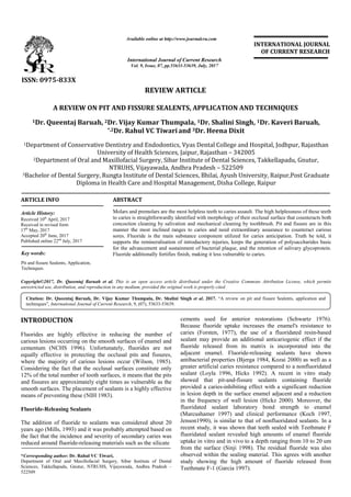
- 1. A REVIEW ON PIT AND FISSURE SEALENTS, APPLICATION AND TECHNIQUES 1Dr. Queentaj Baruah, 2Dr. Vijay Kumar Thumpala, *,2Dr. Rahul VC Tiwari 1Department of Conservative Dentistry and Endodontics, Vyas Dental College and Hospital, Jodhpur, Rajasthan University of Health Sciences, Jaipur, Rajasthan 2Department of Oral and Maxillofacial Surgery, Sibar Institute of Dental Sciences, NTRUHS, Vijayawada, Andhra Pradesh 3Bachelor of Dental Surgery, Rungta Institute of Dental Sciences Diploma in Health Care and Hospital Management, ARTICLE INFO ABSTRACT Molars and premolars are the most helpless teeth to caries assault. The high helplessness of these teeth to caries is straightforwardly identified with morphology of their occlusal surface that counteracts both concoction cleaning by salivation and mechani manner the most inclined ranges to caries and need extraordinary assurance to counteract carious sores. Fluoride is the main substance component utilized for caries anticipation. Truth be told, it supports the remineralisation of introductory injuries, keeps the generation of polysaccharides basic for the advancement and sustainment of bacterial plaque, and the retention of salivary glycoprotein. Fluoride additionally fortifies finish, making it less v Copyright©2017, Dr. Queentaj Baruah et al. This is an open access article distributed under the Creative Commons Att unrestricted use, distribution, and reproduction in any medium, provided the original work is properly cited. INTRODUCTION Fluorides are highly effective in reducing the number of carious lesions occurring on the smooth surfaces of enamel and cementum (NCHS 1996). Unfortunately, fluorides are not equally effective in protecting the occlusal pits and fissures, where the majority of carious lesions occur (Wilson Considering the fact that the occlusal surfaces constitute only 12% of the total number of tooth surfaces, it means that the pits and fissures are approximately eight times as vulnerable as the smooth surfaces. The placement of sealants is a highly effective means of preventing these (NIH 1983). Fluoride-Releasing Sealants The addition of fluoride to sealants was considered about 20 years ago (Mills, 1993) and it was probably attempted based on the fact that the incidence and severity of secondary caries was reduced around fluoride-releasing materials such as the silicate *Corresponding author: Dr. Rahul VC Tiwari, Department of Oral and Maxillofacial Surgery, Sibar Institute of Dental Sciences, Takkellapadu, Gnutur, NTRUHS, Vijayawada, Andhra Pradesh 522509 ISSN: 0975-833X Article History: Received 10th April, 2017 Received in revised form 17th May, 2017 Accepted 20th June, 2017 Published online 22nd July, 2017 Citation: Dr. Queentaj Baruah, Dr. Vijay Kumar Thumpala, Dr. Shalini Singh techniques”, International Journal of Current Research Available online at http://www.journal Key words: Pit and fissure Sealents, Application, Techniques. REVIEW ARTICLE A REVIEW ON PIT AND FISSURE SEALENTS, APPLICATION AND TECHNIQUES Dr. Vijay Kumar Thumpala, 1Dr. Shalini Singh, Dr. Rahul VC Tiwariand 3Dr. Heena Dixit Department of Conservative Dentistry and Endodontics, Vyas Dental College and Hospital, Jodhpur, Rajasthan University of Health Sciences, Jaipur, Rajasthan – 342005 Department of Oral and Maxillofacial Surgery, Sibar Institute of Dental Sciences, NTRUHS, Vijayawada, Andhra Pradesh – 522509 Rungta Institute of Dental Sciences, Bhilai, Ayush University, Raipur, Diploma in Health Care and Hospital Management, Disha College, Raipur ABSTRACT Molars and premolars are the most helpless teeth to caries assault. The high helplessness of these teeth to caries is straightforwardly identified with morphology of their occlusal surface that counteracts both concoction cleaning by salivation and mechanical cleaning by toothbrush. Pit and fissure are in this manner the most inclined ranges to caries and need extraordinary assurance to counteract carious sores. Fluoride is the main substance component utilized for caries anticipation. Truth be told, it ports the remineralisation of introductory injuries, keeps the generation of polysaccharides basic for the advancement and sustainment of bacterial plaque, and the retention of salivary glycoprotein. Fluoride additionally fortifies finish, making it less vulnerable to caries. is an open access article distributed under the Creative Commons Att use, distribution, and reproduction in any medium, provided the original work is properly cited. Fluorides are highly effective in reducing the number of carious lesions occurring on the smooth surfaces of enamel and cementum (NCHS 1996). Unfortunately, fluorides are not equally effective in protecting the occlusal pits and fissures, where the majority of carious lesions occur (Wilson, 1985). Considering the fact that the occlusal surfaces constitute only tal number of tooth surfaces, it means that the pits and fissures are approximately eight times as vulnerable as the smooth surfaces. The placement of sealants is a highly effective addition of fluoride to sealants was considered about 20 and it was probably attempted based on the fact that the incidence and severity of secondary caries was releasing materials such as the silicate Department of Oral and Maxillofacial Surgery, Sibar Institute of Dental Sciences, Takkellapadu, Gnutur, NTRUHS, Vijayawada, Andhra Pradesh – cements used for anterior restorations (Schwartz 1976). Because fluoride uptake increases the enamel's resistance to caries (Forsten, 1977), the use of a fluoridated resin sealant may provide an additional anticariogenic effect if the fluoride released from its matrix is incorporated into the adjacent enamel. Fluoride-releasing sealants have shown antibacterial properties (Bjerga 1984, Kozai 2000) greater artificial caries resistance compared to a nonfluoridated sealant (Loyla 1996, Hicks 1992). A recent in vitro study showed that pit-and-fissure seal provided a caries-inhibiting effect with a significant reduction in lesion depth in the surface enamel adjacent and a reduction in the frequency of wall lesion (Hickz 2000). Moreover, the fluoridated sealant laboratory bond strengt (Marcushamer 1997) and clinical performance (Koch 1997, Jenson1990), is similar to that of nonfluoridated sealants. In a recent study, it was shown that teeth sealed with Teethmate F fluoridated sealant revealed high amounts of enamel fluoride uptake in vitro and in vivo to a depth ranging from 10 to 20 um from the surface (Sinji 1998). The residual fluoride was also observed within the sealing material. This agrees with another study showing the high amount of fluoride released from Teethmate F-1 (Garcia 1997). International Journal of Current Research Vol. 9, Issue, 07, pp.53633-53639, July, 2017 Dr. Queentaj Baruah, Dr. Vijay Kumar Thumpala, Dr. Shalini Singh et al. 2017. “A review on pit and fissure International Journal of Current Research, 9, (07), 53633-53639. Available online at http://www.journalcra.com z A REVIEW ON PIT AND FISSURE SEALENTS, APPLICATION AND TECHNIQUES Dr. Shalini Singh, 1Dr. Kaveri Baruah, Department of Conservative Dentistry and Endodontics, Vyas Dental College and Hospital, Jodhpur, Rajasthan 342005 Department of Oral and Maxillofacial Surgery, Sibar Institute of Dental Sciences, Takkellapadu, Gnutur, Ayush University, Raipur,Post Graduate Disha College, Raipur Molars and premolars are the most helpless teeth to caries assault. The high helplessness of these teeth to caries is straightforwardly identified with morphology of their occlusal surface that counteracts both cal cleaning by toothbrush. Pit and fissure are in this manner the most inclined ranges to caries and need extraordinary assurance to counteract carious sores. Fluoride is the main substance component utilized for caries anticipation. Truth be told, it ports the remineralisation of introductory injuries, keeps the generation of polysaccharides basic for the advancement and sustainment of bacterial plaque, and the retention of salivary glycoprotein. ulnerable to caries. is an open access article distributed under the Creative Commons Attribution License, which permits cements used for anterior restorations (Schwartz 1976). fluoride uptake increases the enamel's resistance to 1977), the use of a fluoridated resin-based sealant may provide an additional anticariogenic effect if the fluoride released from its matrix is incorporated into the releasing sealants have shown antibacterial properties (Bjerga 1984, Kozai 2000) as well as a greater artificial caries resistance compared to a nonfluoridated sealant (Loyla 1996, Hicks 1992). A recent in vitro study fissure sealants containing fluoride inhibiting effect with a significant reduction in lesion depth in the surface enamel adjacent and a reduction in the frequency of wall lesion (Hickz 2000). Moreover, the fluoridated sealant laboratory bond strength to enamel (Marcushamer 1997) and clinical performance (Koch 1997, Jenson1990), is similar to that of nonfluoridated sealants. In a recent study, it was shown that teeth sealed with Teethmate F fluoridated sealant revealed high amounts of enamel fluoride uptake in vitro and in vivo to a depth ranging from 10 to 20 um from the surface (Sinji 1998). The residual fluoride was also observed within the sealing material. This agrees with another study showing the high amount of fluoride released from INTERNATIONAL JOURNAL OF CURRENT RESEARCH A review on pit and fissure Sealents, application and
- 2. The addition of fluoride to the sealants will greatly increase their value in the preventive and restorative use as mentioned above. Fluoride is added to sealants by two methods. The first is by adding a soluble fluoride to the unpolymerized resin. The fluoride can be expected to leach out over a period of time into the adjacent enamel. Eventually the fluoride content of the sealant should be exhausted, but the content of the enamel greatly increased. The second method of incorporating fluoride is by the addition of an organic fluoride compound that is chemically bound to the resin to form an ion exchange resin. As such, when fluoride is low in the saliva, fluoride would be released. Vice versa, when the fluoride in the environment is high, it should bind to the resin to form at least theoretically a continuous reservoir for fluoride release and recharge (Vlankenau 1983). Light-Cured Versus Self-Cured Sealants The main advantage of the light-cured sealant is that the operator can initiate polymerization at any suitable time. Polymerization time is shorter with the light-cured products than with the self-curing sealants. The light-cured process does require the purchase of a light source, which adds to the expense of the procedure. This light, however, is the same one that is used for polymerization of composite restorations, making it available in all dental offices. When using a light- cured sealant in the office, it is prudent to store the product away from bright office lighting, which can sometimes initiate polymerization. Conversely, the self-curing resins do not require an expensive light source. They do, however, have the great disadvantage that once mixing has commenced, if some minor problem is experienced in the operating field, the operator must either continue mixing or stop and make a new mix. For the autopolymerizing resin, the time allowed for sealant manipulation and placement must not be exceeded, even though the material might still appear liquid. Once the hardening begins, it occurs very rapidly, and any manipulation of the material during this critical time jeopardizes retention.The light-cured sealants have a higher compressive strength and a smoother surface (Vlankenau 1983), which is probably caused by air being introduced into the self-cure resins during mixing (CDM 1985). Despite these differences, both the photocured and the autopolymerizing products appear to be equal in retention (Sinji 1998, Waren 2001). Requisites for Sealant Retention For sealant retention the surface of the tooth must (1) have a maximum surface area, (2) have deep, irregular pits and fissures, (3) be clean, and (4) be absolutely dry at the time of sealant placement and uncontaminated with saliva residue. These are the four commandments for successful sealant placement, and they cannot be violated. Preparing the Tooth for Sealant Application The preliminary steps for the light-activated and the autopolymerized resins are similar up to the time of application of the resin to the teeth. After the selected teeth are isolated, they are thoroughly dried for approximately 10 seconds. The 10-second drying period can be mentally estimated by counting off the seconds 1,000, 2,000 until 10,000 has been reached. The liquid etchant is then placed on the tooth with a small resin sponge or cotton pledget held with cotton pliers. Traditionally, the etching solution is gently daubed, not rubbed, on the surface for 1 minute for permanent teeth and for 11/2 minutes for deciduous teeth (ADA 1997, Nordenvall 1980) Other clinical studies, however, have shown that acid etching the enamel of both primary and permanent teeth for only 20 seconds produced similar sealant (ADA 1997) and composite (Eidelman 1988) retention as those etched for 1 and 11/2 minutes. Currently, 20 to 30 seconds enamel-etching time is recommended. Alternatively, acid gels are applied with a supplied syringe and left undisturbed. Another 15 seconds of etching is indicated for fluorosed teeth to compensate for the greater acid resistance of the enamel. The etching period should be timed with a clock. At the end of the etching period, the aspirator tip is positioned with the bevel interposed between the cotton roll and the tooth. For 10 seconds the water from the syringe is flowed over the occlusal surface and thence into the aspirator tip. Again, this 10-second period can be mentally counted. Care should be exercised to ensure that the aspirator tip is close enough to the tooth to prevent any water from reaching the cotton rolls, yet not so close that it diverts the stream of water directly into the aspirator. Following the water flush, the tooth surface is dried for 10 seconds. The air supply needs to be absolutely dry. The dried tooth surface should have a white, dull, frosty appearance. This is because the etching will remove approximately 5 to 10 um of the original surface (Pahlavan 1976) although at times interrod penetrations of up to 100 um may occur. (Silverstone). The etching does not always involve the interrod areas; sometimes the central portion of the rod is etched, and the periphery is unaffected. The pattern on any one tooth is unpredictable (Bozalis 1977). In any event, the surface area is greatly increased by the acid etch. Application of the Sealant With either the light-cured or autopolymerized sealants, the material should first be placed in the fissures where there is the maximum depth. At times penetration of the fissure is negated by the presence of debris, air entrapment, narrow orifices, and excessive viscosity of the sealant (Silverstone 1983). The sealant should not only fill the fissures but should have some bulk over the fissure. After the fissures are adequately covered, the material is then brought to a knife edge approximately halfway up the inclined plane. Following polymerization, the sealants should be examined carefully before discontinuing the dry field. If any voids are evident, additional sealant can be added without the need for any additional etching. The hardened sealant has an oil residue on the surface. This is unreacted monomer that can be either wiped off with a gauze sponge or can be left. If a sealant requires repair at any time after the dry field is discontinued, it is prudent to repeat the same etching and drying procedures as initially used. Because all the commercial sealants both the light-cured and self-cured are of the same Bis-GMA chemical family, they easily bond to one another (Myres 1974) Colored Versus Clear Sealants Both clear and colored sealants are available. They vary from translucent to white, yellow, and pink. Some manufacturers sell both clear and colored sealants in either the light-curing or autopolymerizing form. The selection of a colored versus a clear sealant is a matter of individual preference. The colored products permit a more precise placement of the sealant, with the visual assurance that the periphery extends halfway up the inclined planes. Retention can be more accurately monitored by 53634 Dr. Queentaj Baruah et al. A REVIEW on pit and fissure Sealents, application and techniques
- 3. both the patient and the operator placing the sealant. On the other hand, a clear sealant may be considered more esthetically acceptable. Some clinicians prefer the clear sealants because they are more discrete than white. Others prefer the white sealants as they are easier to monitor at recall appointments. On the other hand, some clinicians seem to prefer the clear sealants because it is possible to see under the sealant if a carious lesion is active or advancing. However, no clinical study has comprehensively compared these issues. Recently, some pit- and-fissure sealants have been introduced that will change color as they are being light-polymerized. This property has not been fully investigated and seems to be only of relative advantage to the dental personnel applying the sealant. Placement of sealants over carious areas Sealing over a carious lesion is important because of the professionals' concern about the possibility of caries progression under the sealant sites. In teeth that have been examined in vivo and later subjected to histologic examination following extraction for orthodontic reasons, it has been found that areas of incipient or overt caries often occur under many fissures, which cannot be detected with the explorer. (Going 1978) In some studies, sealants have been purposely placed over small, overt lesions (Mertz 1992). When compared with control teeth, many of the sealed carious teeth have been diagnosed as sound 3 and 5 years later (Handelman 1976). has indicated that sealants can be considered a viable modality for arrest of pit-and-fissure caries (Jeronimus 1975). In other studies of sealed lesions, the number of bacteria recovered from the sealed area decreased rapidly (Carlsson 1997, Mertz 1992). This decrease in bacterial population is probably due to the integrity of the seal of the resin to the etched tooth surface (Theilade 1977) seal that does not permit the movement of fluids or tracer isotopes between the sealant and the tooth (Jensen 1978).Sealants have been placed over more extensive lesions in which carious dentin is involved (Handleman 1976) Even with these larger lesions, there is a decrease in the bacterial population and arrest of the carious process as a function of time. In another study, clinically detectable lesions into the dentin were covered for 5 years with Nuva-Seal. After that time the bacterial cultures were essentially negative, and an apparent 83% reversal from a caries-active to a caries-inactive state was achieved. (Jordan and Suzuki 1984) sealed small lesions in 300 teeth. During clinical and x-ray observations over a 5-year period, they found no change in size of the carious lesion, so long as the sealant remained intact. More recently (Mertz-Fairhurst and colleagues 1986) demonstrated that sealed lesions became inactive bacteriologically, with the residual carious material suggesting decay cessation. This ability to arrest incipient and early lesions is highlighted by the statement in the 1979 publication of the ADA's Council on Dental Therapeutics: "Studies indicate that there is an apparent reduction in microorganisms in infected dentin covered with sealant. These studies appear to substantiate that there is no hazard in sealing carious lesions." The statements end with the cautionary note: "However, additional long-term studies are required before this procedure can be evaluated as an alternative to traditional restorative procedures (ADT 1982) When sealing incipient lesions, care should be taken to monitor their retention at subsequent recall/annual dental examinations. In addition, there have been reports of sealants being used to achieve penetration of incipient smooth-surface lesions ("white spots") of facial surfaces” (Micik 1972). Sealants Versus Amalgams Comparing sealants and amalgams is not an equitable comparison because sealants are used to prevent occlusal lesions, and amalgam is used to treat occlusal lesions that could have been prevented. Yet, the comparison is necessary. One of the major obstacles to more extensive use of sealants has been the belief that amalgams, and not sealants, should be placed in anatomically defective fissures; this belief stems from misinformation that amalgams can be placed in less time, and that once placed, they are a permanent restoration. Several studies have addressed these suppositions. For instance, sealants require approximately 6 to 9 minutes to place initially, amalgams 13 to 15 minutes (Vurt 1984, Dennison 1987). Many studies on amalgam restorations have indicated a longevity from only a few years to an average life span of 10 years (Sessil 1982, Lavell 1976). Equally perturbing is the fact that in one large study of schoolchildren, 16.2% of all surfaces filled with amalgam had marginal leakage and needed replacement (Robinson 1971) The life span of an amalgam is shorter with younger children than with adults (Hunter 1982). To emphasize the problem of replacement of older restorations, a recent questionnaire study from 91 dentists in Iceland was conducted to determine the cause for replacement of 8,395 restorations. The reason given for the replacement of composites, amalgams, glass-ionomers, and for resin modified glass ionomers was failed restorations (47.2%), primary caries (45.3%) and non- carious defects (7.5%). For every restoration inserted for an overt lesion, there was a need for one to be reinserted previously (Njor 2002). The retention data from the earlier sealant studies were discouraging. In recent years, using later- generation sealants, along with the greater care in technique used for their insertion, much longer retention periods have been reported. In five long-term studies from 3 to 7 years, the average sealant loss per year ranged from 1.3 to 7% (Hassal 2001). If the yearly loss of these studies is extrapolated, the average life of these sealants compares favorably or exceeds that of amalgam (Dennison 1981). When properly placed, sealants are no longer a temporary expedient for prevention; instead, they are the only effective predictable clinical procedure available for preventing occlusal caries. The most frequent cause for sealant replacement is loss of material, which mainly occurs during the first 6 months; the most likely cause for amalgam replacement is marginal decay (Swift 1987), with 4 to 8 years being the average life span (Robinson 1971). To replace the sealant, only resealing is necessary. No damage occurs to the tooth. Amalgam replacement usually requires cutting more tooth structure with each replacement. Even if longevity merits were equal, the sealant has the advantage of being painless to apply and aesthetic, as well as emphasizing the highest objectives of the dental profession prevention and sound teeth. Options for Protecting the Occlusal Surfaces The use of sealants has spawned an entirely different concept of conservation of occlusal tooth structure in the management of deep pits and fissures before, or early in caries involvement. The preventive dentistry restoration embodies the concepts of both prophylactic odontotomy insertion of a restoration and covering the restoration and the connecting fissure system with a resin based sealant. Pain and apprehension are slight, and aesthetics and tooth conservation are maximized (Swift 1987). Several options are now available to protect the occlusal surfaces, with the selection depending on risk and 53635 International Journal of Current Research, Vol. 9, Issue, 07, pp.53633-53639, July, 2017
- 4. professional's judgment (Shaw 2000). The first level of protection is simply to place a conventional sealant over the occlusal fissure system. This sealing preempts future pit-and- fissure caries, as well as arrests incipient or reverses small overt lesions. The second option reported by (Simonsen in 1978) advocated the use of the smallest bur to remove the carious material from the bottom of a pit or fissure and then using an appropriate instrument to tease either sealant or composite into the cavity preparation. This he termed a preventive dentistry restoration. Following insertion of the restoration, sealant was placed over the polymerized material as well as flowed over the remaining fissure system. Aside from protecting the fissures from future caries, it also protects the composite or inserted sealant from abrasion (Bickinson 1988). Third option is use of glass-ionomers material for sealants, which is controversial. Due to their fluoride release and cariostatic effect, glass-ionomers have been used in place of traditional materials, as a pit-and-fissure sealant, however, resin sealants have shown much higher bond strength to enamel than glass-ionomers. Clinical trials(Aranda 1995, Ovrevo 1990) have shown poor retention over periods as short as 6 to 12 months. Though, in vitro studies have suggested that etching previous to application enhances the bonding of glass-ionomer sealant in fissure enamel (De luca 2001, Ereira 2001). One study showed that a conventional silver-reinforced glass- ionomer had superior clinical performance compared to a conventional resin sealant (Forss 1998).Resin-reinforced glass- ionomer cements have been investigated for their effectiveness as pit-and-fissure sealants. The 1-year results revealed that although clinically the glass-ionomer wears at a faster rate than a conventional resin sealant, in the scanning electron microscopic evaluation the material could be seen at the deep recesses of the pits-and-fissures with no carious lesion present. (Ovrevo 1990) A recent study showed that after 3 years the glass-ionomer sealant was completely lost in almost 90% of the teeth compared to less than 10% of the resin sealed teeth; the relative risk of a tooth sealed with glass-ionomer over that of a tooth sealed with resin was higher. Also, the glass-ionomer sealant had poorer retention and less caries protective effect (Poulson 2001).Glass-ionomer does not carry the ADA seal of approval as sealant material. The readers should decide their personal philosophy based on the evidence. A fourth option reported by (Garcia-Godoy 1986) involves the use of a glass-ionomer cement as the preventive glass-ionomer restoration (PGIR). The glass-ionomer cement (conventional or resin-modified) is placed only in the cavity preparation. The occlusal surface is then etched with a gel etchant avoiding, if possible, etching the glass-ionomer. Etching the glass-ionomer may remove some of the glass particles weakening the material. The conventional resin sealant is placed over the glass-ionomer and the entire occlusal fissure system. In the event sealant is lost, the fluoride content of the glass-ionomer helps prevent future primary and secondary caries formation. The same technique has successfully protected the marginal integrity of very small amalgam restoration, as well as providing a protection to the entire fissure system. Each of these options requires a judgment decision by the clinician. That decision can well be based on the criterion that if an overt lesion cannot be visualized, it should be sealed; if it can be visualized, the smallest possible preventive dentistry restoration should be used along with its required sealant "topping." (Mertz-Fairhurst 1987) The first option could provide the preferred model for conservative treatment of incipient and small overt, pit-and-fissure caries. It could also serve as an interim treatment for larger lesions. These options would be especially valuable in areas of the world with insufficient professional dental personnel and where preventive dental auxiliaries have been trained to place sealants. In all cases, the preventive dental filling should be considered as an alternative to the traditional class I amalgam with its accompanying extension for prevention that often includes the entire fissure system. The Sealant as Part of a Total Preventive Package The sealant is used to protect the occlusal surfaces. A major effort should be made to incorporate the use of sealants along with other primary preventive dentistry procedures, such as plaque control, fluoride therapy, and sugar discipline. Whenever a sealant is placed, a topical application of fluoride should follow if at all possible. In this manner the whole tooth can be protected. (Ripa 1987) A 2-year study for children in second and third grades assessing the effectiveness of a 0.2% fluoride mouthrinse used alone compared with a rinse plus sealants. Twenty-four occlusal lesions developed in the 51 rinse subjects, and only 3 in the 84 subjects receiving the rinse plus sealants. The conclusion was that caries could be almost completely eliminated by the combined use of these two preventive procedures. In many public-health programs, however, it is not possible to institute full-scale prevention programs, either because of apathy or lack of time and money. In such cases, there is some consolation in knowing that at least the most vulnerable of all tooth surfaces (the occlusal) is being protected. Use of Pit-and-Fissure Sealants By the mid-1980s most of the answers were available as to the need and effectiveness of Bis-GMA sealants to reduce the incidence of occlusal caries, and the techniques of placement of pit-and-fissure sealants were known. The safety of their placement has been demonstrated by many studies showing that even when placed over incipient and minimally overt caries sites, there was no progression as long as the sealant remained intact (Handelmen 1991) Finally, several clinical studies have pointed out that sealants could be applied by properly trained auxiliaries, thus providing a more economical source of manpower for private and military practices as well as for large school and public health programs. Bis-GMA sealant usage has been strongly supported by the ADA "as a safe and effective means for caries control."The United States Public Health Service, in a request for a proposal for a school pit-and-fissure study, stated "This combination of preventive techniques (combined use of fluoride and sealants) is expected to essentially eliminate caries in teeth erupting after the initiation of the study.Despite the support from the two largest organizations most interested in the dental health of the nation, the rank-and-file of the dental profession have not accepted sealants as a routine method for prevention. In spite of all the knowledge of the properties and successes of the sealants usage has lagged, with about 10% of the posterior teeth of children demonstrating the presence of sealants (Gerlach 1991). For example, a 1994 examination of 117,000 children in North Carolina between the ages of 6 and 17 found that approximately 12% had sealants (Rozier 1994) while the percentage for 927,000 in Tennessee was 10% (Gilchrist 1992) Other states demonstrate similar sealant usage. One study revealed that 88 children did have sealants while 508 did not 53636 Dr. Queentaj Baruah et al. A REVIEW on pit and fissure Sealents, application and techniques
- 5. have needed sealants (Selwitz 1992). For recruits entering the U.S. Air Force, sealants were found on 13.1% of the teeth while there was a need for 47.5% more. In the latter case, it was noted that a third of these personnel had occlusal caries that might have been prevented by the sealants.20 Many barriers exist in meeting the Healthy People 2010 Objective for sealants. In 2001, the State of Alabama was planning how to meet national dental objectives, when 50% of U.S. children are expected to have dental sealants on at least one permanent molar by the age of 14 years (Dasanayake 2001)(Currently, 22% of the children between 12 to 14 years have at least one sealant claim.) A final assessment of the 2010 prospects and the current State's demographics concluded that racial and gender disparities, difficulty in accessing care, the nonavailability of Medicaid-participating dentists in a country, and a lower payment/claim ratio may make the national sealant objective difficult to achieve (Dasanayake 2001). It should be mentioned that in many surveys, children from lower socioeconomic groups had greater sealant needs than those from more affluent neighborhoods. On the other hand, other countries have had marked success with increasing the number of teeth sealed. A study involving 68,704 children living in Lanarkshire, Scotland found approximately 10% of the occlusal surfaces were sealed (Chestnutt 1994). Five years later, in England the percentage of children having sealants dramatically increased between 20 to 50% in several areas (Chestnutt 1994). The placement of sealants is making slow progress. The 1998-99 Ohio State survey of 3rd-grade students in School Based/School Link programs found that in addition to oral-health benefits, "Providing sealant programs in all eligible, high-risk schools could reduce or eliminate racial and economic disparities in the prevalence of dental sealants" (MMWR 2000) Yet, there are problems in examining the number of sealants versus the need for sealants. Other Pit-and-Fissure Initiatives The findings of the following studies must be considered an important extension of the present use of pit-and-fissure sealants, which are used to prevent the development of incipient lesions and to arrest minimal overt lesions. If professional judgment dictates, conservative sealed amalgams or composites could be used to maintain marginal integrity, extend the longevity of the restorative materials, and for achieving a de facto extension for prevention without the need to remove sound tooth structure to extend the restoration over the entire fissure system. These two uses of resins for prevention and restorations without major operative considerations should be of great value in developing countries where professional manpower is at a minimum and the demand for dental care is great. Probably the most important recent research on the use of Bis-GMA sealants and carious lesions (Mertz 1998) in the 10-year study patients with paired permanent molars or premolars with obvious clinical and radiographic class I lesions were selected. The carious lesions extended halfway into the dentin or to the nearest pulp horn. The randomized placement of restorations for each of the tooth pairs consisted of two of the following: (1) a classic amalgam restoration, complete with extension for prevention of all connecting fissures (79 subjects); (2) a conservative amalgam restoration involving only the carious site with a sealant "topping," the latter which was extended into the entire pit-and- fissure system (77 subjects); and (3) with each one of the amalgam restorations, a paired composite restoration placed over the carious tissue with a "topping" of sealant that included all the pits and fissures (156 subjects). In the preparation for the composite, no attempt was made to remove the carious tissue. A 1-millimeter wide, 40- to 60-degree bevel was made in the sound enamel surrounding the lesion. The area was washed, dried, and a bonding agent was placed on the bevel. Hand instruments were used to place the composite, after which rotary instruments were used to shape the occlusal anatomy. Following this step, the occlusal surface was treated as for the placement of the average sealant dry, etch, rinse, and dry before placing the resin over the composite and the entire fissure system. The conclusions of this study after 10 years were: (1) both the sealed composites and the sealed amalgam restorations exhibited superior clinical performance and longevity compared to the unsealed amalgam restorations; (2) bonded and sealed composite restorations placed over the frank cavitated lesions arrested the clinical progress of these lesions for the 10 years of the study. Conclusion The majority of all carious lesions that occur in the mouth occur on the occlusal surfaces. Which teeth will become carious cannot be predicted; however, if the surface is sealed with a pit-and-fissure sealant, no caries will develop as long as the sealant remains in place. Recent studies indicate an approximate 90% retention rate of sealants 1-year after placement. Even when sealants are eventually lost, most studies indicate that the caries incidence for teeth that have lost sealants is less than that of control surfaces that had never been sealed. Research data also indicate that many incipient and small overt lesions are arrested when sealed. Not one report has shown that caries developed in pits or fissures when under an intact sealant. Sealants are easy to apply, but the application of sealants is an extremely sensitive technique. REFERENCES Accepted Dental Therapeutics, 39th ed. American Dental Association, Chicago, Ill. 1982. Allen, D. N. 1977. A longitudinal study of dental restorations. Br Dent J, 143:87-89. Aranda, M., & Garcia-Godoy, F. 1995. Clinical evaluation of a glass ionomer pit-and-fissure sealant. J ClinPediatr Dent, 19:273-7. Bjerga, J. M., & Crall, J. J. 1984. Enamel fluoride uptake and caries-like lesion inhibition in vitro. J Dent Res, 63:239 (Abstr. 618). Blankenau, R. J., Kelsey, W. P., Cavel, W. T., & Blankenau, P. 1983. Wavelength and intensity of seven systems for visible light curing composite resins: A comparison study. JADA, 106:471-74. Bozalis, W. B., & Marshall, G. W. 1977. Acid etching patterns of primary enamel. J Dent Res, 56:185. Burt, B. A. 1984. Fissure sealants: Clinical and economic factors. J Dent Educ, 48 (Suppl.) 2:96-102. Carlsson, A., Patersson, M., & Twetman, S. 1997. 2 year clinical performance of a fluoride-containing fissure sealant in young schoolchildren at caries risk. Am J Dent, 10:3:115-19. Cecil, J. C., Cohen, M. E., Schroeder, D. C., et al. 1982. Longevity of amalgam restorations: A retrospective view. J Dent Res, 61:185. Abstr. 56. Chestnutt, I. G., Shafer, F., Jacobson, A. P., & Stephen, K. W. 1994. The prevalence and effectiveness of fissure sealants in Scottish adolescents (Letter). Br Dent J, 177:125-29. 53637 International Journal of Current Research, Vol. 9, Issue, 07, pp.53633-53639, July, 2017
- 6. Council on Dental Materials, Instruments, and Equipment 1985. Visible light-cured composites and activating units. 110:100-103. Dasanayake, A. P., Li, Y., Philip, S., Kirk, K., Bronstein, J., & Childers, N. K. 2001. Utilization of dental sealants by Alabama Medicaid children barriers in meeting the year 2010 objectives. Pediatr Dent, 23:401-6. De Luca-Fraga, L. R., & FreirePimienta, L. A. 2001. Clinical evaluation of glass-ionomer/ resin-based hybrid materials used as pit-and-fissure sealants. Quintessence Int, 32:6 463-8. Dennison, J. B., & Straffon, L. H. 1981. Clinical evaluation comparing sealant and amalgam 4 years report. J Dent Res, 60(Special Issue A):520. Abstr. 843. Dennison, J. B., & Straffon, L. H. 1984. Clinical evaluation comparing sealant and amalgam after seven years final report. J Dent Res, 1984; 63(Special Issue): 215. Abstr. 401. Dental Sealants ADA Council of Access and Prevention and Interprofessional Relations, 1997. Council on Scientific Affairs JADA, 128:484-88. Dickinson, G., Leinfelder, K. F., & Russell, C. M. 1988. Evaluation of wear by application of a surface sealant. J Dent Res, 67:362. Abstr. 1999. Eidelman, E., Shapira, J., & Houpt, M. 1988. The retention of fissure sealants using twenty-second etching time: Three- year follow-up. J Dent Child, 55:119-20. Forss, H., & Halme, E. 1998. Retention of a glass ionomer cement and resin-based fissure sealant and effect on carious outcome after 7 years. Community Dent Oral Epidemiol, 26:21-25. Forsten, L. 1977. Fluoride release from glass ionomer cement. Scand J Dent Res, 85:503-4. Gandini, M., Vertuan, V., & Davis, J. M. 1991. A comparative study between visible-light-activated and autopolymerizing sealants in relation to retention. ASDC J Dent Child, 58:4 297-9. Garcia-Godoy, F. 1986. Preventive glass-ionomer restorations. Quintessence Int. 17:617-19. Garcia-Godoy, F., Abarzua, I., de Goes, M. F., & Chan, D. C. N. 1997. Fluoride release from fissure sealants. J ClinPediatr Dent, 22:45-49. Gerlach, R. W., & Senning, J. H. 1991. Managing sealant utilization among insured populations: Report from Vermont's "Tooth Fairy" program. ASDC J Dent Child, 58:46-49. Gillcrist, J. A., Collier, D. R., & Wade, G. T. 1992. Dental caries and sealant prevalences in schoolchildren in Tennessee. J Pub Health Dent, 52:69-74. Going, R. E., Loesche, W. J., Grainger, D. A., & Syed, S. A. 1978. The viability of organisms in carious lesions five years after covering with a fissure sealant. JADA, 97:455- 67. Handelman, S. L. 1991. Therapeutic use of sealants for incipient or early carious lesions in children and young adults. Proc Finn Dent Soc, 87:463-75. Handelman, S. L., Washburn, F., & Wopperer, P. 1976. Two year report of sealant effect on bacteria in dental caries. JADA, 93:976-80. Handleman, S. 1982. Effects of sealant placement on occlusal caries progression. Clin Prevent Dent, 4:11-16. Hassal, D. C., & Mellor, A. C. 2001. The sealant restoration: indications, success and clinical technique. Br Dent J, 191:358-62. Hassal, D. C., Mellor, A. C., & Blinkhorn, A. S. 1999. Prevalence and attitudes to fissure sealants in the general dental services in England. Int J Paediatr Dent, 9:243-51. Healey, H. J., & Phillips, R. W. 1949. A clinical study of amalgam failures. J Dent Res, 28:439-46. Hicks, J. M., & Flaitz, C. M. 1992. Caries-like lesion formation around fluoride-releasing sealant and glass ionomer restorations. Am J Dent, 5:329-34. Hicks, M. J., Flaitz, C. M., & Garcia-Godoy, F. 2000. Fluoride-releasing sealant and caries-like enamel lesion formation in vitro. J ClinPediatr Dent, 24:215-9. Hicks, M. J., Flaitz, C. M., & Silverstone, L. M. 1986. Secondary caries formation in vitro around glass ionomer restorations. Quint Int, 17:527-31. Houpt, M., Fuks, A., Shapira, J., Chosack, A., &Eidelman, E. 1987. Autopolymerized versus light-polymerized fissure sealant. J Am Dent Assoc, 115:55-56. Hunter, B. 1982. The life of restorations in children and young adults. J Dent Res, 61:537. Abstr. 18. Jensen, M. E., Wefel, J. S., Triolo, P. T., Hammesfahr, P. D. 1990. Effects of a fluoride-releasing fissure sealant on artificial enamel caries. Am J Dent, 3:75-78. Jensen, O. E., & Handelman, S. L. 1978. In vitro assessment of marginal leakage of six enamel sealants. J Prosthet Dent, 36:304-6. Jensen, O. E., Billings, R. J., & Featherstone, D. B. 1990. Clinical evaluation of FluroShield pit-and-fissure sealant. Clin Prev Dent, 12:24-27. Jeronimus, D. J., Till, M. J., & Sveen, O. B. 1975. Reduced viability of microorganisms under dental sealants. J Dent Child, 42:275-80. Jordan, R. E., & Suzuki, M. 1984. Unpublished report, quoted by Going, R.E. Sealant effect on incipient caries, enamel maturation and future caries susceptibility. J Dent Educ, 48(Suppl.) 2:35-41. Kervanto-Seppala, S., Lavonius, E., Kerosuo, E., &Pietilla, I. 2000. Can glass-ionomer sealants be cost-effective? J Clin Dent, 11:11-3. Koch, M. J., Garcia-Godoy, F., Mayer, T., & Staehle, H. J. 1997. Clinical evaluation of Helioseal-F sealant. Clin Oral Invest, 1:199-202. Kozai, K., Suzuki, J., Okada, M., & Nagasaka N. 2000. In vitro study of antibacterial and antiadhesive activities of fluoride-containing light-cured fissure sealants and a glass ionomer liner/base against oral bacteria. ASDC J Dent Child, 67:117-22. Lavell, C. L. 1976. A cross-sectional, longitudinal survey into the durability of amalgam restorations. J Dent, 4:139-43. Loyola-Rodriguez, J. P., & Garcia-Godoy, F. 1996. Antibacterial activity of fluoride release sealants on mutans streptococci. J ClinPediatr Dent, 20:109-12. Marcushamer, M., Neuman, E., & Garcia-Godoy, F. 1997. Fluoridated and unfluoridated sealants show similar shear strength. Pediatr Dent, 19:289-90. Mertz-Fairhurst, E. J., Call-Smith, K. M., Shuster, G. S., Williams, G. E., Davis, Q. B., Smith, C. D., Bell, R. A., Sherrer, J. D., Myers, D. R., & Morse, P. K. 1987. Clinical performance of sealed composite restorations placed over caries compared with sealed and unsealed amalgam restorations. J Am Dent Assoc, 115:689-94. Mertz-Fairhurst, E. J., Curtis, J. W. Jr., Ergle, J. W., Rueggeberg, F. A., & Adair, S. M. 1998. Ultraconservative and cariostatic sealed restorations: Results at year 10. JADA, 129:55-66. 53638 Dr. Queentaj Baruah et al. A REVIEW on pit and fissure Sealents, application and techniques
- 7. Mertz-Fairhurst, E. J., Richards, E. E., Williams, J. E., Smith, C. D., Mackert, J. R., Schuster, G. S., Sherrer, J. D., O'Dell, N. L., Pierce, K. L., Wenner, K. K., &Ergle, J. W. 1992. Sealed restorations: 5-year results. Am J Dent, 5:5-10. Mertz-Fairhurst, E. J., Shuster, G. S., & Fairhurst, C. W. 1986. Arresting caries by sealants: Results of a clinical study. JADA, 112:194-203. Micik, R. E. Mar 1972. Fate of in vitro Caries-like Lesions Sealed within Tooth Structure. IADR Program, Abstr. 710. Mills, R. W., & Ball, I. A. 1993. A clinical trial to evaluate the retention of a silver cement-ionomer cement used as a fissure sealant. Oper Dent, 18:148-54. Mjor, I. A., Shen, C., Eliasson, S. T., & Richters, S. 2002. Placement and replacement of restorations in general dental practice in Iceland. Oper Dent, 27:117-23. MMWR MorbMor Rep 2000; Aug 31; 50:736-8. Impact of integrated school-based dental sealant programs in reducing racial and economic disparities in sealant prevalence among school children. Myers, C. L., Rossi, F., & Cartz, L. 1974. Adhesive tag-like extensions into acid-etched tooth enamel. J Dent Res, 53:435-41. National Center for Health Statistics (NCHS) 1996. Third National Health and Nutrition Examination Survey (NHANES III) reference manuals and reports. Hyattsville (MD): NCHS, U.S. Department of Health and Human Services, Public Health Service, Centers for Disease Control and Prevention. NIH Consensus Development Conferences Statement 1983. Dental sealant in the prevention of tooth decay, Dec 5-7, 4(11):1-18. Nordenvall, K. J., Brannstrom, M., & Malgrem, O. 1980. Etching of deciduous teeth and young and old permanent teeth. A comparison between 15 and 60 seconds etching. Am J Orthod, 78:99-108. Ovrebo, R. C., & Raadal, M. 1990. Microleakage in fissures sealed with resin or glass ionomer cement. Scand J Dent Res, 98:66-69. Pahlavan, A., Dennison, J. B., & Charbeneau, G. T. 1976. Penetration of restorative resins into acid-etched human enamel. JADA. 1976; 93:1070-76. Pereira, A. C., Pardi, V., Basting, R. T. Menighim, M. C., Pinelli, C., Ambrosano, G. M., & Garcia-Godoy, F. 2001. Clinical evaluation of glass-ionomers used as fissure sealants: twenty four-month results. ASDC J Dent Child, 68:168-74. Poulsen, S., Beiruti, N., & Sadar, N. 2001. A comparison of retention and the effect on caries of fissure sealing with a glass-ionomer and a resin-based sealant. Community Dent Oral Epidemiol, 29:298-301. Ripa, L. W., Leske, G. S., & Forte, F. 1987. The combined use of pit-and-fissure sealants and fluoride mouthrinsing in second and third grade children: Final clinical results after two years. Pediatr Dent, 9:118-20. Robinson, A. D. 1971. The life of a filling. Br Dent J, 130:206-8. Rozier, R. G., Spratt, C. J., Koch, C. G., & Davies, G. M. 1994. The prevalence of dental sealants in North Carolina schoolchildren. J Pub Health Dent, 54:177-83. Selwitz, R. H., Colley, B. J., & Rozier, R. G. 1992. Factors associated with parental acceptance of dental sealants. J Pub Health Dent, 52:137-45. Shaw, L. 2000. Modern thought on fissure sealants. Dent Update, 27:370-4. Shinji, H., Uchimura, N., Ishida, M., Motokawa, W., Miyazaki, K., & Garcia-Godoy, F. 1998. Enamel fluoride uptake from a fluoride releasing sealant. Am J Dent, 11:58- 60. Silverstone, L. M. 1974. Fissure sealants, laboratory studies. Caries Res, 8:2-26. Silverstone, L. M. 1983. Fissure sealants: The enamel-resin interface. J Public Health Dent, 43:205-15. Simonsen, R. J. 1978. Preventive resin restorations. Quintessence Int, 9:69-76. Swartz, M. L., Phillips, R. W., Norman, R. D., et al. 1976. Addition of fluoride to pit-and-fissure sealants: A feasibility study. J Dent Res, 55:757-71. Swift, E. J. 1987. Preventive resin restorations. JADA, 114:819-21. Theilade, E., Fejerskov, O., Migasena, K., &Prachyabrued, W. 1977. Effect of fissure sealing on the microfloral in occlusal fissures of human teeth. Arch Oral Biol, 22:251- 59. Waren, D. P., Infante, N. B., Rice, H. C. et al. 2001. Effect of topical fluoride on retention of pit-and-fissure sealants. J Dent Hyg, 71:21-4. Wilson, I. P. 1985. Preventive dentistry. Dent Dig, 1:70-72. ******* 53639 International Journal of Current Research, Vol. 9, Issue, 07, pp.53633-53639, July, 2017