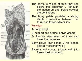
1. The pelvis is region of trunk that lies
below the abdomen . Although
the abdomen and pelvis cavities
are continuous .
The bony pelvis provides a strong
stable connection between the
trunk and lower extremities .
Function
1- body weight
2- support and protect pelvic viscera.
3- Provide attachment of trunk and
lower limb muscles.
Bony pelvis four bones 2 hip bones
(lateral + anterior wall )
Sacrum and coccyx ( back wall ) to
form ( basin shaped).
2. • Content : Parts of intestinal tract , urinary
tract internal reproduction organs and
vessels.
• Above false pelvis
• Below true pelvis
• Pelvis : divided in two part by pelvic brim
• Pelvic brim sacral promontory ( Mar gin
Antierior & upper of first sacral vertebra )
• Poteriorly
• Laterally : leopectineal lines
• Anteriorly symphsis pubis Orientation of
the pelvis relative to the trunk in the
anatomical position . the front of the
symphysis pubis and anterior superior iliac
spines should line in the same vertical
plane this means that pelvic surface of
pubis symphysis faces upward and back
ward and anterior surface sacrum is
directed forward and downward .
3. • False pelvis it is bounded
behind by the lumbar
vertebrae , laterally by the
iliac fossa and the iliacus
muscles & in front by the
lower part of the anterior
abdominal wall . it
supports the obdomnal
content and after third
month of pregnancy helps
support the gravid uterus .
it helps guide the fetus in
to the true pelvis.
4. • True pelvis shape and dimensions of
the female pelvis is of great
importance because it is the bony
canal through which the child passes
during birth.
• Inlet , out let and a cavity. Inlet or
pelvcbrim
• Pelvic outlet is bounded posteriorly
coccyx , laterally by the ischial
tuberosities and anteriorly by the
pubicarch . The pelvic out let does not
present a smooth out line but has
three wide notches.
• Anteriorly pubic arch between
ischiopubicrami .
• Laterlly sciatic notches divided by the
sacratuberous and sacrospinous
ligament in to the greater and lesser
sciatic foramina.
• Sacrotuberous ligament are strong
and relatively inflexible thus , the outlet
is diamond – shaped with
ischiopubicrami and the symphysis
pubis in front , sacrotuberous
ligaments and coccyx behind.
• Pelvic cavity between inlet and out let .
A short curved cancal with shallow
anterior wall and much deeper
posterior wall.
5. • Structure of the pelvic wall:
(Bones and ligament that are
partly lind with muscles covered
with fascia and parietal
peritoneum )
• Anterior pelvic wall : shallow
and formed by the posterior
surfaces of bodies of pubic bones
, the pubicrami and symphysis
pubis.
• Posterior pelvic wall : Deep and
formed by sacrum and coccyx
and piriformis muscles and their
covering of parietal pelvic fasia.
• Inferior pelvic wall , or pelvic
floor is formed by the pelvic
diaphragm the pelvic floor
stretches across the pelvis and
divides it in to main pelvic cavity
above which contains the pelvic
viscera and perineum below.
6. • Pelvic Diaphragm
is formed by
levatores ani
muscles and small
coccygeus muscles
and their covering
fasciae . it is in
complete anteriorly
to allow passage of
urethra in males and
females and in the
female the vagina
also .
7. • Pelvic diaphragm
• Levator Ani muscle, small coccygeus
muscle and their covering fasciae.
• Levator ani muscle: wide thin sheet
that has a linear origion from the back
of the body of the pubis, tendinous
arch, spine of ischium – groups of
fibers sweep down ward and medially
to their insertion as follows:
• 1- Anterior fibers: from a sling
around the prostate or vagina as
levator prostatae or sphincter vaginae
and inserted into mass of fibrous
tissue called perineal body in front of
the anal canal.
• 2- Intermediate fibers form a sling
around the junction of the rectum and
anal canal (puborectalis). The
pubococcygeus posses posteriorly to
be inserted into asmall fibrous mass
(anococcygeal body) between the tip
of the coccyx and anal canal.
• 3- Posterior fibers: iliococcygeus is
inserted into the anococcygeal body
and coccyx.
8. • Action, Nerve supply
• Coccygeus muscle : small triangulor
muscle arises from the spine of the
ischium and is inserted into the lower end
of sacrum and coccyx.
• Action: Assist the levatores ani in
supporting the pelvic viscera.
• Nerve supply: from branch of the 4th and
fifth sacral nerves.
9. • Muscles of the pelvic wall and floor
• 1- piriformis.
• 2- Obturotor internus.
• 3- Levator ani.
• 4- Coccygeus.