6. Investigation of diseases of the
blood
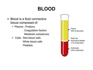
• Full blood count
• Blood film examination
• Bone marrow examination
• Investigation of coagulation
7. 1. History and
examination
2. Blood tests:
• Complete blood count
Total count:WBC
Neutrophils(N)
Lymphocytes(L)
Eosinophil(E)
Basophil(B)
Hb:
Platelets
8. • Correlation of CBC with disease condition .
• What things can u identify?
Hb Anemia. Normal Level:Male:14-18g/dl
Female: 12-16g/dl
Polycythemia
Complete blood count Importance of
Total count :5000-10000 microlitre
High /Low: Malignancy
High: infection
Neutrophil:40-60%: Infection
Lymphocytes: 20-40%:Tuberculosis
Eosinophil:1-4%:Allergies,worm infestations
Monocytes:2-8%
Basophils:0.5-1%
Platelets:1,50,000-4,00,000 microlitre
9.
10. Red cell indices
• MCV(Mean corpuscular volume):Size of red
blood cells
• For Hemoglobin content
• MCH(Mean corpuscular hemoglobin)
• MCHC(Mean corpuscular hemoglobin
concentration)
• Packed cell volume(Hematocrit)
• Red cell Distribution width
11. Packed cell volume(PCV)
• Hematocrit (HCT), also called packed cell volume (PCV), is the
percentage of blood volume occupied by RBCs.
• HCT = ([RBC x MCV]/10).
• Male:41-50%
• Female:36-48%
• Low hematocrit:
Chronic bleeding
Aplastic anemia
Sickle cell anemia
High: smoking
High altitude.
12. Mean corpuscular volume
• Measure of average volume of RBC
• Measured by: Hematocrit(%) X 10/ RBC count
• Measured in femtolitre
• Normal:80-100 fl
• Microcytic: Red cell with reduced volume <80fl
• Macrocytic: Red cell with increase volume:>100 fl
• Causes of microcytic: iron deficiency
• Sideroblastic
• Thalessemia
• Causes of macrocytic: Vit.B12 deficiency
• Folate deficiency
13. Mean corpuscular hemoglobin
concentration
• Average concentration of Hb in deciliter of erythrocytes.
Measured in g/dl.
• Ratio of Hb mass to volume
• MCHC: Hb x100/ Hematocrit
• Normochromic:32-36 g/dl
• Hypochromic:<32 g/dl
• Hyperchromic:>36 g/dl
• Causes of hypochromic: vit B6 def, Iron deficiency
• Causes of hyperchromic: Vit B12,Folate def
14. Mean cell Hemoglobin
• Measurement of individual
weight of Hb in individual
erythrocytes
• Measured in picogram
• MCH: Hb x10/RBC
• Normal value is 28-34 pg
15. Red cell distribution width
• Used along with MCV.
• Used to identify variation in
blood cell size and volume.
• RDW:11.6-14.6 in adult
• Importance: Helps to
identify heterogenous cell
size or presence of two types
of red cell population
• Other RBC indices reflects
RBC values not RBC changes.
19. Prothrombin time
• Measures time it takes plasma to clot when exposed to tissue
factor that assess extrinsic and common pathway
• PT:11-13 seconds
• INR(International normalized ratio):
• Ratio of patients prothrombin time to control prothrombin time
• Dimensionless
Uses: Unexplained bleeding
Patinet under anticoagulant
DIC
20. Activated partial throboplastin time
• measures the time it takes plasma to clot when exposed to
substances that activate the contact factors, which assesses
the intrinsic and common pathways of coagulation .
• Normal range:25-35 seconds
• Uses: Unexplained bleeding
Under heparin therapy
21. Fibrinogen and D-dimer
• Fibrinogen is the precursor to fibrin, the principle component of
a fibrin clot
• Normal value:5—100 mg/dl
• Clinical use: DIC,Liver disease
D-dimer:Fibrin D-dimer is one of the major fibrin degradation
products released upon cleavage of crosslinked fibrin by plasmin.
• Normal plasma levels of D-dimer by ELISA testing are <500
ng/mL
• Uses: Deep vein thrombosis
• Pulmonary embolism
• DIC
22. Peripheral blood smear
• Examination of the peripheral blood smear provides a window
into the functional status of the bone marrow.
• The peripheral smear is indispensable for evaluating the number,
size, and shape of red cells, white cells, and platelets.
• RBC:Review the color, shape, irregular borders, and presence of
nuclei, inclusions, or parasites.
• WBC:Review the presence of early (immature) cells, abnormal
lobulation or nuclear contour, abnormal or absent granulation,
presence of parasites or other inclusions
• Platelets – Review any increase or decrease in platelet number
and size, absence of granules, presence of platelet aggregation,
megakaryocyte fragments.
23.
24.
25.
26.
27.
28. Other tests
• Perpiheral smear always supported by Bone marrow aspiration
and biopsy.
• AML: Myeloperoxidase on peripheral blood smear
• Flow cytometry
• Bone marrow aspiration and biopsy: Usually hypercellular
• Prsence of Auer rod on microscopy
• Cytochemistry: Myeloperoxidase,Sudan black B test.
• ALL: Anemia,neutropenia,thrombocytopenia
• Tests: Peripheral blood smear,Periodic acid shiff test,Non
esterase
• Flow cytometry/cytogenetics.