SlideShare a Scribd company logo
1 of 66
Or
Degenerative Joint Disease
DEFINATION
Osteo : Related to bone system
Arthritis : Arthro + itis
↓ ↓
Joint Inflammation
Condition in which one/ more joints are inflamed
So OA is a condition in which one or more joints are inflamed
involving the loss of cartilage
It is progressive disorder of joints which is caused by gradual loss
of cartilage
resulting in development of bony spurs and crysts at the margins
of the joints.
In addition to damage and loss of articular cartilage, there is
remodelling of subarticular bone, osteophyte formation,
ligamentous laxity, weakening of periarticular muscles, and, in
some cases, synovial inflammation
These changes may occur as a result of an imbalance in the
equilibrium between the breakdown and repair of joint tissue
The most common joints involved
• Distal Interphalangeal joints
• Proximal Interphalangeal joints
• Carpometacarpal joints of thumb
• Weight Bearing joints (Hip, knee)
• Metatarsophalangeal joints of the foot
• Cervicle and lumbar vartebrae
The articular cartilage is
slippery tissue that covers the
end of the bones in the joints
Healthy cartilage
allows bones to glid over
each other
&
helps to absorb shock of
movement
In OA the top layer of cartilage
breaks down and wear away
↓
Rubbing of bones under the cartilage
↓
Due to which there is
Pain
Swelling
Loss of motion of
joints
↓
Over the time joint may lose its
normal shape and also there is
growth of bony spurs on the edge of
the joint
↓
Bits of bones/cartilage can break off
and float inside the joint space
↓
Which cause more pain and damage
So in OA there is progressive distruction of articular cartilage and
also there is involvement of
• Diarthrodial joint
• Synovium
• Capsule
• Subchondral bone
• Surrounding ligaments
• Muscles
Changes in structure and function of this tissues leads to clinical
Osteoarthritis
Which is characterized by
• Joint pain
• Tenderness
• Decrease range of motion
• Weakness
• Joint instability
• Disability
Classification
↙ ↘
Primary OA Secondary OA
(idiopathic) (Due to some other disease)
A. Localised
Hands
Hip
Knee
Spine
B. Generalised
Small joints
Large joints
Mixed
C. Erosive osteoarthritis
i) Congenital and developmental
disorders, bone dysplasias
ii) Post-surgery / injury – meniscectomy
iii) Endocrine –acromegaly,
iv) Metabolic –hemachromatosis,
ochronosis, Marfan syndrome, Ehler-Danlos
syndrome, Paget disease, gout, pseudogout,
Wilson’s disease, Hurler disease, Gaucher
disease
v) Rheumatologic– rheumatoid arthritis
vi) Neurological– Charcot joints.
Epidemiology
OA is one of the core reason for disability
Nearly everyone who lives long is affected by OA at any point of
life, most probably after the age of 55 to 60.
Approximately 15% of population is affected by OA
↙ ↘
↙ ↘
50 % of those 85% of those
over 65 age over 75 age
OA is most widely assessed in studies using the Kellgren
and Lawrence (K&L) score.
The overall grades of severity are determined from 0 to 4
and are related to the presumed sequential appearance of
osteophytes, joint space loss, sclerosis and cysts
The World Health Organization (WHO) adopted these criteria
as the standard for epidemiological studies on OA
Prevalence of osteoarthritis varies with
I. Age
II. Gender
III. Genetics
IV. Ethic Group
V. Specific Joint Involved
VI. Method For Diagnosis
AGE
With increasing age the prevalence OA also increases
Generally for person age 25 – 75,prevalence is estimated
12%
and for those with age over 70, it is 60 – 70% affectet
Hip OA ← ← AGE → → Hand OA
↙ ↘ ↓ ↙ ↘
Age : 30 – 40 Age : >70 Knee OA Age : 40 Age : 80
Pre : 1.6 % Pre : 14% ↙ ↘ Pre: 5% Pre : 65%
Age : >25 Age : 55
Pre : 5% Pre : 12%
Gender
↙ ↘
Age > 50 Age >60
Men are more affacted Women are more
affacted(26%)
Due to higher rate of sports and Due to repeated use of
Injuries weight bearing joints
Genetics
↙ ↘
Pre : 9% Pre : 4%
White population Black & Asian population
Prevalence of OA is 22% to 39% in India
Etiology
The etiology of OA is multifactorial
Many patients have more than one risk factor for developing the
OA.
The most common risk factor for the development of OA includes
• Obesity
• Occupation
• Participation in certain sports (Often)
• History of joint trauma
• Genetic
• Age
• Sex
• Bone density
• Joint location
Obesity
Increased body weight is strongly associated with hip, knee, and
hand OA
Obesity often precedes OA and contributes to its development,
rather than occurring as a result of inactivity from joint pain
In a three-decade Framingham Study, the highest quintile of body
mass was associated with a higher relative risk of knee OA
(relative risk of 1.5 to 1.9 for men and 2.1 to 3.2 for women).
The risk of developing OA increases by about 10% with each
additional kilogram of weight, and in obese persons without OA,
weight loss of even 5 kg decreases the risk of future knee OA by
one-half.
Recent data suggest that OA is associated with the metabolic
syndrome, suggesting a possible common pathogenic mechanism
involving metabolic abnormalities and systemic Inflammation.
It is also likely that vascular disease may both initiate and hasten
disease progression in OA.
This could be due to venous occlusion, stasis or microembolic
disease leading to episodic reduction in blood flow through small
vessels within the subchondral bone.
Subchondral ischaemia may subsequently reduce nutrient
delivery and gas exchange to articular cartilage in addition to
direct deleterious effects on the bone itself.
Occupation
There is increased risk of OA for those who are in occupation
requiring
Prolong Standing
Kneeling
Squatting
Lifting/Moving Heavy Objects
↓
→Miming
Factory Work
Car paintery
Repetitive motion also contributes to hand OA with dominant hand
usually affected
Risk OA depends on type and intensity of physical activity
Sports
Damage to articular cartilage due to sports greatly increase the
risk of OA
Meniscal damage (common in athlete) also increase the risk of
knee OA
1.Because of loss of proper load bearing and shock absorption
2.Increase focal load on cartilage and subchondral bones
Trauma
AGE AT INJURY DOSE MATTER
As older individuals who damage ligaments tends to develope OA
more rapidly than young people with similar injury
Trauma early in life →Increased risk of OA
Genetic Factors
A number of recent studies discovered the presence of over 80
gene mutations involved in the pathogenesis of OA ,among
which the most relevant one is a single nucleotide polymorphism.
This one, called rs143383 and located in the 3' untranslated region
(3'UTR) of the growth and differentiation factor 5 gene (GDF5), is
responsible for the development, maintenance and repair of
synovial joints
Genes for Vitamin D receptors (VDR) and insulin-like growth
factor 1(IGF-1) also seem to be involved in the patho-physiologic
pathways of OA
It also includes genes related to inflaamation,bone
morphogenetic protiens, protease/ its inhibitors
Normal Articular Cartilage
Articular Cartilage Possesses
↓
Viscoelastic Properties
↓Which provides
1. Lubrication With Motion
2. Shock absorbency during rapid movement
3. Load Support
In Synovial joints Articular cartilage is found between synovial
cavity on one side and narrow layer of calcified tissue overlying
subchondral bone on onther side
Charactristics of Articular Cartilage
1.Cartilage is easily compressed lossing up to 40% of its original
hight when load is applied
↓
Compression increase area of contact
↓
Disperse force more evenly to undelying bone,tendon, ligament,
muscles
2.Cartilage is frictionless
Togather with compresibility , this enables smooth movement in
joint and distributes load across joint tissue to prevent damage and
stabilize the joint
Structure of Cartilage
Cartilage is having
↓
1.Strength
2.Low co-efficiant of friction
3.Copressiblity
These all is derived from its unique structure
So,the cartilage is composed of complex, hydrophilic , Extra-
cellular matrix
It contains →75% to 85% Water
2% to 5% Chondrocytes (the only cell in cartilage)
Collagen Protiens
Proteoglycans
Hyaluronic Acid Molecules
Two Major Structure of Cartilage
 Type II Collagen
Tightly woven, triple helical
structure which provides
TENSILE STRENGTH to
cartilage
 Aggrecans
In which there is
Proteoglycan linked with
hyluronic acid, having
negative charge.
The strong electrostatic
repulsion of proteoglycan
gives cartilage the ability to
withstand further
compression
Normal Cartilage turn over
↓
Helps repair and restore cartilage
↓
Respond to usual demand of loading and physical activity
In healthy adult cartilage chondrocyte metabolism is slow with
dynamic balance between anabolic process
Metabolism is premoted by
1. Groth factors
- Bone morphogenetic protien 2
- Insulin like groth factor-1
- tranforming growth factor
2. Catabolism
3. Proteolysis
Stimulated by MMPs, TNF-ᾲ, Interlukein-1, Other cytokines
Joint Protective Mechanisms
1.Muscle Bridging The Joints
2.Sensory receprtors in feedback loops to regulate muscle and
tondon function
3. Supporting ligaments
4. Subchondral bones having shock absorbent properties
Note
Articular cartilge is avascular and aneural and chondrocytes are
nourised by synovial fluid
OA Cartilage
OA begins with damage to articular cartilage, which is due to
1. Trauma or other injury
2. Excess joint loading by obesity / other reason
3. Instability or injury of the joint that causes abnormal loading
Devlopement of OA is due to
1. Local mechanical influence
2. Genetic factor
3. Inflammation
4. Chondrocyte function
Which leads to loss of articular cartilage
When there is damage to articular cartilage
↓
Increase activity of chondrocytes to remove and repair the damage
Depending on degree of damage the balance between breakdown
and resynthesis of cartilage can be lost
↓
Which leads to increase breakdown of cartilage
↓
Ultimately, loss of cartilage
Destruction of aggrecans by proteolytic enzyme is consider to play a
key role
There is also involvement of collagen receptors named DDR-2 ,
located on chodrocyte cell surface
In healthy cartilage ,DDR-2 is inactive ,which is masked by
aggrecan from contact with collegen
Damage to cartilage
↓
Triggers aggrecan destruction
↓
Exposure of DDR-2 to collagen
↓
Active DDR-2 increase activity of MMP-13
↓
Which destroy collagen
Collagen breakdown products further stimulate DDR-2,in which
more collagen is destroyed
Huge research work has been done on genes involved in OA
which shows that
In OA , expression of hundreds of genes of cartilage tissue are
affected which alters chondrocyte phenotype
In addition to articular cartilage there is also role of subchondral
bone in OA
In OA , subchondral bone release vasoactive peptides and MMPs
Neovascularization and subsequent increase in permeability of
the adjacent cartilage occurs and contributes to further cartilage
loss
Substantial loss of cartilage cause joint space narrowing and leads
to painful and deformed joints
The remaining catilage softens and devlopes fibrillation(Verticle
cleft ) and there is splittting and further loss of cartilage and
exposure of underlying bone
As cartilage is destroyed and the adgecent subchondral bone
undergoes pathologic changes, cartilage is eroded completely,
leaving denuded subchondral bone which becomes dence ,
smooth and glistening
A more brittle, stiffer bone results, with decreased weight-bearing
ability and development of sclerosis and microfractures
The joint capsule and synovium also show pathologic changes
in OA.
Inflammation, noted clinically as synovitis, may result
from release of inflammatory mediators from chondrocytes, such
as prostaglandins
Inflammation is localized to the affected joint, in contrast to that
seen in rheumatoid or other inflammatory arthritides.
The pain in OA is not due to distruction of cartilage but arise
from the activation of nociceptive nerve ending within the joint
by mechanical and chemical irritants
So,the slow progressive changes in OA consist of an increase in
water content, loss of PG, and reduction of PG aggregates of
cartilage.
The cartilage is subsequently unable to repair itself.
Alterations in metabolism of subchondral bone adjacent to
articular cartilage appear necessary for continued cartilage
destruction.
Eventually, progressive loss of articular cartilage and increasing
subchondral sclerosis lead to an abnormal and painful joint.
Age
Usually elderly
Gender
Age <45 more common in men
Age >45 more common in women
Symptoms
→Pain
Deep, aching character
Pain on motion
Pain with motion early in disease
Pain with rest late in disease
→ Stiffness in affected joints
Resolves with motion, recurs with rest
Usually <30 minutes’ duration
Often related to weather
→ Limited joint motion
May result in limitations activities
of daily living
Signs, history, and physical
examination
Monarticular or oligoarticular;
asymmetrical involvement
Hands
Distal interphalangeal joints
Heberden’s nodes
Proximal interphalangeal joints
Bouchard’s nodes
First carpometacarpal joint
Osteophytes give characteristic square
appearance of the hand
Knees
Patellofemoral compartment involvement
Pain related to climbing stairs
Transient joint effusions
Typically noninflammatory synovial fluid
Clinical Represntation
Hips
Pain during weight-bearing activities
Stiffness, especially after inactivity
Limited joint motion
Spine
L3 and L4 involvement is most common in
the lumbar spine
Signs and symptoms of nerve root
compression
Radicular pain
Paresthesias
Loss of reflexes
Muscle weakness associated with
the affected nerve root
Feet
Typically involves the first
metatarsophalangeal
joint
Other sites, less commonly involved
Shoulder, elbow, acromioclavicular,
sternoclavicular, and temporomandibular
joints
Observation on joint examination
Bony proliferation or occasional synovitis
Local tenderness
Muscle atrophy
Limited motion with passive/active
movement
Deformity
Radiologic evaluation
Early mild OA
Radiographic changes often absent
Progression of OA
Joint space narrowing
Subchondral bone sclerosis
Marginal osteophytes
Late OA
Abnormal alignment of joints
Effusions
Characteristics of synovial fluid
High viscosity
Mild leukocytosis
Laboratory values
ESR
Treatment
Goals
(1) to educate the patient, caregivers, and relatives;
(2) to relieve pain and stiffness;
(3) to maintain or improve joint mobility;
(4) to limit functional impairment; and
(5) to maintain or improve quality of life
Treatment of OA
Non-pharmacological Treatment
1.Diet
2.Physical And Occupational Therapy
3.Surgery
Pharmacological Treatment
1.Analgesics
Oral
Topical
2.NSAIDs
3. Glucosamine &Chondroitin
4.Hyluronate Injection
5.Narcotic Analgesics
6.Disease Modifying Drugs
Diet
Excess weight increases the biomechanical load on weight-
bearing joints, and is the single best predictor of need for
eventual joint replacement.
Weight loss is associated with decreased symptoms and
Disability
Even 3-5 kg of weight loss can decrease the biomechanical force
on a weight-bearing joint.
Although dietary intervention for overweight OA patients is
reasonable, weight loss usually requires a motivated patient and a
structured weight-loss program.
Diet with low fat (calories) and high fibre as well as balanced diet
should be taken to decrease weight
Physical And Occupational Therapy
Physical therapy—with heat or cold treatments and an exercise
program Which helps to maintain and restore joint range of
motion and reduce pain and muscle spasms.
Warm baths or warm water soaks may decrease pain and
stiffness.
Exercise programs and quadriceps strengthening can improve
physical functioning and can decrease disability, pain, and
analgesic use by OA patients.
The decision about whether to encourage walking should be
made on an individual basis.
With weak or deconditioned muscles,the load is transmitted
excessively to the joints, so weight-bearing activities
can exacerbate symptoms.
However, avoidance of activity by those with hip or knee OA leads
to further deconditioning or weight gain.
A program of patient education, muscle stretching and
strengthening,and supervised walking can improve physical
function and decrease pain in patients with knee OA.
Surgery
Surgery can be recommended for OA patients with functional
disability and/or severe pain that is unresponsive to conservative
Patients with mild knee OA, an osteotomy may correct the
misalignment of knee
Knee arthroscopy or lavage have been recommended for short-
term relief of pain, a recent study showed these two procedures to
be equivalent to surgery
Experimental but potentially restorative approaches involve
Soft tissue grafts,
Penetration of subchondral bone,
Cell transplantation, and
Use of growth factors
Pharmacological Therapy
Things to be taken in concern
1.Adverse effect of the drug given for OA
2.As OA is usually seen in old age people with some
other conditions they Pharmacological treatment
should be done with so much causion
3.Always combine non pharmacological therapy
with pharmacological one for more improvement
Analgesics
Acetaminophen is used as first-line drug therapy for pain
management in OA, due to its relative safety, efficacy, and lower
cost compared to NSAIDs
Pain relief with acetaminophen can be similar to that obtained
with NSAIDs, although some patients will respond better to
NSAIDs.
Some studies have shown comparable efficacy for acetaminophen
and NSAIDS, others have reported
that patients experienced better pain control with NSAIDs than
with acetaminophen, and that OA patients preferred NSAIDs to
acetaminophen
Mechanism Of Action
Acetaminophen is thought to work within the central nervous
system to inhibit the synthesis of prostaglandins, agents that
enhance pain sensations.
Acetaminophen prevents prostaglandin synthesis by blocking
the action of central cyclooxygenase.
Kinetics
Acetaminophen is well absorbed after oral administration
(bioavailability is 60% to 98%), achieves peak concentrations
within 1 to 2 hours, is inactivated in the liver by conjugation with
sulfate or glucuronide, and its metabolites are renally excreted
Efficacy
Comparable relief of mild to moderate OA pain has been
demonstrated for acetaminophen at 2.6 to 4 g/day
ADR
Hepatotoxicity, and
Possibly renal toxicity
Drug – Drug Intrection
Isoniazid
can increase the risk of hepatotoxicity.
Chronic ingestion of maximal doses of acetaminophen may
intensify the anticoagulant effect in patients taking warfarin,
Food decreases the maximum serum concentration
of acetaminophen by approximately one-half.
Non Steroidal Anti - Iflammatory Drugs
MOA
Blockade of prostaglandin synthesis through inhibition of
cyclooxygenase (bothCOX-1 andCOX-2 enzymes) is the principal
mechanism by which NSAIDs relieve pain and inflammation
Kinetics
High oral availability, high protein binding, and absorption as
active drugs (except for sulindac and nabumetone, which require
hepatic conversion for activity).
The most important difference in NSAIDs is a serum half-life
ranging from 1 hour for tolmetin to 50 hours for piroxicam,
impacting the frequency of dosing and potentially, compliance
with therapy.
Elimination of NSAIDs largely depends on hepatic inactivation,
with a small fraction of active drug being renally excreted.
NSAIDs penetrate joint fluid, reaching about 60% of blood levels
ADR
Minor complaints—nausea, dyspepsia, anorexia, abdominal
pain, flatulence, and diarrhea
To minimize these symptoms, NSAIDs should be taken with food
or milk, except for enteric-coated products, which should not be
taken with milk or antacids
All NSAIDs have the potential to cause GI bleeding.
Possible Mechanism For this
Unionized NSAIDs enter gastric mucosal cells, release hydrogen
ions, and are concentrated (“ion trapped”) within cells, with cell
death or damage.
Gastric mucosal injury can also result from NSAID inhibition
of gastroprotective prostaglandins.
COX-2 inhibitors pose a decreased risk of GI toxicity compared to
nonspecific NSAIDs, an especially important consideration when
treating those at risk for clinically significant GI adverse effects.
NSAIDs may cause kidney diseases,
Acute renal insufficiency,
Hyperkalemia, and
Renal papillary necrosis
Clinical features of these NSAID-induced renal syndromes
Increased serum creatinine
Increased blood urea nitrogen,
Hyperkalemia,
Elevated blood pressure,
Peripheral edema, and
Weight gain.
Mechanisms of NSAID injury include
direct toxicity,
and inhibition of local prostaglandins that promote vasodilation
of renal blood vessels
and preserve renal blood flow.
COX-2 inhibitors also have potential for renal toxicity;
COX-2 activity has been demonstrated in a variety of sites in the
kidney and is upregulated in salt-depleted states
Importantly,COX-2 inhibitors decrease urinary prostaglandins
and cause sodium and potassium retention, as do nonspecific
NSAIDs
Drug Intrection
Lithium,
Warfarin,
Oral hypoglycemics,
High-dose methotrexate,
Antihypertensives,
Angiotensin-converting enzyme inhibitors,
β-blockers, and diuretics.
Topical Therapies
Topical products can be used alone or in combination with oral
analgesics or NSAIDs.
Capsaicin, isolated from hot peppers, releases and ultimately
depletes substance P from afferent nociceptive nerve fibers.
Substance P has been implicated in the transmission of pain in
arthritis, and capsaicin cream has been shown in four controlled
studies to provide pain relief in OA when applied over affected
joints
To be effective, capsaicin must be used regularly, and it may
take up to 2 weeks to work. It is well tolerated, except that some
patients experience a temporary burning sensation at the site of
application.
Although use is recommended four times a day, a twice-daily
application may enhancelong-term adherence and edequate effet
GLUCOSAMINE AND CHONDROITIN
These substances stimulate proteoglycan synthesis from articular
cartilage in vitro
Chondroitin and glucosamine are superior to placebo in
alleviating pain from knee or hip OA.
In studies, chondroitin sulfate showed similar analgesic
benefits to diclofenac
A recent meta-analysis of glucosamine and chondroitin
indicated that both agents had efficacy in reducing pain
and improving mobility, and that glucosamine reduced
joint space narrowing further more were associated with
slower loss of cartilage than placebo, in knees affected by
OA
CORTICOSTEROIDS
Intra-articular glucocorticoid injections can provide excellent
pain relief, particularly when a joint effusion is present
Aspiration of the effusion and injection of glucocorticoid are
carried out aseptically,and examination of the aspirate to exclude
crystalline arthritis or infection is recommended.
The therapy is generally limited to three or four injections per
year because of the potential systemic effects of steroids, and
because the need for more frequent injections indicates
little response to the therapy.
Systemic corticosteroid therapy is not recommended in OA,
given the lack of proven benefit and the well-known adverse
effects with long-term use.
HYALURONATE INJECTIONS
Agents containing hyaluronic acid (HA) (sodium hyaluronate)
are available for intra-articular injection for treatment of knee
OA.
High-molecular-weight HA is an important constituent of
normal cartilage, with viscoelastic properties providing
lubrication with motion and shock absorbency during rapid
movements
Endogenous HA also may have anti-inflammatory effects because
the concentration and
molecular size of synovial HA decrease in OA, administration of
exogenous HA products has been studied, with the theory that
this could reconstitute synovial fluid and reduce symptoms.
Injections temporarily and modestly increase viscosity.
HA products are injected once weekly for either 3 or 5 weeks.
Injections are well tolerated, although acute joint swelling and
local skin reactions, including rash, ecchymoses, and pruritus
have been reported.
HA injections may be beneficial for patients unresponsive to
other therapies.
These agents are expensive because the treatment includes both
drug costs and administration costs.
As a result, HA injections are often used after less expensive
therapies have demonstrated lack of efficacy
DISEASE-MODIFYING DRUGS
Disease-modifying drugs are targeted not at pain relief but at
preventing, retarding, or reversing damage to articular cartilage
Most products have been tested in animal models, and limited
human data are available.
Thus far, OA is a disease whose symptoms can be alleviated,
but whose progress is impossible to stop because of this,
clinicians were very interested to learn that both chondroitin and
glucosamine show disease-modifying potential in patients with
OA
Another approach is that of pharmacologic agents that could
mimic TIMPs and thus potentially decrease cartilage destruction.
Heparinoid products that contain glycosaminoglycans, sodium
pentosan polysulfate, and calcium pentosan polysulfate show
promise in preliminary work limited largely to animal models and
in vitro studies
There is also recent interest in the potential of cyclooxygenase-
inhibiting nitric oxide–donor compounds to relieve OA while
sparing GI adverse effects.
NARCOTIC ANALGESICS
Low-dose narcotic analgesics may be very useful in patients who
experience no relief with acetaminophen, NSAIDs, intra-articular
injections, or topical therapy.
These agents are particularly useful in patients who cannot take
NSAIDs due to renal failure, or for patients in whom all other
treatment options have failed and who are at high
surgical risk, precluding joint arthroplasty.
Low-dose narcotics are the initial intervention, usually given in
combination with acetaminophen.
Sustained-release compounds usually offer better pain control
throughout the day, and are used when simple narcotics are
ineffective.
If pain is intolerable and limits activities of daily living, and the
patient has sufficiently good cardiopulmonary health to undergo
a major surgery, joint replacement may be preferable to continued
reliance on narcotics.
Guided By
Samir Rabadiya, Associate Profesor ,Department Of
Pharmaceutical Sciences
Acknowledgement
Sachin Parmar,Associate Profesor ,Department Of
Pharmaceutical Sciences
Payal Bhalodia,Associate Profesor ,Department Of
Pharmaceutical Sciences
Rahul Solanki,BE
References
Pharmacotherapy ,A Pathophysiological Approch
Joseph T. DiPiro, PharmD, FCCP
Professor and Executive Dean, South Carolina College of Pharmacy,
University of South Carolina, Columbia, and Medical University of South Carolina,
Charleston
Robert L. Talbert, PharmD, FCCP, BCPS
Professor, College of Pharmacy, University of Texas at Austin;
Professor, Departments of Medicine and Pharmacology, University of Texas Health Science
Center at San Antonio, Texas
Gary C. Yee, PharmD, FCCP
Professor and Chair, Department of Pharmacy Practice, College of Pharmacy,
University of Nebraska Medical Center, Omaha, Nebraska
Gary R. Matzke, PharmD, FCP, FCCP
Professor, Department of Pharmacy and Therapeutics, School of Pharmacy,
Renal-Electrolyte Division, School of Medicine,
University of Pittsburgh, Pittsburgh, Pennsylvania
Barbara G. Wells, PharmD, FASHP, FCCP, BCPP
Dean and Professor, School of Pharmacy, The University of Mississippi, University,
Mississippi
L. Michael Posey, BS Pharm
President, PENS Pharmacy Editorial and News Services, Athens, Georgia
Arthritis Research UK
Obesity Action Coalition
Anna Litwic,
MD [Specialist Registrar in Rheumatology]
Giuseppe Musumeci ,
Department of Biomedical and Biotechnological Sciences, Human Anatomy and Histology
Section, School of Medicine, University of Catania, Via S. Sofia 87, 95123 Catania, Italy;
F. Berenbaum yz
Department of Rheumatology, AP-HP Saint-Antoine Hospital, 75012 Paris, France
V Tandon

More Related Content

What's hot (20)

RA Rheumatoid Arthritis
RA Rheumatoid ArthritisRA Rheumatoid Arthritis
RA Rheumatoid Arthritis
 
Osteoarthritis ppt
Osteoarthritis pptOsteoarthritis ppt
Osteoarthritis ppt
 
Ankylosing spondylitis
Ankylosing spondylitisAnkylosing spondylitis
Ankylosing spondylitis
 
Osteoarthritis
OsteoarthritisOsteoarthritis
Osteoarthritis
 
Osteoarthritis
OsteoarthritisOsteoarthritis
Osteoarthritis
 
Osteoarthritis
OsteoarthritisOsteoarthritis
Osteoarthritis
 
Osteoarthritis slideshare
Osteoarthritis slideshareOsteoarthritis slideshare
Osteoarthritis slideshare
 
Osteoarthritis ppt
Osteoarthritis pptOsteoarthritis ppt
Osteoarthritis ppt
 
Osteoarthritis
OsteoarthritisOsteoarthritis
Osteoarthritis
 
Rheumatoid Arthritis
Rheumatoid ArthritisRheumatoid Arthritis
Rheumatoid Arthritis
 
Osteoarthritis
OsteoarthritisOsteoarthritis
Osteoarthritis
 
Arthritis ppt dr ankit
Arthritis ppt dr ankitArthritis ppt dr ankit
Arthritis ppt dr ankit
 
Osteoarthritis Diagnosis and management
Osteoarthritis Diagnosis and managementOsteoarthritis Diagnosis and management
Osteoarthritis Diagnosis and management
 
Osteoarthritis
OsteoarthritisOsteoarthritis
Osteoarthritis
 
Osteoarthritis
OsteoarthritisOsteoarthritis
Osteoarthritis
 
Plantar fasciitis
Plantar fasciitisPlantar fasciitis
Plantar fasciitis
 
Osteoarthritis Presentation
Osteoarthritis PresentationOsteoarthritis Presentation
Osteoarthritis Presentation
 
Ankylosing spondylitis pathogenesis
Ankylosing spondylitis pathogenesisAnkylosing spondylitis pathogenesis
Ankylosing spondylitis pathogenesis
 
Osteoarthritis
OsteoarthritisOsteoarthritis
Osteoarthritis
 
Osteoarthritis lecture
Osteoarthritis lectureOsteoarthritis lecture
Osteoarthritis lecture
 

Viewers also liked

Viewers also liked (7)

Osteoarthritis
OsteoarthritisOsteoarthritis
Osteoarthritis
 
The pathophysiology of osteoarthritis
The pathophysiology of osteoarthritisThe pathophysiology of osteoarthritis
The pathophysiology of osteoarthritis
 
Pathophysiology+of+Osteoarthritis
Pathophysiology+of+OsteoarthritisPathophysiology+of+Osteoarthritis
Pathophysiology+of+Osteoarthritis
 
Updates in OA 2011 Post Grad
Updates in OA 2011 Post GradUpdates in OA 2011 Post Grad
Updates in OA 2011 Post Grad
 
Osteoarthritis
OsteoarthritisOsteoarthritis
Osteoarthritis
 
Osteoarthritis
OsteoarthritisOsteoarthritis
Osteoarthritis
 
Osteoarthritis
OsteoarthritisOsteoarthritis
Osteoarthritis
 

Similar to Osteoarthritis general

Osteoarthritis
OsteoarthritisOsteoarthritis
OsteoarthritisZeelNaik2
 
osteoarthritis
osteoarthritisosteoarthritis
osteoarthritisdeddytri26
 
Osteoarthritis In elderly
Osteoarthritis In elderlyOsteoarthritis In elderly
Osteoarthritis In elderlyDoha Rasheedy
 
Visco supplymentation in osteoarthrosis of knee and autologous copy
Visco supplymentation in osteoarthrosis of knee and autologous   copyVisco supplymentation in osteoarthrosis of knee and autologous   copy
Visco supplymentation in osteoarthrosis of knee and autologous copypunithpc605
 
Musculoskeletal disorders: Osteoarthritis,.pptx
Musculoskeletal disorders: Osteoarthritis,.pptxMusculoskeletal disorders: Osteoarthritis,.pptx
Musculoskeletal disorders: Osteoarthritis,.pptxraviapr7
 
OSTEOPOROSIS & PAGET’S DISEASE.pptx
OSTEOPOROSIS & PAGET’S DISEASE.pptxOSTEOPOROSIS & PAGET’S DISEASE.pptx
OSTEOPOROSIS & PAGET’S DISEASE.pptxRITIKARana18
 
Knee Osteoarthritis
Knee OsteoarthritisKnee Osteoarthritis
Knee Osteoarthritismrinal joshi
 
Disesases of locomotor system
Disesases of locomotor systemDisesases of locomotor system
Disesases of locomotor systemDrRavi Jain
 
OSTEOARTHRITIS
OSTEOARTHRITISOSTEOARTHRITIS
OSTEOARTHRITISULVAN OZAD
 
Musculoskeletal disorders pdf
Musculoskeletal disorders pdfMusculoskeletal disorders pdf
Musculoskeletal disorders pdfGetenet Dessie
 
All you need to learn about osteoporosis
All you need to learn about osteoporosisAll you need to learn about osteoporosis
All you need to learn about osteoporosisMedical and Health
 
Facts and figures about osteoporosis in rural india11
Facts and figures about osteoporosis in rural india11Facts and figures about osteoporosis in rural india11
Facts and figures about osteoporosis in rural india11sigedar.prakash2
 
Presentation on Joints (Medical)
Presentation on Joints (Medical)Presentation on Joints (Medical)
Presentation on Joints (Medical)ABDUL WADOOD ☑
 

Similar to Osteoarthritis general (20)

Osteoarthritis
Osteoarthritis Osteoarthritis
Osteoarthritis
 
Osteoarthritis
OsteoarthritisOsteoarthritis
Osteoarthritis
 
osteoarthritis
osteoarthritisosteoarthritis
osteoarthritis
 
Osteoarthritis In elderly
Osteoarthritis In elderlyOsteoarthritis In elderly
Osteoarthritis In elderly
 
U 634
U 634U 634
U 634
 
Osteoarthritis
OsteoarthritisOsteoarthritis
Osteoarthritis
 
Visco supplymentation in osteoarthrosis of knee and autologous copy
Visco supplymentation in osteoarthrosis of knee and autologous   copyVisco supplymentation in osteoarthrosis of knee and autologous   copy
Visco supplymentation in osteoarthrosis of knee and autologous copy
 
U 634
U 634U 634
U 634
 
OA for undergraduates: diagnosis & treatment.
OA for undergraduates: diagnosis & treatment.OA for undergraduates: diagnosis & treatment.
OA for undergraduates: diagnosis & treatment.
 
Musculoskeletal disorders: Osteoarthritis,.pptx
Musculoskeletal disorders: Osteoarthritis,.pptxMusculoskeletal disorders: Osteoarthritis,.pptx
Musculoskeletal disorders: Osteoarthritis,.pptx
 
OSTEOPOROSIS & PAGET’S DISEASE.pptx
OSTEOPOROSIS & PAGET’S DISEASE.pptxOSTEOPOROSIS & PAGET’S DISEASE.pptx
OSTEOPOROSIS & PAGET’S DISEASE.pptx
 
Osteoarthritis
OsteoarthritisOsteoarthritis
Osteoarthritis
 
Knee Osteoarthritis
Knee OsteoarthritisKnee Osteoarthritis
Knee Osteoarthritis
 
Disesases of locomotor system
Disesases of locomotor systemDisesases of locomotor system
Disesases of locomotor system
 
OSTEOARTHRITIS
OSTEOARTHRITISOSTEOARTHRITIS
OSTEOARTHRITIS
 
Musculoskeletal disorders pdf
Musculoskeletal disorders pdfMusculoskeletal disorders pdf
Musculoskeletal disorders pdf
 
All you need to learn about osteoporosis
All you need to learn about osteoporosisAll you need to learn about osteoporosis
All you need to learn about osteoporosis
 
Facts and figures about osteoporosis in rural india11
Facts and figures about osteoporosis in rural india11Facts and figures about osteoporosis in rural india11
Facts and figures about osteoporosis in rural india11
 
Presentation on Joints (Medical)
Presentation on Joints (Medical)Presentation on Joints (Medical)
Presentation on Joints (Medical)
 
Fracture
FractureFracture
Fracture
 

More from Vijay Kevlani

Pharamcokinetics: Distribution
Pharamcokinetics: Distribution Pharamcokinetics: Distribution
Pharamcokinetics: Distribution Vijay Kevlani
 
Pharmacology of Semi synthetic Penicillins
Pharmacology of Semi synthetic Penicillins Pharmacology of Semi synthetic Penicillins
Pharmacology of Semi synthetic Penicillins Vijay Kevlani
 
Pharmacology of Penicillin G
Pharmacology of Penicillin G Pharmacology of Penicillin G
Pharmacology of Penicillin G Vijay Kevlani
 
Introduction of Penicillin
Introduction of Penicillin Introduction of Penicillin
Introduction of Penicillin Vijay Kevlani
 
Pharmacology of Cephalosporins
Pharmacology of CephalosporinsPharmacology of Cephalosporins
Pharmacology of CephalosporinsVijay Kevlani
 
Histamine: Turnover, Release and Receptor
Histamine: Turnover, Release and Receptor   Histamine: Turnover, Release and Receptor
Histamine: Turnover, Release and Receptor Vijay Kevlani
 
Drug Distribution & Factors Affecting Distribution
Drug Distribution & Factors Affecting DistributionDrug Distribution & Factors Affecting Distribution
Drug Distribution & Factors Affecting DistributionVijay Kevlani
 
Introduction to Drug Distribution
Introduction to Drug Distribution   Introduction to Drug Distribution
Introduction to Drug Distribution Vijay Kevlani
 
Autacoids: Introduction and classification
Autacoids: Introduction and classificationAutacoids: Introduction and classification
Autacoids: Introduction and classificationVijay Kevlani
 
Pharmacokinetics: Absorption & Bioavaibility & Bioequivalence
Pharmacokinetics: Absorption & Bioavaibility & Bioequivalence  Pharmacokinetics: Absorption & Bioavaibility & Bioequivalence
Pharmacokinetics: Absorption & Bioavaibility & Bioequivalence Vijay Kevlani
 
Pharmacokinetics: Absorption (Transport mechanism)
Pharmacokinetics: Absorption (Transport mechanism)Pharmacokinetics: Absorption (Transport mechanism)
Pharmacokinetics: Absorption (Transport mechanism)Vijay Kevlani
 
Introduction to General pharmacology
Introduction to General pharmacologyIntroduction to General pharmacology
Introduction to General pharmacologyVijay Kevlani
 
Anatomy and Physiology: Brain Stem
Anatomy and Physiology: Brain StemAnatomy and Physiology: Brain Stem
Anatomy and Physiology: Brain StemVijay Kevlani
 

More from Vijay Kevlani (20)

Pharamcokinetics: Distribution
Pharamcokinetics: Distribution Pharamcokinetics: Distribution
Pharamcokinetics: Distribution
 
Pharmacology of Semi synthetic Penicillins
Pharmacology of Semi synthetic Penicillins Pharmacology of Semi synthetic Penicillins
Pharmacology of Semi synthetic Penicillins
 
Pharmacology of Penicillin G
Pharmacology of Penicillin G Pharmacology of Penicillin G
Pharmacology of Penicillin G
 
Introduction of Penicillin
Introduction of Penicillin Introduction of Penicillin
Introduction of Penicillin
 
Pharmacology of Cephalosporins
Pharmacology of CephalosporinsPharmacology of Cephalosporins
Pharmacology of Cephalosporins
 
Histamine: Turnover, Release and Receptor
Histamine: Turnover, Release and Receptor   Histamine: Turnover, Release and Receptor
Histamine: Turnover, Release and Receptor
 
Drug Distribution & Factors Affecting Distribution
Drug Distribution & Factors Affecting DistributionDrug Distribution & Factors Affecting Distribution
Drug Distribution & Factors Affecting Distribution
 
Introduction to Drug Distribution
Introduction to Drug Distribution   Introduction to Drug Distribution
Introduction to Drug Distribution
 
Autacoids: Introduction and classification
Autacoids: Introduction and classificationAutacoids: Introduction and classification
Autacoids: Introduction and classification
 
Pharmacokinetics: Absorption & Bioavaibility & Bioequivalence
Pharmacokinetics: Absorption & Bioavaibility & Bioequivalence  Pharmacokinetics: Absorption & Bioavaibility & Bioequivalence
Pharmacokinetics: Absorption & Bioavaibility & Bioequivalence
 
Pharmacokinetics: Absorption (Transport mechanism)
Pharmacokinetics: Absorption (Transport mechanism)Pharmacokinetics: Absorption (Transport mechanism)
Pharmacokinetics: Absorption (Transport mechanism)
 
Quinolones
QuinolonesQuinolones
Quinolones
 
Cotrimoxazole
CotrimoxazoleCotrimoxazole
Cotrimoxazole
 
Introduction to General pharmacology
Introduction to General pharmacologyIntroduction to General pharmacology
Introduction to General pharmacology
 
Anatomy and Physiology: Brain Stem
Anatomy and Physiology: Brain StemAnatomy and Physiology: Brain Stem
Anatomy and Physiology: Brain Stem
 
Mid brain
Mid brainMid brain
Mid brain
 
Pons
PonsPons
Pons
 
Medulla Oblongata
Medulla OblongataMedulla Oblongata
Medulla Oblongata
 
Alzheimer's disease
Alzheimer's diseaseAlzheimer's disease
Alzheimer's disease
 
CNS Disorders
CNS DisordersCNS Disorders
CNS Disorders
 

Recently uploaded

Best Call Girls In Sector 29 Gurgaon❤️8860477959 EscorTs Service In 24/7 Delh...
Best Call Girls In Sector 29 Gurgaon❤️8860477959 EscorTs Service In 24/7 Delh...Best Call Girls In Sector 29 Gurgaon❤️8860477959 EscorTs Service In 24/7 Delh...
Best Call Girls In Sector 29 Gurgaon❤️8860477959 EscorTs Service In 24/7 Delh...lizamodels9
 
Behavioral Disorder: Schizophrenia & it's Case Study.pdf
Behavioral Disorder: Schizophrenia & it's Case Study.pdfBehavioral Disorder: Schizophrenia & it's Case Study.pdf
Behavioral Disorder: Schizophrenia & it's Case Study.pdfSELF-EXPLANATORY
 
Analytical Profile of Coleus Forskohlii | Forskolin .pdf
Analytical Profile of Coleus Forskohlii | Forskolin .pdfAnalytical Profile of Coleus Forskohlii | Forskolin .pdf
Analytical Profile of Coleus Forskohlii | Forskolin .pdfSwapnil Therkar
 
Scheme-of-Work-Science-Stage-4 cambridge science.docx
Scheme-of-Work-Science-Stage-4 cambridge science.docxScheme-of-Work-Science-Stage-4 cambridge science.docx
Scheme-of-Work-Science-Stage-4 cambridge science.docxyaramohamed343013
 
Manassas R - Parkside Middle School 🌎🏫
Manassas R - Parkside Middle School 🌎🏫Manassas R - Parkside Middle School 🌎🏫
Manassas R - Parkside Middle School 🌎🏫qfactory1
 
(9818099198) Call Girls In Noida Sector 14 (NOIDA ESCORTS)
(9818099198) Call Girls In Noida Sector 14 (NOIDA ESCORTS)(9818099198) Call Girls In Noida Sector 14 (NOIDA ESCORTS)
(9818099198) Call Girls In Noida Sector 14 (NOIDA ESCORTS)riyaescorts54
 
Harmful and Useful Microorganisms Presentation
Harmful and Useful Microorganisms PresentationHarmful and Useful Microorganisms Presentation
Harmful and Useful Microorganisms Presentationtahreemzahra82
 
TOPIC 8 Temperature and Heat.pdf physics
TOPIC 8 Temperature and Heat.pdf physicsTOPIC 8 Temperature and Heat.pdf physics
TOPIC 8 Temperature and Heat.pdf physicsssuserddc89b
 
STOPPED FLOW METHOD & APPLICATION MURUGAVENI B.pptx
STOPPED FLOW METHOD & APPLICATION MURUGAVENI B.pptxSTOPPED FLOW METHOD & APPLICATION MURUGAVENI B.pptx
STOPPED FLOW METHOD & APPLICATION MURUGAVENI B.pptxMurugaveni B
 
Pests of castor_Binomics_Identification_Dr.UPR.pdf
Pests of castor_Binomics_Identification_Dr.UPR.pdfPests of castor_Binomics_Identification_Dr.UPR.pdf
Pests of castor_Binomics_Identification_Dr.UPR.pdfPirithiRaju
 
Recombinant DNA technology( Transgenic plant and animal)
Recombinant DNA technology( Transgenic plant and animal)Recombinant DNA technology( Transgenic plant and animal)
Recombinant DNA technology( Transgenic plant and animal)DHURKADEVIBASKAR
 
zoogeography of pakistan.pptx fauna of Pakistan
zoogeography of pakistan.pptx fauna of Pakistanzoogeography of pakistan.pptx fauna of Pakistan
zoogeography of pakistan.pptx fauna of Pakistanzohaibmir069
 
RESPIRATORY ADAPTATIONS TO HYPOXIA IN HUMNAS.pptx
RESPIRATORY ADAPTATIONS TO HYPOXIA IN HUMNAS.pptxRESPIRATORY ADAPTATIONS TO HYPOXIA IN HUMNAS.pptx
RESPIRATORY ADAPTATIONS TO HYPOXIA IN HUMNAS.pptxFarihaAbdulRasheed
 
Is RISC-V ready for HPC workload? Maybe?
Is RISC-V ready for HPC workload? Maybe?Is RISC-V ready for HPC workload? Maybe?
Is RISC-V ready for HPC workload? Maybe?Patrick Diehl
 
Environmental Biotechnology Topic:- Microbial Biosensor
Environmental Biotechnology Topic:- Microbial BiosensorEnvironmental Biotechnology Topic:- Microbial Biosensor
Environmental Biotechnology Topic:- Microbial Biosensorsonawaneprad
 
BIOETHICS IN RECOMBINANT DNA TECHNOLOGY.
BIOETHICS IN RECOMBINANT DNA TECHNOLOGY.BIOETHICS IN RECOMBINANT DNA TECHNOLOGY.
BIOETHICS IN RECOMBINANT DNA TECHNOLOGY.PraveenaKalaiselvan1
 
BUMI DAN ANTARIKSA PROJEK IPAS SMK KELAS X.pdf
BUMI DAN ANTARIKSA PROJEK IPAS SMK KELAS X.pdfBUMI DAN ANTARIKSA PROJEK IPAS SMK KELAS X.pdf
BUMI DAN ANTARIKSA PROJEK IPAS SMK KELAS X.pdfWildaNurAmalia2
 
Call Us ≽ 9953322196 ≼ Call Girls In Lajpat Nagar (Delhi) |
Call Us ≽ 9953322196 ≼ Call Girls In Lajpat Nagar (Delhi) |Call Us ≽ 9953322196 ≼ Call Girls In Lajpat Nagar (Delhi) |
Call Us ≽ 9953322196 ≼ Call Girls In Lajpat Nagar (Delhi) |aasikanpl
 
Twin's paradox experiment is a meassurement of the extra dimensions.pptx
Twin's paradox experiment is a meassurement of the extra dimensions.pptxTwin's paradox experiment is a meassurement of the extra dimensions.pptx
Twin's paradox experiment is a meassurement of the extra dimensions.pptxEran Akiva Sinbar
 

Recently uploaded (20)

Best Call Girls In Sector 29 Gurgaon❤️8860477959 EscorTs Service In 24/7 Delh...
Best Call Girls In Sector 29 Gurgaon❤️8860477959 EscorTs Service In 24/7 Delh...Best Call Girls In Sector 29 Gurgaon❤️8860477959 EscorTs Service In 24/7 Delh...
Best Call Girls In Sector 29 Gurgaon❤️8860477959 EscorTs Service In 24/7 Delh...
 
Behavioral Disorder: Schizophrenia & it's Case Study.pdf
Behavioral Disorder: Schizophrenia & it's Case Study.pdfBehavioral Disorder: Schizophrenia & it's Case Study.pdf
Behavioral Disorder: Schizophrenia & it's Case Study.pdf
 
Analytical Profile of Coleus Forskohlii | Forskolin .pdf
Analytical Profile of Coleus Forskohlii | Forskolin .pdfAnalytical Profile of Coleus Forskohlii | Forskolin .pdf
Analytical Profile of Coleus Forskohlii | Forskolin .pdf
 
Hot Sexy call girls in Moti Nagar,🔝 9953056974 🔝 escort Service
Hot Sexy call girls in  Moti Nagar,🔝 9953056974 🔝 escort ServiceHot Sexy call girls in  Moti Nagar,🔝 9953056974 🔝 escort Service
Hot Sexy call girls in Moti Nagar,🔝 9953056974 🔝 escort Service
 
Scheme-of-Work-Science-Stage-4 cambridge science.docx
Scheme-of-Work-Science-Stage-4 cambridge science.docxScheme-of-Work-Science-Stage-4 cambridge science.docx
Scheme-of-Work-Science-Stage-4 cambridge science.docx
 
Manassas R - Parkside Middle School 🌎🏫
Manassas R - Parkside Middle School 🌎🏫Manassas R - Parkside Middle School 🌎🏫
Manassas R - Parkside Middle School 🌎🏫
 
(9818099198) Call Girls In Noida Sector 14 (NOIDA ESCORTS)
(9818099198) Call Girls In Noida Sector 14 (NOIDA ESCORTS)(9818099198) Call Girls In Noida Sector 14 (NOIDA ESCORTS)
(9818099198) Call Girls In Noida Sector 14 (NOIDA ESCORTS)
 
Harmful and Useful Microorganisms Presentation
Harmful and Useful Microorganisms PresentationHarmful and Useful Microorganisms Presentation
Harmful and Useful Microorganisms Presentation
 
TOPIC 8 Temperature and Heat.pdf physics
TOPIC 8 Temperature and Heat.pdf physicsTOPIC 8 Temperature and Heat.pdf physics
TOPIC 8 Temperature and Heat.pdf physics
 
STOPPED FLOW METHOD & APPLICATION MURUGAVENI B.pptx
STOPPED FLOW METHOD & APPLICATION MURUGAVENI B.pptxSTOPPED FLOW METHOD & APPLICATION MURUGAVENI B.pptx
STOPPED FLOW METHOD & APPLICATION MURUGAVENI B.pptx
 
Pests of castor_Binomics_Identification_Dr.UPR.pdf
Pests of castor_Binomics_Identification_Dr.UPR.pdfPests of castor_Binomics_Identification_Dr.UPR.pdf
Pests of castor_Binomics_Identification_Dr.UPR.pdf
 
Recombinant DNA technology( Transgenic plant and animal)
Recombinant DNA technology( Transgenic plant and animal)Recombinant DNA technology( Transgenic plant and animal)
Recombinant DNA technology( Transgenic plant and animal)
 
zoogeography of pakistan.pptx fauna of Pakistan
zoogeography of pakistan.pptx fauna of Pakistanzoogeography of pakistan.pptx fauna of Pakistan
zoogeography of pakistan.pptx fauna of Pakistan
 
RESPIRATORY ADAPTATIONS TO HYPOXIA IN HUMNAS.pptx
RESPIRATORY ADAPTATIONS TO HYPOXIA IN HUMNAS.pptxRESPIRATORY ADAPTATIONS TO HYPOXIA IN HUMNAS.pptx
RESPIRATORY ADAPTATIONS TO HYPOXIA IN HUMNAS.pptx
 
Is RISC-V ready for HPC workload? Maybe?
Is RISC-V ready for HPC workload? Maybe?Is RISC-V ready for HPC workload? Maybe?
Is RISC-V ready for HPC workload? Maybe?
 
Environmental Biotechnology Topic:- Microbial Biosensor
Environmental Biotechnology Topic:- Microbial BiosensorEnvironmental Biotechnology Topic:- Microbial Biosensor
Environmental Biotechnology Topic:- Microbial Biosensor
 
BIOETHICS IN RECOMBINANT DNA TECHNOLOGY.
BIOETHICS IN RECOMBINANT DNA TECHNOLOGY.BIOETHICS IN RECOMBINANT DNA TECHNOLOGY.
BIOETHICS IN RECOMBINANT DNA TECHNOLOGY.
 
BUMI DAN ANTARIKSA PROJEK IPAS SMK KELAS X.pdf
BUMI DAN ANTARIKSA PROJEK IPAS SMK KELAS X.pdfBUMI DAN ANTARIKSA PROJEK IPAS SMK KELAS X.pdf
BUMI DAN ANTARIKSA PROJEK IPAS SMK KELAS X.pdf
 
Call Us ≽ 9953322196 ≼ Call Girls In Lajpat Nagar (Delhi) |
Call Us ≽ 9953322196 ≼ Call Girls In Lajpat Nagar (Delhi) |Call Us ≽ 9953322196 ≼ Call Girls In Lajpat Nagar (Delhi) |
Call Us ≽ 9953322196 ≼ Call Girls In Lajpat Nagar (Delhi) |
 
Twin's paradox experiment is a meassurement of the extra dimensions.pptx
Twin's paradox experiment is a meassurement of the extra dimensions.pptxTwin's paradox experiment is a meassurement of the extra dimensions.pptx
Twin's paradox experiment is a meassurement of the extra dimensions.pptx
 

Osteoarthritis general

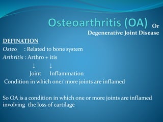
  • 1. Or Degenerative Joint Disease DEFINATION Osteo : Related to bone system Arthritis : Arthro + itis ↓ ↓ Joint Inflammation Condition in which one/ more joints are inflamed So OA is a condition in which one or more joints are inflamed involving the loss of cartilage
  • 2. It is progressive disorder of joints which is caused by gradual loss of cartilage resulting in development of bony spurs and crysts at the margins of the joints. In addition to damage and loss of articular cartilage, there is remodelling of subarticular bone, osteophyte formation, ligamentous laxity, weakening of periarticular muscles, and, in some cases, synovial inflammation These changes may occur as a result of an imbalance in the equilibrium between the breakdown and repair of joint tissue
  • 3. The most common joints involved • Distal Interphalangeal joints • Proximal Interphalangeal joints • Carpometacarpal joints of thumb • Weight Bearing joints (Hip, knee) • Metatarsophalangeal joints of the foot • Cervicle and lumbar vartebrae
  • 4. The articular cartilage is slippery tissue that covers the end of the bones in the joints Healthy cartilage allows bones to glid over each other & helps to absorb shock of movement
  • 5. In OA the top layer of cartilage breaks down and wear away ↓ Rubbing of bones under the cartilage ↓ Due to which there is Pain Swelling Loss of motion of joints ↓ Over the time joint may lose its normal shape and also there is growth of bony spurs on the edge of the joint ↓ Bits of bones/cartilage can break off and float inside the joint space ↓ Which cause more pain and damage
  • 6.
  • 7. So in OA there is progressive distruction of articular cartilage and also there is involvement of • Diarthrodial joint • Synovium • Capsule • Subchondral bone • Surrounding ligaments • Muscles Changes in structure and function of this tissues leads to clinical Osteoarthritis Which is characterized by • Joint pain • Tenderness • Decrease range of motion • Weakness • Joint instability • Disability
  • 8. Classification ↙ ↘ Primary OA Secondary OA (idiopathic) (Due to some other disease) A. Localised Hands Hip Knee Spine B. Generalised Small joints Large joints Mixed C. Erosive osteoarthritis i) Congenital and developmental disorders, bone dysplasias ii) Post-surgery / injury – meniscectomy iii) Endocrine –acromegaly, iv) Metabolic –hemachromatosis, ochronosis, Marfan syndrome, Ehler-Danlos syndrome, Paget disease, gout, pseudogout, Wilson’s disease, Hurler disease, Gaucher disease v) Rheumatologic– rheumatoid arthritis vi) Neurological– Charcot joints.
  • 9. Epidemiology OA is one of the core reason for disability Nearly everyone who lives long is affected by OA at any point of life, most probably after the age of 55 to 60. Approximately 15% of population is affected by OA ↙ ↘ ↙ ↘ 50 % of those 85% of those over 65 age over 75 age OA is most widely assessed in studies using the Kellgren and Lawrence (K&L) score. The overall grades of severity are determined from 0 to 4 and are related to the presumed sequential appearance of osteophytes, joint space loss, sclerosis and cysts
  • 10. The World Health Organization (WHO) adopted these criteria as the standard for epidemiological studies on OA Prevalence of osteoarthritis varies with I. Age II. Gender III. Genetics IV. Ethic Group V. Specific Joint Involved VI. Method For Diagnosis
  • 11. AGE With increasing age the prevalence OA also increases Generally for person age 25 – 75,prevalence is estimated 12% and for those with age over 70, it is 60 – 70% affectet Hip OA ← ← AGE → → Hand OA ↙ ↘ ↓ ↙ ↘ Age : 30 – 40 Age : >70 Knee OA Age : 40 Age : 80 Pre : 1.6 % Pre : 14% ↙ ↘ Pre: 5% Pre : 65% Age : >25 Age : 55 Pre : 5% Pre : 12%
  • 12. Gender ↙ ↘ Age > 50 Age >60 Men are more affacted Women are more affacted(26%) Due to higher rate of sports and Due to repeated use of Injuries weight bearing joints Genetics ↙ ↘ Pre : 9% Pre : 4% White population Black & Asian population Prevalence of OA is 22% to 39% in India
  • 13.
  • 14. Etiology The etiology of OA is multifactorial Many patients have more than one risk factor for developing the OA. The most common risk factor for the development of OA includes • Obesity • Occupation • Participation in certain sports (Often) • History of joint trauma • Genetic • Age • Sex • Bone density • Joint location
  • 15. Obesity Increased body weight is strongly associated with hip, knee, and hand OA Obesity often precedes OA and contributes to its development, rather than occurring as a result of inactivity from joint pain In a three-decade Framingham Study, the highest quintile of body mass was associated with a higher relative risk of knee OA (relative risk of 1.5 to 1.9 for men and 2.1 to 3.2 for women). The risk of developing OA increases by about 10% with each additional kilogram of weight, and in obese persons without OA, weight loss of even 5 kg decreases the risk of future knee OA by one-half.
  • 16. Recent data suggest that OA is associated with the metabolic syndrome, suggesting a possible common pathogenic mechanism involving metabolic abnormalities and systemic Inflammation. It is also likely that vascular disease may both initiate and hasten disease progression in OA. This could be due to venous occlusion, stasis or microembolic disease leading to episodic reduction in blood flow through small vessels within the subchondral bone. Subchondral ischaemia may subsequently reduce nutrient delivery and gas exchange to articular cartilage in addition to direct deleterious effects on the bone itself.
  • 17. Occupation There is increased risk of OA for those who are in occupation requiring Prolong Standing Kneeling Squatting Lifting/Moving Heavy Objects ↓ →Miming Factory Work Car paintery Repetitive motion also contributes to hand OA with dominant hand usually affected Risk OA depends on type and intensity of physical activity
  • 18. Sports Damage to articular cartilage due to sports greatly increase the risk of OA Meniscal damage (common in athlete) also increase the risk of knee OA 1.Because of loss of proper load bearing and shock absorption 2.Increase focal load on cartilage and subchondral bones Trauma AGE AT INJURY DOSE MATTER As older individuals who damage ligaments tends to develope OA more rapidly than young people with similar injury Trauma early in life →Increased risk of OA
  • 19. Genetic Factors A number of recent studies discovered the presence of over 80 gene mutations involved in the pathogenesis of OA ,among which the most relevant one is a single nucleotide polymorphism. This one, called rs143383 and located in the 3' untranslated region (3'UTR) of the growth and differentiation factor 5 gene (GDF5), is responsible for the development, maintenance and repair of synovial joints Genes for Vitamin D receptors (VDR) and insulin-like growth factor 1(IGF-1) also seem to be involved in the patho-physiologic pathways of OA It also includes genes related to inflaamation,bone morphogenetic protiens, protease/ its inhibitors
  • 20.
  • 21. Normal Articular Cartilage Articular Cartilage Possesses ↓ Viscoelastic Properties ↓Which provides 1. Lubrication With Motion 2. Shock absorbency during rapid movement 3. Load Support In Synovial joints Articular cartilage is found between synovial cavity on one side and narrow layer of calcified tissue overlying subchondral bone on onther side
  • 22. Charactristics of Articular Cartilage 1.Cartilage is easily compressed lossing up to 40% of its original hight when load is applied ↓ Compression increase area of contact ↓ Disperse force more evenly to undelying bone,tendon, ligament, muscles 2.Cartilage is frictionless Togather with compresibility , this enables smooth movement in joint and distributes load across joint tissue to prevent damage and stabilize the joint
  • 23. Structure of Cartilage Cartilage is having ↓ 1.Strength 2.Low co-efficiant of friction 3.Copressiblity These all is derived from its unique structure So,the cartilage is composed of complex, hydrophilic , Extra- cellular matrix It contains →75% to 85% Water 2% to 5% Chondrocytes (the only cell in cartilage) Collagen Protiens Proteoglycans Hyaluronic Acid Molecules
  • 24. Two Major Structure of Cartilage  Type II Collagen Tightly woven, triple helical structure which provides TENSILE STRENGTH to cartilage  Aggrecans In which there is Proteoglycan linked with hyluronic acid, having negative charge. The strong electrostatic repulsion of proteoglycan gives cartilage the ability to withstand further compression
  • 25. Normal Cartilage turn over ↓ Helps repair and restore cartilage ↓ Respond to usual demand of loading and physical activity In healthy adult cartilage chondrocyte metabolism is slow with dynamic balance between anabolic process Metabolism is premoted by 1. Groth factors - Bone morphogenetic protien 2 - Insulin like groth factor-1 - tranforming growth factor 2. Catabolism 3. Proteolysis Stimulated by MMPs, TNF-ᾲ, Interlukein-1, Other cytokines
  • 26. Joint Protective Mechanisms 1.Muscle Bridging The Joints 2.Sensory receprtors in feedback loops to regulate muscle and tondon function 3. Supporting ligaments 4. Subchondral bones having shock absorbent properties Note Articular cartilge is avascular and aneural and chondrocytes are nourised by synovial fluid
  • 27.
  • 28.
  • 29. OA Cartilage OA begins with damage to articular cartilage, which is due to 1. Trauma or other injury 2. Excess joint loading by obesity / other reason 3. Instability or injury of the joint that causes abnormal loading Devlopement of OA is due to 1. Local mechanical influence 2. Genetic factor 3. Inflammation 4. Chondrocyte function Which leads to loss of articular cartilage
  • 30. When there is damage to articular cartilage ↓ Increase activity of chondrocytes to remove and repair the damage Depending on degree of damage the balance between breakdown and resynthesis of cartilage can be lost ↓ Which leads to increase breakdown of cartilage ↓ Ultimately, loss of cartilage Destruction of aggrecans by proteolytic enzyme is consider to play a key role
  • 31. There is also involvement of collagen receptors named DDR-2 , located on chodrocyte cell surface In healthy cartilage ,DDR-2 is inactive ,which is masked by aggrecan from contact with collegen Damage to cartilage ↓ Triggers aggrecan destruction ↓ Exposure of DDR-2 to collagen ↓ Active DDR-2 increase activity of MMP-13 ↓ Which destroy collagen Collagen breakdown products further stimulate DDR-2,in which more collagen is destroyed
  • 32. Huge research work has been done on genes involved in OA which shows that In OA , expression of hundreds of genes of cartilage tissue are affected which alters chondrocyte phenotype In addition to articular cartilage there is also role of subchondral bone in OA In OA , subchondral bone release vasoactive peptides and MMPs Neovascularization and subsequent increase in permeability of the adjacent cartilage occurs and contributes to further cartilage loss
  • 33. Substantial loss of cartilage cause joint space narrowing and leads to painful and deformed joints The remaining catilage softens and devlopes fibrillation(Verticle cleft ) and there is splittting and further loss of cartilage and exposure of underlying bone As cartilage is destroyed and the adgecent subchondral bone undergoes pathologic changes, cartilage is eroded completely, leaving denuded subchondral bone which becomes dence , smooth and glistening A more brittle, stiffer bone results, with decreased weight-bearing ability and development of sclerosis and microfractures The joint capsule and synovium also show pathologic changes in OA.
  • 34. Inflammation, noted clinically as synovitis, may result from release of inflammatory mediators from chondrocytes, such as prostaglandins Inflammation is localized to the affected joint, in contrast to that seen in rheumatoid or other inflammatory arthritides. The pain in OA is not due to distruction of cartilage but arise from the activation of nociceptive nerve ending within the joint by mechanical and chemical irritants
  • 35. So,the slow progressive changes in OA consist of an increase in water content, loss of PG, and reduction of PG aggregates of cartilage. The cartilage is subsequently unable to repair itself. Alterations in metabolism of subchondral bone adjacent to articular cartilage appear necessary for continued cartilage destruction. Eventually, progressive loss of articular cartilage and increasing subchondral sclerosis lead to an abnormal and painful joint.
  • 36.
  • 37. Age Usually elderly Gender Age <45 more common in men Age >45 more common in women Symptoms →Pain Deep, aching character Pain on motion Pain with motion early in disease Pain with rest late in disease → Stiffness in affected joints Resolves with motion, recurs with rest Usually <30 minutes’ duration Often related to weather → Limited joint motion May result in limitations activities of daily living Signs, history, and physical examination Monarticular or oligoarticular; asymmetrical involvement Hands Distal interphalangeal joints Heberden’s nodes Proximal interphalangeal joints Bouchard’s nodes First carpometacarpal joint Osteophytes give characteristic square appearance of the hand Knees Patellofemoral compartment involvement Pain related to climbing stairs Transient joint effusions Typically noninflammatory synovial fluid Clinical Represntation
  • 38. Hips Pain during weight-bearing activities Stiffness, especially after inactivity Limited joint motion Spine L3 and L4 involvement is most common in the lumbar spine Signs and symptoms of nerve root compression Radicular pain Paresthesias Loss of reflexes Muscle weakness associated with the affected nerve root Feet Typically involves the first metatarsophalangeal joint Other sites, less commonly involved Shoulder, elbow, acromioclavicular, sternoclavicular, and temporomandibular joints Observation on joint examination Bony proliferation or occasional synovitis Local tenderness Muscle atrophy Limited motion with passive/active movement Deformity Radiologic evaluation Early mild OA Radiographic changes often absent Progression of OA Joint space narrowing Subchondral bone sclerosis Marginal osteophytes Late OA Abnormal alignment of joints Effusions Characteristics of synovial fluid High viscosity Mild leukocytosis Laboratory values ESR
  • 39. Treatment Goals (1) to educate the patient, caregivers, and relatives; (2) to relieve pain and stiffness; (3) to maintain or improve joint mobility; (4) to limit functional impairment; and (5) to maintain or improve quality of life Treatment of OA Non-pharmacological Treatment 1.Diet 2.Physical And Occupational Therapy 3.Surgery Pharmacological Treatment 1.Analgesics Oral Topical 2.NSAIDs 3. Glucosamine &Chondroitin 4.Hyluronate Injection 5.Narcotic Analgesics 6.Disease Modifying Drugs
  • 40. Diet Excess weight increases the biomechanical load on weight- bearing joints, and is the single best predictor of need for eventual joint replacement. Weight loss is associated with decreased symptoms and Disability Even 3-5 kg of weight loss can decrease the biomechanical force on a weight-bearing joint. Although dietary intervention for overweight OA patients is reasonable, weight loss usually requires a motivated patient and a structured weight-loss program. Diet with low fat (calories) and high fibre as well as balanced diet should be taken to decrease weight
  • 41. Physical And Occupational Therapy Physical therapy—with heat or cold treatments and an exercise program Which helps to maintain and restore joint range of motion and reduce pain and muscle spasms. Warm baths or warm water soaks may decrease pain and stiffness. Exercise programs and quadriceps strengthening can improve physical functioning and can decrease disability, pain, and analgesic use by OA patients. The decision about whether to encourage walking should be made on an individual basis. With weak or deconditioned muscles,the load is transmitted excessively to the joints, so weight-bearing activities can exacerbate symptoms.
  • 42. However, avoidance of activity by those with hip or knee OA leads to further deconditioning or weight gain. A program of patient education, muscle stretching and strengthening,and supervised walking can improve physical function and decrease pain in patients with knee OA.
  • 43. Surgery Surgery can be recommended for OA patients with functional disability and/or severe pain that is unresponsive to conservative Patients with mild knee OA, an osteotomy may correct the misalignment of knee Knee arthroscopy or lavage have been recommended for short- term relief of pain, a recent study showed these two procedures to be equivalent to surgery Experimental but potentially restorative approaches involve Soft tissue grafts, Penetration of subchondral bone, Cell transplantation, and Use of growth factors
  • 44. Pharmacological Therapy Things to be taken in concern 1.Adverse effect of the drug given for OA 2.As OA is usually seen in old age people with some other conditions they Pharmacological treatment should be done with so much causion 3.Always combine non pharmacological therapy with pharmacological one for more improvement
  • 45. Analgesics Acetaminophen is used as first-line drug therapy for pain management in OA, due to its relative safety, efficacy, and lower cost compared to NSAIDs Pain relief with acetaminophen can be similar to that obtained with NSAIDs, although some patients will respond better to NSAIDs. Some studies have shown comparable efficacy for acetaminophen and NSAIDS, others have reported that patients experienced better pain control with NSAIDs than with acetaminophen, and that OA patients preferred NSAIDs to acetaminophen
  • 46. Mechanism Of Action Acetaminophen is thought to work within the central nervous system to inhibit the synthesis of prostaglandins, agents that enhance pain sensations. Acetaminophen prevents prostaglandin synthesis by blocking the action of central cyclooxygenase. Kinetics Acetaminophen is well absorbed after oral administration (bioavailability is 60% to 98%), achieves peak concentrations within 1 to 2 hours, is inactivated in the liver by conjugation with sulfate or glucuronide, and its metabolites are renally excreted Efficacy Comparable relief of mild to moderate OA pain has been demonstrated for acetaminophen at 2.6 to 4 g/day
  • 47. ADR Hepatotoxicity, and Possibly renal toxicity Drug – Drug Intrection Isoniazid can increase the risk of hepatotoxicity. Chronic ingestion of maximal doses of acetaminophen may intensify the anticoagulant effect in patients taking warfarin, Food decreases the maximum serum concentration of acetaminophen by approximately one-half.
  • 48. Non Steroidal Anti - Iflammatory Drugs MOA Blockade of prostaglandin synthesis through inhibition of cyclooxygenase (bothCOX-1 andCOX-2 enzymes) is the principal mechanism by which NSAIDs relieve pain and inflammation
  • 49. Kinetics High oral availability, high protein binding, and absorption as active drugs (except for sulindac and nabumetone, which require hepatic conversion for activity). The most important difference in NSAIDs is a serum half-life ranging from 1 hour for tolmetin to 50 hours for piroxicam, impacting the frequency of dosing and potentially, compliance with therapy. Elimination of NSAIDs largely depends on hepatic inactivation, with a small fraction of active drug being renally excreted. NSAIDs penetrate joint fluid, reaching about 60% of blood levels
  • 50. ADR Minor complaints—nausea, dyspepsia, anorexia, abdominal pain, flatulence, and diarrhea To minimize these symptoms, NSAIDs should be taken with food or milk, except for enteric-coated products, which should not be taken with milk or antacids All NSAIDs have the potential to cause GI bleeding. Possible Mechanism For this Unionized NSAIDs enter gastric mucosal cells, release hydrogen ions, and are concentrated (“ion trapped”) within cells, with cell death or damage. Gastric mucosal injury can also result from NSAID inhibition of gastroprotective prostaglandins.
  • 51. COX-2 inhibitors pose a decreased risk of GI toxicity compared to nonspecific NSAIDs, an especially important consideration when treating those at risk for clinically significant GI adverse effects. NSAIDs may cause kidney diseases, Acute renal insufficiency, Hyperkalemia, and Renal papillary necrosis Clinical features of these NSAID-induced renal syndromes Increased serum creatinine Increased blood urea nitrogen, Hyperkalemia, Elevated blood pressure, Peripheral edema, and Weight gain.
  • 52. Mechanisms of NSAID injury include direct toxicity, and inhibition of local prostaglandins that promote vasodilation of renal blood vessels and preserve renal blood flow. COX-2 inhibitors also have potential for renal toxicity; COX-2 activity has been demonstrated in a variety of sites in the kidney and is upregulated in salt-depleted states Importantly,COX-2 inhibitors decrease urinary prostaglandins and cause sodium and potassium retention, as do nonspecific NSAIDs
  • 53.
  • 54. Drug Intrection Lithium, Warfarin, Oral hypoglycemics, High-dose methotrexate, Antihypertensives, Angiotensin-converting enzyme inhibitors, β-blockers, and diuretics.
  • 55. Topical Therapies Topical products can be used alone or in combination with oral analgesics or NSAIDs. Capsaicin, isolated from hot peppers, releases and ultimately depletes substance P from afferent nociceptive nerve fibers. Substance P has been implicated in the transmission of pain in arthritis, and capsaicin cream has been shown in four controlled studies to provide pain relief in OA when applied over affected joints To be effective, capsaicin must be used regularly, and it may take up to 2 weeks to work. It is well tolerated, except that some patients experience a temporary burning sensation at the site of application. Although use is recommended four times a day, a twice-daily application may enhancelong-term adherence and edequate effet
  • 56. GLUCOSAMINE AND CHONDROITIN These substances stimulate proteoglycan synthesis from articular cartilage in vitro Chondroitin and glucosamine are superior to placebo in alleviating pain from knee or hip OA. In studies, chondroitin sulfate showed similar analgesic benefits to diclofenac A recent meta-analysis of glucosamine and chondroitin indicated that both agents had efficacy in reducing pain and improving mobility, and that glucosamine reduced joint space narrowing further more were associated with slower loss of cartilage than placebo, in knees affected by OA
  • 57. CORTICOSTEROIDS Intra-articular glucocorticoid injections can provide excellent pain relief, particularly when a joint effusion is present Aspiration of the effusion and injection of glucocorticoid are carried out aseptically,and examination of the aspirate to exclude crystalline arthritis or infection is recommended. The therapy is generally limited to three or four injections per year because of the potential systemic effects of steroids, and because the need for more frequent injections indicates little response to the therapy. Systemic corticosteroid therapy is not recommended in OA, given the lack of proven benefit and the well-known adverse effects with long-term use.
  • 58. HYALURONATE INJECTIONS Agents containing hyaluronic acid (HA) (sodium hyaluronate) are available for intra-articular injection for treatment of knee OA. High-molecular-weight HA is an important constituent of normal cartilage, with viscoelastic properties providing lubrication with motion and shock absorbency during rapid movements Endogenous HA also may have anti-inflammatory effects because the concentration and molecular size of synovial HA decrease in OA, administration of exogenous HA products has been studied, with the theory that this could reconstitute synovial fluid and reduce symptoms. Injections temporarily and modestly increase viscosity. HA products are injected once weekly for either 3 or 5 weeks.
  • 59. Injections are well tolerated, although acute joint swelling and local skin reactions, including rash, ecchymoses, and pruritus have been reported. HA injections may be beneficial for patients unresponsive to other therapies. These agents are expensive because the treatment includes both drug costs and administration costs. As a result, HA injections are often used after less expensive therapies have demonstrated lack of efficacy
  • 60. DISEASE-MODIFYING DRUGS Disease-modifying drugs are targeted not at pain relief but at preventing, retarding, or reversing damage to articular cartilage Most products have been tested in animal models, and limited human data are available. Thus far, OA is a disease whose symptoms can be alleviated, but whose progress is impossible to stop because of this, clinicians were very interested to learn that both chondroitin and glucosamine show disease-modifying potential in patients with OA Another approach is that of pharmacologic agents that could mimic TIMPs and thus potentially decrease cartilage destruction.
  • 61. Heparinoid products that contain glycosaminoglycans, sodium pentosan polysulfate, and calcium pentosan polysulfate show promise in preliminary work limited largely to animal models and in vitro studies There is also recent interest in the potential of cyclooxygenase- inhibiting nitric oxide–donor compounds to relieve OA while sparing GI adverse effects.
  • 62. NARCOTIC ANALGESICS Low-dose narcotic analgesics may be very useful in patients who experience no relief with acetaminophen, NSAIDs, intra-articular injections, or topical therapy. These agents are particularly useful in patients who cannot take NSAIDs due to renal failure, or for patients in whom all other treatment options have failed and who are at high surgical risk, precluding joint arthroplasty. Low-dose narcotics are the initial intervention, usually given in combination with acetaminophen. Sustained-release compounds usually offer better pain control throughout the day, and are used when simple narcotics are ineffective.
  • 63. If pain is intolerable and limits activities of daily living, and the patient has sufficiently good cardiopulmonary health to undergo a major surgery, joint replacement may be preferable to continued reliance on narcotics.
  • 64. Guided By Samir Rabadiya, Associate Profesor ,Department Of Pharmaceutical Sciences Acknowledgement Sachin Parmar,Associate Profesor ,Department Of Pharmaceutical Sciences Payal Bhalodia,Associate Profesor ,Department Of Pharmaceutical Sciences Rahul Solanki,BE
  • 65. References Pharmacotherapy ,A Pathophysiological Approch Joseph T. DiPiro, PharmD, FCCP Professor and Executive Dean, South Carolina College of Pharmacy, University of South Carolina, Columbia, and Medical University of South Carolina, Charleston Robert L. Talbert, PharmD, FCCP, BCPS Professor, College of Pharmacy, University of Texas at Austin; Professor, Departments of Medicine and Pharmacology, University of Texas Health Science Center at San Antonio, Texas Gary C. Yee, PharmD, FCCP Professor and Chair, Department of Pharmacy Practice, College of Pharmacy, University of Nebraska Medical Center, Omaha, Nebraska Gary R. Matzke, PharmD, FCP, FCCP Professor, Department of Pharmacy and Therapeutics, School of Pharmacy, Renal-Electrolyte Division, School of Medicine, University of Pittsburgh, Pittsburgh, Pennsylvania Barbara G. Wells, PharmD, FASHP, FCCP, BCPP Dean and Professor, School of Pharmacy, The University of Mississippi, University, Mississippi L. Michael Posey, BS Pharm President, PENS Pharmacy Editorial and News Services, Athens, Georgia
  • 66. Arthritis Research UK Obesity Action Coalition Anna Litwic, MD [Specialist Registrar in Rheumatology] Giuseppe Musumeci , Department of Biomedical and Biotechnological Sciences, Human Anatomy and Histology Section, School of Medicine, University of Catania, Via S. Sofia 87, 95123 Catania, Italy; F. Berenbaum yz Department of Rheumatology, AP-HP Saint-Antoine Hospital, 75012 Paris, France V Tandon

Editor's Notes

  1. , aspirin 650 mg four times daily, ibuprofen at 1200 or 2400 mg daily, and naproxen 750 mg/day
  2. Efficacy Choosing of cox inhibitior Valdecoxib new drug Cvs toxisity