
Recommended
More Related Content
What's hot
What's hot (20)
Similar to Peptic Ulcer Disease.pptx
Similar to Peptic Ulcer Disease.pptx (20)
More from TwinkleJoshi4
More from TwinkleJoshi4 (15)
Recently uploaded
Recently uploaded (20)
Peptic Ulcer Disease.pptx
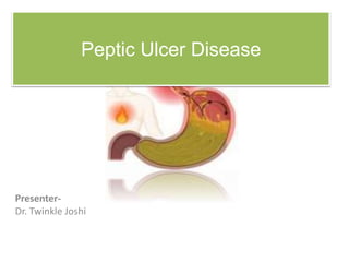
- 1. Peptic Ulcer Disease Presenter- Dr. Twinkle Joshi
- 2. Introduction • Peptic ulcer disease (PUD) refers to ulceration of the mucosa anywhere in the GI tract exposed to acid and pepsin. • Ulcers : breaks in the mucosal surface >5 mm, with depth to the sub-mucosa. • They can range in size from a few millimeters to a few centimeters • The 2 most common forms/locations of PUD are – Duodenal ulcer – Gastric ulcer Site • Lower esophagus • Stomach • Duodenum
- 3. Types Acute • Superficial erosion • Minimal erosion Chronic • Muscular wall erosion with formation of fibrous tissue • Present continuously for many months or intermittently
- 4. Aggressive factors Hydrochloric acid and pepsin destroy gastric and duodenal mucosa. Defensive factors Mucus and bicarbonate ion secretions protect mucosa Na bicarbonate is considered as a buffer so it inhibits auto digestion of the stomach wall Prostaglandins (PGE2 & PGI2) protect mucosa by inhibiting acid secretion increasing mucus and bicarbonate production enhancing mucosal blood flow. • Imbalance between aggressive factors (acid &pepsin) and defensive factors (e.g. prostaglandins, mucus & bicarbonate layer). • However, nowadays, it seems that H. pylori is a major cause Pathophysiology One of the main action of Prostaglandins is increase the turnover and regeneration of damaged cells
- 7. Etiology • Common causes of PUD – Helicobacter pylori (H.pylori) infection – Nonsteroidal Anti-inflammatory Drugs (NSAIDs) – Critical illness (stress-related mucosal damage) • Uncommon causes of PUD – Idiopathic (non-H.pylori, non- NSAID) – Hypersecretion of gastric acid (e.g. Zollinger Ellison syndrome) – Viral infections – Radiation therapy – Chemotherapy
- 8. • Helicobacter pylori (HP) is a spiral shaped, gram negative, flagellated bacteria first associated with PUD in the early 1980’s • Found in most people with duodenal and gastric ulcers – About 95% of those with duodenal ulcers – About 80% of those with gastric ulcers • HP is primarily spread through the fecal to oral route • People are most often infected during Helicobacter pylori
- 9. • Mechanisms by which HP causes mucosal injury are not entirely clear but occurs through a combination of the following mechanisms: – HP catalyzes urea ammonia is produced ammonia erodes the mucous barrier and causes epithelial damage – HP produces cytotoxins – HP produces mucolytic enzymes
- 10. NSAIDs • In long-term NSAID users, there is a 10% - 20% prevalence of gastric ulcers and a 2% - 5% prevalence of duodenal ulcers • Mechanisms for NSAID-induced ulceration – NSAIDs are weak acids and are non-ionized at gastric pH • Diffuse freely across the mucous barrier into gastric epithelial cells H+ ions are liberated and cause cellular damage • Aspirin is the most ulcerno-genic of all NSAIDs.
- 11. • NSAIDs inhibit cyclooxygenase activity and therefore decrease prostaglandin production which results in a: • Reduction in gastric and mucosal blood flow • Decrease in mucous and bicarbonate secretion • Decrease in cellular repair and replication – Greater than 60 years old – With a prior history of PUD
- 12. Zollinger Ellison Syndrome • ZES is characterized by gastric acid hypersecretion and recurrent peptic ulcers that result from a gastrin- producing tumor – More than 50% of gastrinomas are malignant • ZES is suspected for patients with multiple ulcers and recurrent or refractory PUD often accompanied by esophagitis or ulcer complications • Only accounts for 0.1% to 1% of those with duodenal ulcer
- 13. Other factors • Cigarette smoking – Impairs ulcer healing and increases the risk of recurrence • Psychological stress – Stress may induce behavioral risks such as smoking and the use of NSAIDs or may alter the inflammatory response or resistance to HP infection • Dietary factors – Certain foods (e.g. coffee, tea, carbonated beverages, beer, milk, spices) may cause dyspepsia but do not increase the risk of developing PUD
- 14. Signs and symptoms • Symptoms depend on ulcer location, ulcer etiology, and patient age • Many patients, particularly the elderly, have few or even no symptoms • NSAID-induced ulcers are often silent – Complications such as bleeding and perforation are often the initial presentation
- 15. • Pain localized to the epigastrium is the most common symptom • The pain is described as burning, gnawing, cramping, or hunger • A typical nocturnal pain that wakes the patient from sleep (especially between 12 and 3am) • The severity of ulcer pain varies from patient to patient and may be seasonal, occurring more often in the spring or fall
- 16. • Episodes of pain usually occur in clusters, lasting up to a few weeks followed by a pain-free period or remission lasting weeks to years • Changes in the character of pain may suggest the presence of complications • Pyrosis (heartburn), belching, and bloating may accompany the pain
- 18. Gastric Ulcers • Less common than duodenal ulcers – Especially in chronic NSAID use • Most commonly located in the lesser curvature of the antrum of the stomach • More common in people greater than 60 years old Characterized by • A normal to low secretion of gastric acid • Back diffusion of acid is greater (chronic ) • Critical pathologic process is amount of acid able to penetrate mucosal barrier • H pylori is present in 50% to 70% • Drugs --- Aspirin, corticosteroids, N SAIDs, reserpine, Chronic alcohol abuse, chronic gastritis
- 19. Duodenal Ulcers • Most common form of PUD – It is 3 times more common than gastric ulcers • Usually located in the duodenal bulb of the small intestine • Most commonly occurs in people between the ages of 30 and 50 • Associated with ↑HCl acid secretion • H.pylori associated in 9 0- 9 5 % of cases • Diseases with ↑risk of duodenal ulcers COPD, cirrhosis of liver, chronic pancreatitis, hyperparathyroidism, chronic renal failure
- 20. Complications • 3 major complications Hemorrhage - 15% of patients with active PUD Perforation - 7% of patients with active PUD Gastric outlet obstruction • Initially treated conservatively • May require surgery at any time during course of therapy
- 21. Diagnostic Studies • Endoscopy procedure – Determines degree of ulcer healing after treatment – Tissue specimens can be obtained to identify H. pylori and to rule out gastric cancer • Tests for H.pylori – Noninvasive tests • Serum or whole blood antibody tests – Immunoglobin G (I g G) • Urea breath test • C 14 breath test • Fecal antigen test – Invasive tests • Biopsy of stomach • Rapid urease test
- 22. Rapid Urease Testing – Rapid urease tests detect the presence of ammonia in the biopsy sample – The ammonia is generated by H.pylori urease activity – Test of choice at endoscopy – Greater than 90% sensitive and specific – Easily performed with rapid results – Tests for active HP infection
- 23. Urea Breath Test • Detects the exhalation of radioactive CO2 following ingestion of 13C or 14C radiolabeled urea • H. pylori hydrolysis of the radiolabeled urea results in radiolabeled CO2 production • 97% sensitivity and 95% specificity
- 24. Fecal Antigen Test • Polyclonal antibody test that detects the presence of H.pylori antigen in the stool • Sensitivity and specificity similar to urea breath test • Patients may have a reluctance to collect stool samples
- 25. • The urea breath and fecal antigen tests may be falsely negative in patients who have recently taken – Antibiotics (up to 4 weeks) – Bismuth compounds (up to 4 weeks) – Antisecretory agents (up to 2 weeks)
- 26. • Barium contrast studies – Widely used • X- ray studies – Ineffective in differentiating a peptic ulcer from a malignant tumor
- 27. Treatment Medical regimen consists of – Adequate rest – Dietary modification – Drug therapy – Elimination of smoking – Long-term follow-up care Aim of treatment program – ↓ degree of gastric acidity – Enhance mucosal defense mechanisms – Minimize harmful effects on mucosa
- 28. Drug Therapy • Antacids • H2 receptor blockers • PPIs • Antibiotics • Anticholinergics • Cytoprotective therapy
- 29. Histamine receptor blocks (H2 R blockers)- famotidine,cimetidine Used to manage peptic ulcer disease Block action of histamine on H2 receptors ↓ HCl acid secretion ↓ conversion of pepsinogen to pepsin ↑ ulcer healing Proton pump inhibitors – pantoprazole, rabeprazole – Block ATPase enzyme that is important for secretion of HCl acid Antibiotic therapy – Eradicate H. pylori infection – No single agents have been effective in eliminating H. pylori
- 30. • Antacids – calcium carbinate, MgOH – Used as adjunct therapy for peptic ulcer disease – ↑ gastric pH by neutralizing acid • Anticholinergic drugs- Dicyclomine – Occasionally ordered for treatment – ↓ cholinergic stimulation of HCl acid • Bismuth preparations • Agents – Bismuth subsalicylate – Bismuth exhibits antimicrobial activity against bacterial and viral gastrointestinal pathogens
- 31. • Standard triple therapy regimen contains – Amoxicillin 1000mg twice day + Clarithromycin 500mg twice a day + a PPI dosed once to twice a day – Given for 10 to 14 days • 14 day regimens are generally preferred as 14 day regimens significantly increases the eradication rate • Bismuth-based quadruple-therapy contains – Tetracycline 500mg 4 times day +Metronidazole 250-500mg 4 times a day + Bismuth subsalicylate 525mg 4 times a day + a PPI once or twice a day OR H2-receptor antagonist twice a day
- 32. Refractory Ulcers • When symptoms, ulcers, or both persist beyond 8 to 12 weeks despite conventional treatment as previously described or when several courses of H. pylori eradication therapy fail • Patient should undergo an upper endoscopy to assess the situation • Treatment depends on cause and may include additional H. pylori eradication attempts, higher PPI dosages, or surgery
- 33. Nutritional therapy • Dietary modifications may be necessary so that foods and beverages irritating to patient can be avoided or eliminated • Nonirritating or bland diet consisting of 6 small meals a day during symptomatic phase • Protein considered best neutralizing food – Stimulates gastric secretions • Carbohydrates and fats are least stimulating to HCl acid secretion – Do not neutralize well
- 34. Surgical Treatment • < 20% of patients with ulcers need surgical intervention • Indications for surgical interventions Intractability History of hemorrhage, ↑ risk of bleeding Prepyloric or pyloric ulcers Multiple ulcer sites Drug-induced ulcers Possible existence of a malignant ulcer Obstruction
- 35. Surgical procedures Gastroduodenostomy Gastrojejunostomy Vagotomy Pyloroplasty
- 36. A. Billroth I Procedure B. Billroth II Procedure
- 37. Thank you