1. This work was supported by the University of Massachusetts Lowell Co-op Scholars Program
Control of Vascular Endothelial Growth Factor
Binding to Its Receptor
Surenna Pecchia, Divyabharathy Tsiros, Matthew A. Nugent, Ph.D.
Department of Biological Sciences, University of Massachusetts Lowell
Objective
Background
Approach
Angiogenesis is the process of growing new blood vessels from
pre-existing blood vessels2
. This process involves the proliferation
and maintenance of endothelial cells, and serves as the main
method of transporting oxygen and nutrients to cells throughout
the body1
. The angiogenic signal plays a crucial role in the
maintenance of homeostasis—a poor signal leads to deficiencies
in regeneration and healing, while an excessive signal can serve
to fuel tumor growth2
(Figure 1). Tumors require a constant blood
supply in order to grow to a substantial size; therefore, tumors
stimulate angiogenesis by either transmitting chemical signals, or
by stimulating normal cells nearby to secrete angiogenesis
signaling molecules1
. One such signaling molecule is vascular
endothelial growth factor (VEGF). VEGF is a key protein regulator
of angiogenesis, and is present in both normal and cancerous
cells2
. Two VEGF receptors (VEGFR1 and VEGFR2) are located
on the endothelial cell surface, and initiate an angiogenic signal
upon the binding of VEGF to one of its receptors2
(Figure 2). The
VEGF+VEGFR2 complex is made more secure by the additional
binding of heparan sulfate proteoglycans (HSPGs), which are also
located on endothelial cell surfaces3
. These HSPGs consist of a
core protein, with heparan sulfate molecules branching off3
(Figure
3). Heparan sulfates are long, sugar-chain molecules with a
variable structure, which allows for extensive protein binding sites
on its surface2
. HSPGs can modulate the transport and distribution
of proteins bound to the heparan sulfate chains to various
intracellular locations4
. On the endothelial cell surface, HSPGs
and VEGFR2 in close proximity can result in both complexes
binding VEGF molecules to create a high affinity signaling
complex4
. Previous studies suggest that VEGF bound to both
HSPGs and VEGFR2 induces a stronger angiogenic signal than
that produced by VEGF-VEGFR2 complexes alone.
My research project was focused on understanding the
interactions between several different molecules involved in
angiogenesis. I explored how different combinations of vascular
endothelial growth factor (VEGF), VEGF receptor 2, and
heparin/heparan sulfates bound to each other, as well as which
combinations yielded the strongest binding affinites. Another focus
of mine was to explore the mechanism by which VEGF, VEGFR2,
and heparin bound to each other.
References
A 96-well Heparin Binding Plate was used in each
binding assay. The bottom of each well is pre-coated with
positive charges in order to ensure the binding of the
negatively charged heparin/heparan sulfates.
Heparin/Heparan sulfates are negatively-
charge sugar chain molecules. They contain
multiple binding sites and are good
facilitators of proteins and other molecules
into cells. Because they are negatively
charged, they’re able to bind to the bottom
of each well.
VEGF Receptor 2 was added to
each well containing heparin.
VEGF molecules were also
added to each well
containing heparin and
VEGF Receptor 2.
Binding occurred between VEGF, R2, and heparin.
Any molecules that
were not bound to the
plate were washed
away with buffers.
A Donkey anti-human HRP-
linked antibody was added to
each well. It bound to the Fc
region of the VEGFR2
chimera.
The antibody contains a linked
HRP region, which interacted
with the TMB substrate solution
to create a yellow pigment.
TMB
substrate
Colorchange
(yellow)
Conclusions
1. http://www.cancer.gov/about-cancer/treatment/types/immunotherapy/angiogenesis-inhibitors-fact-
sheet
2. Teran, M., & Nugent, M. A. (2015). Synergistic binding of vascular endothelial growth factor-A and
its receptors to heparin selectively modulates complex affinity. Journal of Biological Chemistry,
290(26), 16451-16462.
3. Lin, X. (2004). Functions of heparan sulfate proteoglycans in cell signaling during development.
Development, 131(24), 6009-6021.
4. Bernfield, M., Götte, M., Park, P. W., Reizes, O., Fitzgerald, M. L., Lincecum, J., & Zako, M. (1999).
Functions of cell surface heparan sulfate proteoglycans. Annual review of biochemistry, 68(1), 729-
777.
5. http://polysac3db.cermav.cnrs.fr/discover_GAGs.html
6. Shibuya, M. (2003). Vascular endothelial growth factor receptor 2: its unique signaling and specific‐
ligand, VEGF E.‐ Cancer science, 94(9), 751-756.
PBST-B
(Blank)
One Hour
PBST-B
(Blank)
PBST-B
(Blank)
PBST-B
(Blank)
One Hour
Treatment – First Addition
Treatment – Second Addition
VEGF Plays a Critical Role in the Binding of R2 to HeparinVEGF R2
Donkey anti-human
HRP-linked secondary
antibody
VEGF+R2
In order to investigate the mechanisms by which heparin, VEGF, and VEGFR2 bind to each other, the sequence by which these proteins were added to heparin-coated wells was varied.
All wells were coated with heparin and then incubated with VEGF or R2 in PBST-B, or with PBST-B alone for 1 hour and then each solution was removed from the wells, the wells were
washed, and the second addition of R2 or R2+VEGF were added and allowed to incubate for an additional hour. Of the wells that had been incubated with VEGF only, three were given
R2 only, and three were given PBST-B. Of the wells that had been incubated with R2 only, three were given VEGF only, and three were given PBST-B. Three of the wells that had been
previously incubated with PBST-B, were given VEGF only, three were given R2 only, three more were given VEGF+R2, and the rest were given PBST-B as a blank. The wells treated
with PBST-B first, and VEGF+R2 second, or with VEGF first, and R2 second showed similar high levels of binding. These results suggest that in order for there to be a strong binding
affinity, VEGF must bind to heparin first, and R2 can bind the VEGF afterwards.
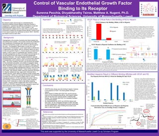
In order to investigate the differences in binding affinities between VEGF and
R2, heparin coated and uncoated wells within 96-well plates were exposed to
solutions containing: VEGF (10nM), VEGFR2 (1 nM), or VEGF (10 nM) and
R2 (1 nM) in triplicate. The amount of R2 bound was measured using an ELISA
detecting the Fc portion of the VEGFR2-Fc chimera protein, and the average ±
S.D. are shown for each condition. The greatest amount of R2 binding was
observed when VEGF and R2 were incubated with heparin coated plates. There
appeared to be a small amount of binding of R2 to heparin in the absence of
VEGF. There was virtually no signal in heparin coated and uncoated wells when
R2 was not included in the incubation (i.e., VEGF alone or binding buffer
containing bovine serum albumin without any additions).
Modified Heparins Result in Different Binding Affinities with VEGF and R2
2O-DS: The
sulfate on the 2-
carbon ring is
removed and
replaced with a
hydrogen
molecule.
6O-DS: The sulfate on
the 6-carbon ring is
removed and replaced
with a hydrogen
molecule.DOS: The sulfates on
both the 2-carbon ring
and the 6-carbon ring
are removed and
replaced with hydrogen
molecules.
NDS: The sulfate on the
nitrogen is removed and
replaced with a hydrogen
molecule.
NAc: The sulfate on the
nitrogen is removed and
replaced with an acetyl-
group.
• VEGFR2 alone shows very low binding to heparin; however,
in the presence of VEGF, R2 shows greater binding to
heparin.
• When heparin is treated with VEGF first, and R2 second,
there is a very strong binding affinity, while heparin treated
with R2 first and VEGF second results in relatively little
binding. This indicates that VEGF is a necessary facilitator
of R2 binding heparin.
• By understanding how VEGF and VEGFR2 interact with
each other, we can investigate therapies to either stimulate
or inhibit angiogenesis. Stimulating this process would likely
allow for tissue repair, while inhibiting the process could
slow, or stop tumor growth.
VEGF165
VEGFR-2
Figure 2. Vascular endothelial growth
factor (VEGF) is a protein dimer. VEGFR2,
is a dimer as well, and exists as a
transmembrane protein. This receptor is
characterized by a tyrosine kinase structure,
as well as several immunoglobin domains
located in the extracellular matrix6
.
Modified from Teran, (2015) Boston U.
In this assay, several different modified heparins were
used to coat a well plate and compared to heparin as a
control. Unlike un-modified heparin, which contains a
sulfate group at the N and 6-O position of the
glucosamine residues and on the 2-O position of the
uronic acid residues, the modified heparins have had
specific sulfate groups selectively removed (Figure 4).
Most of the conditions displayed a high binding affinity
when treated with R2+VEGF, except for the DOS
heparin, which is devoid in 6-O and 2-O sulfation. This
indicates that O-sulfation is critical for binding to occur,
even though both the 2-O and 6-O desulfated heparins
(2OS and 6OS) were able to support a significant amount
of binding. N-desulfated (NDS) and N-acetylated (NAc)
heparin showed an intermediate binding response,
indicating that sulfation on the N-group is somewhat
important for binding to occur. Concentration dependent
variability is shown with heparin and heparin derivatives
in its ability to form ternary complexes with VEGF and
VEGFR-2.
Modified from PolySac Database5
Figure 1. Angiogenesis differs from
vasculogenesis in that the former is the
process of growing new blood vessels from
pre-existing ones. This process can be
manipulated positively, for enhanced wound
healing, or negatively, in the case of cancers
and other diseases2
.
Modified from Teran, (2015) Boston U.
Heparin
Figure 4.
Figure 3. Heparan sulfate
proteoglycans consist of a core
trans-membrane protein, with sugar
chains branching off into the
extracellular matrix. Heparan
sulfates contain many protein
binding sites, which allows them to
bind VEGF and aid in stimulating
angiogenesis3
.
Modified from Teran, (2015) Boston U.