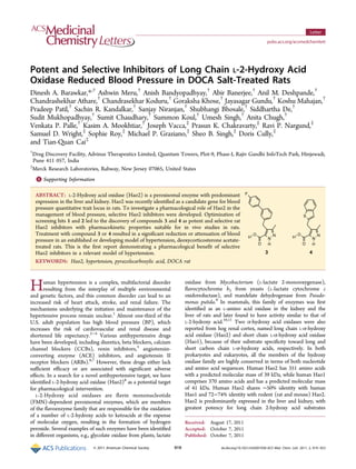
2. Figure 1. Structures of screening hits (1 and 2) and optimized leads (3 and 4). (A) Cartoon representation of docked compound 3 in the active site
of rat Hao2 (Protein Data Bank entry 1TB3). The FMN ring is colored light blue. (B) Close-up view of docked compound 3 (yellow) in the active
site of the enzyme. The FMN ring is colored light pink. The putative hydrogen bonds are shown as dashed lines.
Table 1. PK Properties of Lead Compounds 3 and 4a
a
NA means not available. iv (1 mg/kg), po (10 mg/kg); vehicle: iv [dimethylacetamide (2%), crempohor (2%), PEG400 (2%), and Milli Q water
(94%)], po [Tween 80 (0.5%) and 0.5% CMC (99.5%)].
ACS Medicinal Chemistry Letters Letter
dx.doi.org/10.1021/ml2001938|ACS Med. Chem. Lett. 2011, 2, 919−923920
3. (displays the highest activity toward 2-hydroxypalmitic acid).
Hao1 is expressed primarily in liver and pancreas and shows
greatest potency for the two-carbon 2-hydroxy acid substrate
(glycolic acid) but also displays activity on long chain 2-hydroxy
fatty acids. Both Hao2 and Hao1 are capable of oxidizing
2-hydroxy fatty acids, but the endogenous physiological
substrates remain to be identified.
Hao2 has been identified as a candidate gene for the systolic
BP quantitative trait locus (QTL) in rats.12−14
Genome-wide
linkage analysis in humans locates a BP QTL in a defined
region containing Hao2 (located in Ch. 1 at 119.6 cM), thus
supporting a potential link between Hao2 and hypertension in
humans, as well.15
To establish a pharmacological validation of Hao2 in blood
pressure regulation, potent and selective rat Hao2 inhibitors
were developed. Pyrazole carboxylic acids, 1 and 2 (Figure 1),
were first identified as inhibitors of Hao2 by a focused enzyme
screen of our compound collection. Subsequent optimization of
these hits resulted in the discovery of compounds 3 and 4 as
potent inhibitors of rat Hao2, each exhibiting an IC50 value of
0.3 μM.16
These two compounds were further characterized in
a spectrum of assays, including intervention studies in a well-
established deoxycorticosterone acetate (DOCA) salt hyper-
tension model.
Molecular modeling studies using the rat Hao2 crystal
structure (Protein Data Bank entry 1TB3)17
were used to
understand the potential binding mode of the inhibitor (3) in the
enzyme active site. The modeling suggests that the carboxylic
acid moiety binds in the active site by forming salt bridge
interactions with basic residues R250 and R164 (Figure 1). The
interaction of the carboxylic acid moiety with these residues
has also been observed in homologous enzymes of Hao2.18,19
The NH group of the pyrazole moiety forms putative hydrogen
bonds with the catalytic H247 and nearby Y129 residues. The
biphenyl moiety resides in the hydrophobic pocket and is
surrounded by residues such as F79, A185, L172, L174, E188,
F23, and L161.
The model suggests that a one-carbon linker between the
pyrazole and aromatic moiety (3 and 4) is optimal for filling the
available space and placing the aromatic moiety in an
appropriate orientation. A longer and flexible carbon linker
may increase the conformational entropy of these structures
(1 and 2), thereby decreasing their activity. Deletion of the
carbon linker makes the molecules very rigid and leads to poor
occupancy of the phenyl moiety in the active site. Hence,
compound 5 without any linker shows substantially lower
activity (Figure 1).
To assess selectivity and to demonstrate compounds 3 and 4
are selective inhibitors of rat Hao2, we profiled these two
Figure 2. (A and B) Lowering of SBP and (C and D) change in SBP from baseline by Hao2 inhibitors 3 and 4, respectively, in a DOCA model (n =
6−8). Compared to the control group on each day, *P < 0.05, **P < 0.01, and ***P < 0.001. Two-way analysis of variance (ANOVA) followed by a
Bonferroni post test.
ACS Medicinal Chemistry Letters Letter
dx.doi.org/10.1021/ml2001938|ACS Med. Chem. Lett. 2011, 2, 919−923921
4. compounds in a range of in vitro assays. First, we tested their
potential cross reactivity against a closely related enzyme,
Hao1.16
Compound 3 inhibited rat Hao1 with an IC50 of 45.7
μM, while compound 4 was completely inactive at 10 μM,
indicating that compounds 3 and 4 have a minimum 150-fold
selectivity against rat Hao1. Compounds 3 and 4 were further
profiled against a panel of 125 targets (MDS PanLab Drug Matrix
Screen), which includes most targets known to regulate blood
pressure, and were shown to be completely inactive at 10 μM.
Additionally, compounds 3 and 4 were screened against the
GPR109a20
receptor agonist assay, as they are structurally similar
to the known high-affinity agonists 3-methylpyrazole-5-carboxylic
acid and 3-n-butylpyrazole-5-carboxylic acid, and were found to be
completely inactive at 10 μM.
The in vitro pharmacokinetic (PK) properties of compounds 3
and 4 are summarized in Table 1. Both leads showed low rates
of oxidative metabolism in rat liver microsomes (RLM) and no
cytotoxicity in the HepG2 cell line. They neither inhibit nor
induce cytochrome P450 at 10 μM. Both were highly (>99%)
bound to rat plasma protein.
The in vivo PK properties of 3 and 4 were examined in rats
(Table 1). Both compounds exhibited low systemic plasma
clearance (8 mL/min/kg for 3 and 0.52 mL/min/kg for 4) and
elimination half-lives (1.7 h for 3 and 3.7 h for 4). At an oral
dose of 10 mg/kg, compound 4 showed much higher plasma
exposure (Cmax = 134 μM) than compound 3 (Cmax = 24 μM)
with an AUC0−8 h values of 517 and 27 μM h−1
for compounds
4 and 3, respectively. Compounds 3 and 4 exhibited oral
bioavailability of 38 and 53%, respectively. Thus, both leads
were selected for investigation of their antihypertensive effect in
the well-studied DOCA salt model of hypertension in
treatment as well as prevention of hypertension mode.
The effect of Hao2 inhibitors in a DOCA salt rat model of
hypertension (treatment mode) was examined. To investigate
the potential effect of Hao2 inhibitors on blood pressure regula-
tion, compounds 3 and 4 along with atenolol (as a positive
control) were studied in a well-established rat model of
hypertension, DOCA salt-treated Wistar rats.16
Treatment with
atenolol resulted in a significant reduction in systolic blood
pressure (SBP) (Figure 2). Treatment with compound 3 or 4
reduced ∼30 mmHg of SBP from the baseline at a dose of
30 mg/kg. Compound 3 exhibited a similar reduction in blood
pressure at a dose of 3 mg/kg, but the scale of blood pressure
reduction by compound 4 was lower at a dose of 3 mg/kg.
To demonstrate the role of Hao2 inhibition in the observed
lowering of SBP, the degree of inhibition of enzyme activity in
vivo (target engagement) was assessed. Rats were sacrificed at
the end of the treatment period, and their plasma and kidneys
were collected 1 and 12−15 h postdosing for estimation of drug
concentration and ex vivo inhibition of Hao2 activity.
Appropriate drug concentrations for Hao2 inhibition were
observed (Table 2). The ex vivo Hao2 activity was studied by
monitoring the conversion of [3
H]-2-hydroxyoctanoic acid into
2-ketooctanoic acid with the release of [3
H]H2O using kidney
slices.16
Both compounds (3 and 4) showed target engagement
1 h postdosing at 3 and 30 mg/kg, whereas 12−15 h postdosing,
target engagement was observed at only the 30 mg/kg dose
(Table 3). Demonstration of appropriate drug concentration
(compound 3 or 4) and target engagement data clearly indicate
the role of Hao2 inhibition in the observed lowering of SBP.
The effect of Hao2 inhibitors in a DOCA salt rat model of
hypertension (prevention mode) was examined. Compound 3
was further tested in a hypertension prevention model along
with atenolol. In this study, DOCA salt (25 mg/kg, twice a
week) was administered in uninephrectomized male Wistar
rats continuously for 35 days.21,22
A sham control group
(surgery with no nephrectomy and without DOCA
administration) was also included in this study. Compound
treatment23
was initiated on day 14 of DOCA administration
and continued for the next 21 days. Blood pressure was
measured in conscious rats on days 0, 7, 14, 28, 30, and 35 of
DOCA administration by tail-cuff plethysmography 12−15 h
after the last dose of compound had been administered.
Treatment with compound 3 at 30 mg/kg significantly
attenuated DOCA salt-induced BP elevation (Figure 3) from
Table 2. Drug Levels of Compounds 3 and 4 in Plasma and Kidney
concn 1 h postdose (n = 3) concn 12−15 h postdose (n = 3)
compd dose (mg/kg) plasma (μM) kidney (μmol/g) plasma (μM) kidney (μmol/g) ΔSBP (mmHg) on day 15
3 3 2.2 ± 0.5 10 ± 1 0.2 ± 0.1 3 ± 1 32 ± 7
30 8 ± 2 24 ± 3 1.9 ± 0.8 7 ± 2 31 ± 4
4 3 48 ± 11 34 ± 5 12 ± 2 13 ± 12 27 ± 8
30 157 ± 9 113 ± 36 86 ± 13 NAa
33 ± 6
a
Not available.
Table 3. Target Engagement Study
Conversion of [3
H]-2-Hydroxyoctanoate to [3
H]H2O (% inhibition vs
vehicle)
3 4
dose (mg/
kg)
1 h
postdose
12−15 h
postdose
1 h
postdose
12−15 h
postdose
3 29 ± 15 0 ± 12 75 ± 5 0 ± 55
30 38 ± 13 28 ± 16 93 ± 3 42 ± 9
Figure 3. Prevention of DOCA-induced hypertension (n = 6).
Compared to the control group on each day, *P < 0.05 and ***P <
0.001. Two-way analysis of variance (ANOVA) followed by a
Bonferroni post test.
ACS Medicinal Chemistry Letters Letter
dx.doi.org/10.1021/ml2001938|ACS Med. Chem. Lett. 2011, 2, 919−923922
5. day 14 to day 35, suggesting its role in preventing kidney
damage.
In summary, we have successfully identified pyrazole-3-
carboxylic acids, lead compounds 3 and 4, as potent and
selective inhibitors of rat Hao2. These compounds are
metabolically stable, with good PK profiles, and demonstrate
in vivo efficacy in the DOCA model of hypertension and for the
first time validated that inhibition of Hao2 leads to lowering of
blood pressure in a well-established rat hypertension model.
We hope the development of such inhibitors will facilitate
studies in understanding the mechanism of Hao2 in blood
pressure regulation.
■ ASSOCIATED CONTENT
*S Supporting Information
Experimental procedures, analytical data for compounds 3−5,
expression and purification of recombinant proteins, in vitro
screening protocol, protocol for the DOCA model, blood
pressure measurement, and target engagement assay. This
material is available free of charge via the Internet at http://
pubs.acs.org.
■ AUTHOR INFORMATION
Corresponding Author
*Telephone: 91-20-66539630. Fax: 91-20-6653 9620. E-mail:
dinesh.barawkar@advinus.com.
■ ACKNOWLEDGMENTS
This research was part of collaborative program between
Advinus Therapeutics and Merck Research Laboratories. We
thank Dr. Mahesh Mone for analytical support and Dr. Anup
Ranade for managing intellectual property. We thank all the
members of the team, business alliance leaders, and senior
management from both organizations. Advinus Publication
ADV-A-015.
■ REFERENCES
(1) Biaggioni, I. Sympathetic control of the circulation in
hypertension: Lessons from antonomic disorders. Curr. Opin. Nephrol.
Hypertens. 2003, 12, 175−180.
(2) Martiniuk, A. L.; Lee, C. M.; Lawes, C. M.; Ueshima, H.; Suh, I.;
Lam, T. H.; Gu, D.; Feigin, V.; Jamrozik, K.; Ohkubo, T.; Woodward,
M. Hypertension: Its prevalence and population attributable fraction
for mortality from cardiovascular disease in the Asia-Pacific region. J.
Hypertens. 2007, 25, 73−79.
(3) Burt, V. L.; Whelton, P.; Roccella, E. J.; Brown, C.; Cutler, J. A.;
Higgins, M.; Horan, M. J.; Labarthe, D. Prevalence of hypertension in
the US adult population. Results from the Third National Health and
Nutrition Examination Survey, 1988−1991. Hypertension 1995, 25,
305−313.
(4) Hajjar, I.; Theodore, A.; Kotchen, T. A. Trends in prevalence,
awareness, treatment, and control of hypertension in the United
States, 1988−2000. JAMA, J. Am. Med. Assoc. 2003, 290, 199−206.
(5) Webb, R. L.; Schiering, N.; Sedrani, R.; Maibaum, J. Direct Renin
Inhibitors as a New Therapy for Hypertension. J. Med. Chem. 2010, 53,
7490−7520.
(6) Williams, B. Drug treatment of hypertension. Br. Med. J. 2003,
326, 61−62.
(7) Chobanian, A. V.; Bakris, G. L.; Black, H. R.; Cushman, W. C.;
Green, L. A.; Izzo, J. L. Jr.; Jones, D. W.; Materson, B. J.; Oparil, S.;
Wright, J. T. Jr.; Roccella, E. J. The seventh report of the Joint
National Committee on Prevention, Detection, Evaluation, and
Treatment of High Blood Pressure: The JNC 7 report. JAMA, J.
Am. Med. Assoc. 2003, 289, 2560−2572.
(8) Jones, J. M.; Morrell, J. C.; Gould, S. J. Identification and
characterization of HAOX1, HAOX2, and HAOX3, three human
peroxisomal 2-hydroxy-acid oxidases. J. Biol. Chem. 2000, 275, 12590.
(9) Diep Le, K. H.; Florence, L. Amino acid sequence of long chain
α-hydroxy acid oxidase from rat kidney, a member of the family of
FMN-dependent α-hydroxy acid-oxidizing enzymes. J. Biol. Chem.
1991, 266, 20877−20881.
(10) Blanchard, M.; Green, D. E. Isolation of L-amino acid oxidase. J.
Biol. Chem. 1945, 161, 583−597.
(11) Robinson, J. C.; Keay, L.; Molinari, R.; Sizer, I. W. L-α-Hydroxy
acid oxidases of hog renal cortex. J. Biol. Chem. 1962, 237, 2001−2010.
(12) Lee, S. J.; Liu, J.; Qi, N.; Guarnera, R. A.; Lee, S. Y.; Cicila, G. T.
Use of a Panel of Congenic Strains to Evaluate Differentially Expressed
Genes as Candidate Genes for Blood Pressure Quantitative Trait Loci.
Hypertens. Res. 2003, 26, 75−87.
(13) Rice, T.; Rankinen, T.; Province, M. A.; Chagnon, Y. C.;
Pérusse, L.; Borecki, I. B.; Bouchard, C.; Rao, D. C. Genome-wide
linkage analysis of systolic and diastolic blood pressure: The Québec
family study. Circulation 2000, 102, 1956−1963.
(14) Unpublished result from Merck Research Laboratories.
(15) Rice, T.; Rankinen, T.; Michael, A.; Province, M. A.; Chagnon,
Y. C.; Pérusse, L. Quantitative trait loci for maximal exercise capacity
phenotypes and their responses to training in the HERITAGE Family
Study. Physiol. Genomics 2004, 16, 256−260.
(16) See the Supporting Information.
(17) Cumane, L. M.; Barton, J. D.; Chen, Z-w.; Diep Le, K. H.;
David, A.; Lederer, F.; Mathews, F. S. Crystal structure analysis of
recombinant rat kidney long chain hydroxy acid oxidase. Biochemistry
2005, 44, 1521−1531.
(18) Xia, Z.-x.; Mathews, F. S. Molecular structure of Flavocyto-
chrome b2 at 2.4 Å resolution. J. Mol. Biol. 1990, 212, 837−863.
(19) Stenberg, K.; Lindqvist, Y. Three-dimensional structures of
glycolate oxidase with bound active-site inhibitors. Protein Sci. 1997, 6,
1009−1015.
(20) Gharbaoui, T.; Skinner, P. J.; Shin, Y. J.; Averbuj, C.; Jung, J. K.;
Johnson, B. R.; Duong, T.; Decaire, M.; Uy, J.; Cherrier, M. C.; Webb,
P. J.; Tamura, S. Y.; Zou, N.; Rodriguez, N.; Boatman, P. D.; Saga,
C. R.; Lindstrom, A.; Xu, J.; Schrader, T. O.; Smith, B. M.; Chen, R.;
Richman, J. G.; Connolly, D. T.; Colletti, S. L.; Tata, J. R.; Semple, G.
Agonist lead identification for the high affinity niacin receptor
GPR109a. Bioorg. Med. Chem. Lett. 2007, 17, 4914−4919.
(21) Scott, C. Calcitonin gene related peptide protects against
hypertension induced heart and kidney damage. Hypertension 2005,
45, 109−114.
(22) Loch, D.; Hoey, A.; Morisseau, C.; Hammock, B. O.; Brown, L.
Prevention of hypertension in DOCA salt rats by an inhibitor of
soluble epoxide hydrolase. Cell Biochem. Biophys. 2007, 47, 87−98.
(23) Compound 3 was orally dosed with the same vehicle (0.5%
Tween 80 and 0.5% sodium methylcellulose) at 3, 10, and 30 mg/kg,
twice a day. Atenonol was used as the positive control in this study,
with a dose of 10 mg/kg, twice a day.
ACS Medicinal Chemistry Letters Letter
dx.doi.org/10.1021/ml2001938|ACS Med. Chem. Lett. 2011, 2, 919−923923
![Figure 1. Structures of screening hits (1 and 2) and optimized leads (3 and 4). (A) Cartoon representation of docked compound 3 in the active site
of rat Hao2 (Protein Data Bank entry 1TB3). The FMN ring is colored light blue. (B) Close-up view of docked compound 3 (yellow) in the active
site of the enzyme. The FMN ring is colored light pink. The putative hydrogen bonds are shown as dashed lines.
Table 1. PK Properties of Lead Compounds 3 and 4a
a
NA means not available. iv (1 mg/kg), po (10 mg/kg); vehicle: iv [dimethylacetamide (2%), crempohor (2%), PEG400 (2%), and Milli Q water
(94%)], po [Tween 80 (0.5%) and 0.5% CMC (99.5%)].
ACS Medicinal Chemistry Letters Letter
dx.doi.org/10.1021/ml2001938|ACS Med. Chem. Lett. 2011, 2, 919−923920](data:image/gif;base64,R0lGODlhAQABAIAAAAAAAP///yH5BAEAAAAALAAAAAABAAEAAAIBRAA7)