The document describes research into the function of the putative helix-turn-helix protein gp73 in mycobacteriophage HelDan. The researchers created an epitope-tagged version of gp73 and found that it may bind to HelDan genomic DNA based on crosslinking experiments. Future work aims to purify gp73 under native conditions in HelDan-infected M. smegmatis to identify interacting protein partners via mass spectrometry and examine gp73 expression during infection by RT-PCR.
ShRNA-specific regulation of FMNL2 expression in P19 cellsYousefLayyous
This video encompasses all the steps and data produced for my graduation project in BSc in Biopharmaceutical science. During the course of the project we modified mammalian cells using Short Hairpin RNA to inhibit the correct function of the cytoskelleton. In this way we studied the importance of FMNL2 for the activation and regulation of actin fibers. Among the methods used are Flourescent microscopy, mamallian cell culture, cloning and flow cytometry.
Epigenetic silencing of MGMT (O6-methylguanine DNA methyltransferase) gene in...arman170701
O6–methylgunine-DNA methyltransferace (MGMT) is a DNA binding protein that is involved in repairing mutations.
MGMT gene - a tumor suppressor gene that codes MGMT (O6-methylguanine DNA methyltransferase) protein.
The MGMT protein removes mutagenic methyl groups from guanines through the methyltransferase activity.
Improved vector design eases cell line development workflow in the CHOZN GS-/...Merck Life Sciences
This poster was presented at ESACT meeting in 2017 in Lausanne, Switzerland. Cell line development for production of monoclonal antibody therapeutics requires an expression vector encoding both the heavy and light chains of the antibody. When expression of the heavy and lights chains is driven by the same promoter, the sequence redundancy can be problematic for verifying the vector sequence, copy number and insertion site in the host cell genome. This poster describes the work done to identify an expression vector that maintains a high level of antibody expression but lacks the sequence similarities, easing the cell line development workflow.
DNA construct instability in bacteria used for Agrobacterium mediated plant t...iosrjce
The use of plasmid in the production of genetically modified (GM) crops is highly essential in
research and in commercial production of GM plants. However plasmid instability constitutes a major problem
in the use of recombined microorganisms in the production of GM crops. In this study we evaluated the stability
of p8114 carrying a gene coding for a transcription factor (TFIIIA) driven by Cassava Vein Mosaic Virus
(CsVMV) promoter and an nptII selectable marker driven by 35S promoter in the T-DNA. The plasmid was
amplified in E.coliDH5α strain on Luria Broth (LB)agar supplemented with 100 µg/ml kanamycin. The colonies
were confirmed by Restriction Fragment Length Analysis (RFLA) and by DNA sequencing. The confirmed
colonies were stored as glycerol stock at -80
0C and as DNA extracts in TE buffer at 40C. Agrobacterium strains
LBA4404, EHA 105 and AGL1 were also transformed with DNA from the confirmed colonies. Plasmid stability
was evaluated after 3 months. Sixteen to hundred percent level of instability was observed in E.colicolonies
stored at -80
0C and 50% level of instability in plasmid transformed into Agrobacterium strain LBA4404.
Agrobacterium strain LBA4404 showed a higher level of stability 75% compared to EHA 105 (0%) and AGL1 (50%).
This is Part 2 of a presentation on Genetic Toxicology that was given by Dr. David Kirkland to scientific staff at Health Canada in Nov. 2010. Part 1 is availabile here in ppt and as a webinar at the LinkedIn DABT CE group link
Purification of G-Protein Coupled Receptor from Membrane Cell of Local Strain...iosrjce
The aim of this study to purify GPCR from a local strain of S. cerevisiae using gel filtration
chromatography techniques , by packing materials for columns which will be chosen of low cost comparing to
the already used in published researches, which depend on the costly affinity chromatography and other
expensive methods of purification. Local strain of S. cerevisiae chosen for extraction and purification of Gprotein
coupled receptor (GPCR) .The strains were obtained from biology department in Al- Mosul University,
Iraq. The isolated colony was activated on Yeast Extract Pepton Dextrose Broth (YEPDB) and incubated at 30
C˚ for 24 h .Loop fully of the yeast culture was transferred to (10ml) of yeast extract peptone glucose agar
(YEPGA) slant , then incubated at 30C˚for 24h , after that it was stored at 4C˚ ,the yeast cultures were
reactivated and persevered after each two weeks period. S.cerevisiae was identified by morphological,
microscopic characterization and biochemical test . The GPCR that extract from membrane of S.cerevisiae was
purified by gel filtration chromatography in two steps using Sepharose 6B. The optical density for each fraction
was measured at 280 nm by UV-VS spectrophotometer then the GPCR concentration was determined by using
ELISA Kit . The fractions which gave the highest absorbance and concentration of GPCR were collected .The
molecular weight of GPCR was determined by gel filtration chromatography using blue dextrin solution.
Standard curve was plotted between log of molecular weight for standard protein and the ratio of Ve/Vo of
GPCR . The purity of the GPCR that extracted and purified from whole cell of S, cerevisiae were carried out by
using SDS-PAGE electrophoresis In the first step 5ml of crude extract was applied on sepharose 6B column
(1.6x 96 cm) which previously equilibrated with 50 mM phosphate buffer saline pH= 7.4 . Multiple proteins
peaks appeared after elution with elution buffer (PBS PH= 7.4 containing 0. 5 % DDM). One peak only give
positive result with GPCR assay, fractions representing GPCR were collected , pooled and concentrated by
sucrose. In the second step five active fractions from the previous step were collected and applied once again on
the same column and same conditions. This step gave a single peak that was identical with the peak of GPCR
concentration ,maximum concentration of GPCR that observed in the fractions (34-38) was 18.541 (ng/ml) . The
specific activity for these fractions was 261.14 (ng/mg) protein with yield of 47.717%. The present study a chive
a relatively high purification of GPCR from membrane fraction of a local strain S. cerevisiae with fold
purification 5.094 and a yield of 47.717%. and molecular weight about~55KD.
The Matrix metalloproteinase-9 is involved in several pathologies. Its strong presence in ocular pathologies explains our interest for its genetic variation in cataract, glaucoma and retinoblastoma in Senegal. MMP9 is highly polymorphic with cataract and glaucoma. 77 mutations were noted with 21 haplotypes for the entire population. The haplotype diversity Hd is 0.831 and the nucleotide diversity Pi is 0.016; k = 17.395. The polymorphism of the Matrix metalloproteinase-9 gene is associated with all three diseases and SNP 3918249 is found in both cataract and glaucoma.
Lab: Differential Expression Differential gene expression provides the ability for a cell or
organism to respond to a constantly changing external environment. The specific constellation of
proteins required for optimal function and growth varies with cellular age and environmental
context. Thus, protein production is carefully regulated by multiple mechanisms that modulate
both transcriptional and translational pathways. Control of transcription initiation by RNA
polymerase is a predominant mechanism for regulating expression of specific proteins,
presumably because it provides maximal conservation of energy for the cell. We can often
observe the consequence of differential transcription due to the presence or absence of particular
proteins or the growth in particular environments. Control can also occur at translation; the
mRNA is synthesized, but only in certain circumstances is it translated. Control can also occur at
the level of protein function; the protein is inactive, or activity is not observed due to the lack of
the substrate. In this lab we will observe differential expression of two different genes encoded
on plasmids. We will analyze transcriptional activity, translational activity, and protein function.
Plasmids are extra-chromosomal DNA. Bacteria often have plasmids and will replicate the
plasmid and pass it to daughter cells (vertical transmission) and to neighboring cells (horizontal).
Plasmids are a mechanism of gene diversity. In order to stably retain the plasmid, there needs to
be some type of metabolic reason for the bacteria to maintain the plasmid. In other words, the
plasmid confers an advantage. Plasmids contain: 1. Ori: the plasmid may present is low or high
copy number. 2. Lab generated plasmids typically also contain a selectable marker (antibiotic
resistance), 3. Additional gene for ease of visual screening 4. Multiple cloning site
pUC19 is one of a series of plasmid cloning vectors created by Joachim Messing and co-workers.
The designation "pUC" is derived from the classical "p" prefix (denoting "plasmid") and the
abbreviation for the University of California, where early work on the plasmid series had been
conducted. It is a circular double stranded DNA and has 2686 base pairs. pUC19 is one of the
most widely used vector molecules as the recombinants, or the cells into which foreign DNA has
been introduced, can be easily distinguished from the non-recombinants based on color
differences of colonies on growth media. pUC18 is similar to pUC19, but the MCS region is
reversed. - pUC 19 has an origin of replication and is maintained at a high copy number. -
pUC19 encodes for an ampicillin resistance gene (amopR), via a -lactamase enzyme that
functions by degrading ampicillin and reducing its toxicity to the host. - It has an N-terminal
fragment of -galactosidase (lacZ) gene of E. coli which allows for visual screening of
recombinant plasmids. The transformed cells containing the plasmid with the gene of interest ca.
ONLY THE LAST QUESTION IS THE POINT OF POST. THE OTHER PAGES ARE B.pdfamzonknr
ONLY THE LAST QUESTION IS THE POINT OF POST. THE OTHER PAGES ARE
BACKGROUND CONTEXT Lab: Differential Expression Differential gene expression provides
the ability for a cell or organism to respond to a constantly changing external environment. The
specific constellation of proteins required for optimal function and growth varies with cellular
age and environmental context. Thus, protein production is carefully regulated by multiple
mechanisms that modulate both transcriptional and translational pathways. Control of
transcription initiation by RNA polymerase is a predominant mechanism for regulating
expression of specific proteins, presumably because it provides maximal conservation of energy
for the cell. We can often observe the consequence of differential transcription due to the
presence or absence of particular proteins or the growth in particular environments. Control can
also occur at translation; the mRNA is synthesized, but only in certain circumstances is it
translated. Control can also occur at the level of protein function; the protein is inactive, or
activity is not observed due to the lack of the substrate. In this lab we will observe differential
expression of two different genes encoded on plasmids. We will analyze transcriptional activity,
translational activity, and protein function. Plasmids are extra-chromosomal DNA. Bacteria often
have plasmids and will replicate the plasmid and pass it to daughter cells (vertical transmission)
and to neighboring cells (horizontal). Plasmids are a mechanism of gene diversity. In order to
stably retain the plasmid, there needs to be some type of metabolic reason for the bacteria to
maintain the plasmid. In other words, the plasmid confers an advantage. Plasmids contain: 1. Ori:
the plasmid may present is low or high copy number. 2. Lab generated plasmids typically also
contain a selectable marker (antibiotic resistance), 3. Additional gene for ease of visual screening
4. Multiple cloning site
pUC19 is one of a series of plasmid cloning vectors created by Joachim Messing and co-workers.
The designation "pUC" is derived from the classical "p" prefix (denoting "plasmid") and the
abbreviation for the University of California, where early work on the plasmid series had been
conducted. It is a circular double stranded DNA and has 2686 base pairs. pUC19 is one of the
most widely used vector molecules as the recombinants, or the cells into which foreign DNA has
been introduced, can be easily distinguished from the non-recombinants based on color
differences of colonies on growth media. pUC18 is similar to pUC19, but the MCS region is
reversed. - pUC 19 has an origin of replication and is maintained at a high copy number. -
pUC19 encodes for an ampicillin resistance gene (amopR), via a -lactamase enzyme that
functions by degrading ampicillin and reducing its toxicity to the host. - It has an N-terminal
fragment of -galactosidase (lacZ) gene of E. coli which allows for visual screening of
recombinant.
ONLY THE LAST QUESTION IS THE POINT OF POST. THE OTHER PAGES ARE BAC.pdfamzonknr
ONLY THE LAST QUESTION IS THE POINT OF POST. THE OTHER PAGES ARE
BACKGROUND CONTEXT Lab: Differential Expression Differential gene expression provides
the ability for a cell or organism to respond to a constantly changing external environment. The
specific constellation of proteins required for optimal function and growth varies with cellular
age and environmental context. Thus, protein production is carefully regulated by multiple
mechanisms that modulate both transcriptional and translational pathways. Control of
transcription initiation by RNA polymerase is a predominant mechanism for regulating
expression of specific proteins, presumably because it provides maximal conservation of energy
for the cell. We can often observe the consequence of differential transcription due to the
presence or absence of particular proteins or the growth in particular environments. Control can
also occur at translation; the mRNA is synthesized, but only in certain circumstances is it
translated. Control can also occur at the level of protein function; the protein is inactive, or
activity is not observed due to the lack of the substrate. In this lab we will observe differential
expression of two different genes encoded on plasmids. We will analyze transcriptional activity,
translational activity, and protein function. Plasmids are extra-chromosomal DNA. Bacteria often
have plasmids and will replicate the plasmid and pass it to daughter cells (vertical transmission)
and to neighboring cells (horizontal). Plasmids are a mechanism of gene diversity. In order to
stably retain the plasmid, there needs to be some type of metabolic reason for the bacteria to
maintain the plasmid. In other words, the plasmid confers an advantage. Plasmids contain: 1. Ori:
the plasmid may present is low or high copy number. 2. Lab generated plasmids typically also
contain a selectable marker (antibiotic resistance), 3. Additional gene for ease of visual screening
4. Multiple cloning site
pUC19 is one of a series of plasmid cloning vectors created by Joachim Messing and co-workers.
The designation "pUC" is derived from the classical "p" prefix (denoting "plasmid") and the
abbreviation for the University of California, where early work on the plasmid series had been
conducted. It is a circular double stranded DNA and has 2686 base pairs. pUC19 is one of the
most widely used vector molecules as the recombinants, or the cells into which foreign DNA has
been introduced, can be easily distinguished from the non-recombinants based on color
differences of colonies on growth media. pUC18 is similar to pUC19, but the MCS region is
reversed. - pUC 19 has an origin of replication and is maintained at a high copy number. -
pUC19 encodes for an ampicillin resistance gene (amopR), via a -lactamase enzyme that
functions by degrading ampicillin and reducing its toxicity to the host. - It has an N-terminal
fragment of -galactosidase (lacZ) gene of E. coli which allows for visual screening of
recombinant.
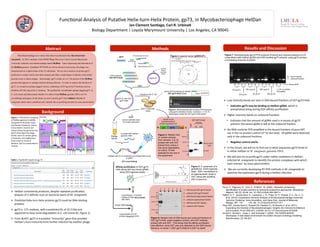
1. Func%onal
Analysis
of
Puta%ve
Helix-‐turn-‐Helix
Protein,
gp73,
in
Mycobacteriophage
HelDan
Jan
Clement
San+ago,
Carl
R.
Urbina+
Biology
Department
|
Loyola
Marymount
University
|
Los
Angeles,
CA
90045
Abstract
Background
Results
and
Discussion
References
Mycobacteriophages are viruses that infect mycobacteria like Mycobacterium
smegmatis. In 2010, students of the HHMI Phage Discovery Lab at Loyola Marymount
University isolated a mycobacteriophage named HelDan. Upon sequencing and annotation of
the HelDan genome (GenBank JF957058) as well as electron microscopy, the phage was
characterized as a siphoviridae of the A3 subcluster. We set out to analyze its protein gp73,
predicted to contain a helix-turn-helix domain and share a high degree of identity with similar
proteins from A-cluster phages. Interestingly, gp73 resides on a 4.1 kb portion of the HelDan
genome that appears to undergo deletion during infection. In order to analyze the function of
gp73, we created an epitope-tagged version, containing a GST-tag at the N-terminus and six
histidine (6X-His) tag at the C-terminus. We purified the recombinant epitope-tagged gp73 in
E. coli extract and determined whether it is able to bind HelDan genomic DNA via UV-
crosslinking techniques. In the future we aim to purify gp73 from HelDan-‐infected M.
smegmatis under native conditions and identify the co-purifying proteins by mass spectrometry.
Pucci, P., Pagnozzi, D., Orrù, S., & Monti , M. (2005). Interaction proteomics:
Identification of protein partners by funtcional proteomics approaches. Bioscience
Reports, 25(1/2), 45-56. doi: 10.1007/s10540-005-2847-z
Hatfull, G. F., Jacobs-Sera, D., Lawrence, J. G., Pope, W. H., Russel, D. A., Ko, C. C.,
et al. (2010) Comparative Genomic Analysis of 60 Mycobacteriophage Genomes:
Genome Clustering, Gene Acquisition, and Gene Size. Journal of Molecular
Biology, 397, 119- 143. doi: 10.1016/j.jmb.2010.01.011
Pope WH, Jacobs-Sera D, Russell DA, Peebles CL, Al-Atrache Z, et al. (2011)
Expanding the Diversity of Mycobacteriophages: Insights into Genome Architecture
and Evolution. PLoS ONE 6(1): e16329. doi:10.1371/journal.pone.0016329
Arnold K., Bordoli L., Kopp J., and Schwede T. (2006). The SWISS-MODEL
Workspace: A web-based environment for protein structure homology modelling.
Bioinformatics, 22,195-201.
Figure
1.
Phamerator
mapping
of
HelDan
genome
(middle)
compared
to
Rockstar
(top)
and
Norbert
(boPom).
Genes
transcribed
in
reverse
are
shown
below
the
genome
line.
Gp73
show
high
homology
(violet
color)
to
certain
genes
in
Rockstar,
and
slightly
less
(blue
color)
to
those
in
Norbert.
Gp73
is
transcribed
in
reverse.
Table
1.
Top
BLAST
results
for
gp
73
Ø Low
intensity
bands
are
seen
in
GSH-‐bound
frac%ons
of
GST-‐gp73-‐His6
• Indicates
gp73
may
be
binding
to
HelDan
gDNA,
which
is
precipitated
along
during
GSH
affinity
purifica%on
Ø Higher
intensity
bands
on
unbound
frac%ons
• Indicates
that
the
amount
of
gDNA
used
is
in
excess
of
gp73
present;
the
excess
gDNA
is
le[
at
the
unbound
frac%on
Ø No
DNA
could
be
PCR
amplified
in
the
bound
frac%ons
of
pure
GST,
nor
in
the
no
protein
control
(3rd
to
last
lane)
.
All
gDNA
were
detected
only
in
the
unbound
frac%ons.
• Nega+ve
control
works
Ø In
the
future,
we
will
try
to
find
out
to
what
sequences
gp73
binds
to
in
either
HelDan
or
M. smegmatis genomic DNA
Ø We
will
also
try
to
purify
gp73
under
na%ve
condi%ons
in
HelDan-‐
infected
M.
smegma)s
to
iden%fy
the
protein
complexes
with
which
each
interact
by
mass
spectrometry
Ø
We
are
currently
developing
RT-‐PCR
methods
in
M.
smegma)s
to
examine
the
expression
gp73
during
a
HelDan
infec%on
100
kDa
50
kDa
25
kDa
GST
GST-‐GP73
-‐His6
#2
#15
GST
*
GST-‐GP73-‐His6
Clones
pGEX-2T
gp73
GST extended primer (f)
His6 extended primer (r)
PCR
PCR products:
GST-GP73-His6
ligate
Transform into E. coli
EcoRI &
BamHI
restriction
digest
pGEX-2T with GST-
gp73-His6
grow in ampicillin
media
colony PCR screening (to identify
GST-gp73-His6 clones) as primers
Figure 2. plasmid vector (pGEX-2T):
Screen E.coli
transformants for
protein expression of:
GST
GP73
His6
N
-‐
-‐
C
Methods
1 kb 1 2 3 4 5 6 7 8 9 10 11 12 13 14 15
ladder
Culture selected clone
for protein extraction
Radiate with UV to covalently link proteins that bound
to gDNA
(UV cross-linking)
UV
Incubate with HelDan
genomic DNA
Affinity purification of GST-gp73-
His6 (along with any bound gDNA)
using GSH-agarose beads
Elute GSH beads
Liquid fraction of random
proteins & DNA (don’t interact
with GSH)
Liquid fraction of GST
proteins (bound to GSH)
Ø HelDan
consistently
produces,
despite
repeated
purifica%on,
plaques
of
2
definite
sizes
on
bacterial
lawns
of
M.
smegma)s
Ø Predicted
helix-‐turn-‐helix
proteins
gp73
could
be
DNA-‐binding
proteins
Ø gp73
is
171
residues,
with
a
predicted
Mr
of
22.3
kDa
and
appeared
to
have
some
degrada%on
in
E.
coli
(clone
#2,
Figure
1)
Ø From
BLAST,
gp73
is
a
puta%ve
“immunity”
gene
that
provides
HelDan’s
host
immunity
from
further
infec%on
by
another
phage
Figure 3. Electrophoresis gel of colony PCR products,
using gp73 extended primers. Colonies 2, 3, 4 & 15
presumably contain the GST-gp73-His6 inserts.
Figure 4. Western blot
of protein extracts
from colonies 2 & 15,
probed with anti-GST.
Extract from colony 2
has some degradation,
so colony 15 was
selected for the rest of
the experiment.
Figure 5. A schematic of a
glutathione (GSH) agarose
bead. GSH, immobilized to
an agarose bead, binds to
GST, along with anything
GST is bound to.
Figure 7. Electrophoresis gel of PCR products of bound and unbound fractions of UV
cross-linked (with HelDan gDNA) and GSH-purified gp73 extracts, using gp73 primers,
in increasing amounts of protein.
1 2 3 4 5 6
100
kDa
50
kDa
25
kDa
1 – GSH-bound GST-gp73 fraction
2 – unbound GST-gp73 fraction
3 – GSH-bound yeast extract fraction
4 – unbound yeast extract fraction
5 – GSH-bound GST fraction
6 – unbound GST fraction
Figure 6. Western blot of GSH-bound and unbound fractions of
GST-gp73-His6, yeast (negative control), and GST extracts,
probed with anti-GST. This shows that the affinity purification
was effective, since GST was only detected on the GSH-bound
fractions, on lanes 1 (GST-gp73-His6) & 5 (GST by itself)
B U B U B U B U B U B U B U (+) ctrl, Heldan gDNA
B – bound fraction
U – unbound fraction
20 μg
gp73 100 μg
gp73 500 μg
gp73
8.8 μg
GST 44 μg
GST 220 μg
GST
(-) ctrl, no protein,
only gDNA