1. Using the BCA assay, we determined that we
had obtained 118.316 ug/mL of SDF and
1.754 ug/mL of sRAGE., We then decided to
use gel electrophoresiswith transcribed RNA to
determine if using different plasmids or
restriction enzymes would increase the yield of
sRAGE. sRAGE yielded depended on the
plasmid and not the enzyme. Bacterial colonies
5 and 10, which were transfected with
Plasmid 1, providedthe highest yield and were
thereby used to produce sRAGE.
The high glucose environment impairs the ability of the
HL-60 cells and white blood cells to migrate towards SDF-
1, ultimately impairing cell migration. The addition of
sRAGE reverses the SDF-1 impairment. Current
challenges include the inability to produce a high yield of
sRAGE protein. This could possibly be because of the
condition in which the sRAGE bacteria is grown.
Changing the temperature or the pH of the media in which
sRAGE is being grown could possibly increase the yield of
sRAGE. Thereby, future work includes adjusting these
conditions of the procedure to increase expression and
yield of sRAGE. Adding additional ELP repeats onto the
fusion protein proposes the possibility of lowering the
transition temperature making it close to body
temperature. Furthermore, future direction also includes
designing a nanoparticle that contains both sRAGE and
SDF-1. Because sRAGE potentiates SDF-1 activity,
packaging them both in one nanoparticle would increase
the efficiency of our design and decrease wound closing
time. Future directions also include in vivo animal studies
to analyze the wound healing kinetics of sRAGE in
various conditions. From this, the stability of sRAGE
nanoparticle in wound fluid will be measured and
realistically compared to existing solutions.
We would like to acknowledge our mentor Dr. Berthiaume who has been involved in our project every step of the way and
provided the moral support to push through the most difficult times. We would also like to thank our graduate student advisor
Hwan June Kang who has guided us through all the cell studies. Thank you to Dr. Cohen who helped us with all the protein
work and our student mentor Sagar Nisraiyya for guiding us through the different experiments our first semester. We would also
like to thank Agnes Yeboaha for being around to answer any of our impromptu questions and provide any supplemental
materials we needed. Thank you to the National Institute of Health for funding this senior design project.
Diabetic individuals have a decreased wound healing ability
that causes them to suffer from chronic wounds such as
foot ulcers. An important signaling pathway, SDF-1, that
contributes to cell migration and cell proliferation is
downregulated in a diabetic individuals. There is an
increased production of advanced glycated end-products
(AGEs). AGEs have a receptor RAGE and when AGE-
RAGE binding occurs, there is an increase inflammation
and infection at the wound site. To interfere with the AGE-
RAGE binding and restore the SDF-1 pathway, sRAGE, the
soluble isoform of RAGE is used. It competes with RAGE
to bind to AGE instead and when the SDF-1 pathway
recovers, cell migration and cell proliferation recovers to
heal the wound at a normal rate.
In the United States alone, 29 million are diagnosed with
diabetes and of the 15% of people who develop foot ulcers,
20% of them are non-healing.This amounts to over 870,000
patients with chronic foot ulcers. These chronic wounds take
over $30,000 to treat and many have to face amputation as
their only option. Current solutions include hyperbaric
chambers, vacuum-assisted wound closure devices
however these require multiple visits to a physician’s office
and can be painful. Current wound dressings serve as a
microbial and moisturizing agent but do not alter the healing
process. A dressing that contains an active that will enhance
the cellular process behind wound healing is very needed.
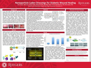
In order to produce the recombinant sRAGE
protein, we took advantage of the ELP motif
we attached to the protein. ELP causes the
protein to self-assemble into nanoparticles
below a certain transition temperature. Above
this temperature, the proteins will be in
solution. We transfected bacteria with a
plasmid containing the sRAGE-ELP DNA.
After allowing the bacteria to grow, we
centrifuged and lysed them.
We then took this lysate and used temperature inversion purification, as described in the figure, to
purify sRAGE-ELP. To determine the amount of purified protein, we used a BCA assay and then
measured the absorbance of our samples at 595nm to determine the concentration of protein in our
samples. We evaluated the efficacy of the produced sRAGE protein using a transwell migration assay.
HL60 cells, which were used as model responder cells, were incubated in environments of varying
glucose concentrations for one day. We used a 5mM glucose environment to represent non-diabetic
conditions, while a 50mM glucose environment simulated highly diabetic conditions. Cells were then
transferred to the migration wells, and they were given either no bioactive molecules, SDF-1 alone, or
both SDF-1 and sRAGE. After 70 minutes, we counted the number of cells that had migrated out of
each of the wells
In order to test the efficacy of the produced
sRAGE, we performed transwell migration assays.
The control condition, which contained 5 mM glucose
and no bioactive proteins, showed virtually no cell
migration (only 4.6875%). Wells that contained 5mM
glucose with SDF exhibited an increased cell
migration rate of 26.875%, proving that SDF does
indeed lead to high cellular migration. The high
glucose condition (50 mM) with SDF showed a slight
decline in cellular migration to 19.165%. The final
experimental diabetic condition, which contained
50mM glucose with a combination of SDF and
sRAGE, displayed an increased cellular migration
rate of 22.9%.
Melissa Ann Olekson’s Dissertation: STRATEGIES FOR IMPROVING GROWTH FACTOR FUNCTION IN DIABETIC WOUNDS
Faulknor, R.A., Mesenchymal stromal cells in alginate dressings to enhance chronic wound healing. 2015, Rutgers University-Graduate School-New Brunswick.
Oliveira, M.I.A., et al., RAGE receptor and its soluble isoforms in diabetes mellitus complications. Jornal Brasileiro de Patologia e Medicina Laboratorial, 2013. 49(2):
p. 97-108.
Schmidt, Ann Marie, et al. "The multiligand receptor RAGE as a progression factor amplifying immune and inflammatory responses." Journal of Clinical Investigation
108.7 (2001): 949
Figure: (Above) Diagram depicting the packaging of our nanoparticle. Once our nanoparticle is
developed, we will experiment with different polymers and pore size, and package the nanoparticle in a
combination that provides for the most mobility.