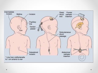
Ventriculoperitoneal
Shunt Placement
Stephanie Bartkowicz
CSULA/CSMC
The Patient
• Baby Girl
• 8 days old (born 5/15)
o Born at 38 weeks
• No Known Allergies
• Admitted to NICU after birth
• Diagnosis: Hydrocephalus
• History: VP shunt placement 5/17
The Patient
• BG’s diagnosis was confirmed by:
o Ultrasound
o Symptoms:
• Enlarged head circumference
• Irritability
• Vomiting
• Parents waiting in NICU
Hydrocephalus
• Excess accumulation of CSF in the
ventricles
• Results in an abnormal widening and
expansion of the ventricular system
• This widening creates potentially harmful
pressure on the tissues of the brain
• Normally, CSF flows through the
ventricles, and exits into cisterns
(reservoirs) at the base of the brain
• CSF is then reabsorbed into the
bloodstream
Ventriculoperitoneal
Shunt
• A catheter is placed into the ventricle
• It is then advanced, subcutaneously,
behind the ear, down the neck, and
through to the abdomen
• The excess CSF is released and
absorbed by the peritoneal cavity
• There is typically a valve which
prevents the fluid from moving in the
wrong direction and only lets fluid drain
when the pressure is too high
Pediatric Considerations
• Room temperature: 79.5° F
o Gaymar heating pad
o Heat lamp
• Patient’s weight on the whiteboard: 5lbs 9ounces (2.5kg)
o Medications
o Bovie pad (smallest size)
o Implants (VP catheters)
• Always remain by the patient’s side
o Especially during intubation!
Anesthesia and Medication
• General Anesthesia
o Weight confirmed by 3 team members
o Dose calculated by Anesthesia attending and resident
o Propofol for induction and maintenance
o Desflurane inhalation maintenance
• Intubated
• Ancef
o Intravenous
o 30 minutes prior to incision
o Antibiotic prophylaxis
• Bupivacaine with Epinephrine
o Diluted with 0.9% NaCl
o 10ml/10ml
Equipment and Instrumentation
Equipment:
• Gaymar heating pad
• Heat lamp
• Suction
• Bovie
o Monopolar: 18 cut/18 coag
• Grounded left abdomen
o Bipolar: 25
• Fluid warmer
o 0.9% NaCl irrigation
Instrumentation:
• Basic Craniotomy tray
• Curette tray
Implants:
• Ventricular catheter
• Peritoneal catheter
• Delta valve
Positioning
• OR bed
o Leg board unlatched and kept down
o Turned 90°
• Anesthesia at lateral side, surgeon at head,
resident on opposing lateral side
• Modified Supine
o Shoulder roll
o Head turned to opposing side with donut
o Arms at sides
o Secured with foam and tape
Prepping
• Clippers to remove hair surrounding burr hole site
o From right ear to crown
• Betadine scrub and paint
o Head
o Abdomen
Concluding the Procedure
• Procedure length: 1 hour 10 minutes
• All counts correct
• Closing sutures: 3-0 Vicryl PS2
o Purse string suture in peritoneum to secure catheter
• Steri Strips
• Specimen:
o Previous VP shunt reservoir removed  Pathology
o CSF  Microbiology
• Patient was not extubated
o Discharged to NICU
o Still under sedation when transferred
o Report given to NICU nurse (incision sites noted)
Post-Operative Considerations
• Infection, infection, infection!
• Bowel perforation
• Bladder perforation
• CSF leaks
• Over drainage of CSF from ventricles
• Assess and monitor developmental milestones
References
Alexander, E. L., Rothrock, J. C., & McEwen, D. R.
(2015). Alexander's care of the patient in surgery
(15th ed.). St. Louis, MO: Mosby/Elsevier.
Hammon, W. (n.d.). Evaluation and use of the
ventriculoperitoneal shunt in hydrocephalus.
Journal of Neurosurgery, 34(6), 792-795.
Keucher, T., & Mealey, J., (2009) Long-term results after
ventriculoatrial and ventriculoperitoneal shunting for
infantile hydrocephalus. Journal of Neurosurgery
50(2), 179-186.

VP shunt OR

  • 1.
  • 2.
    The Patient • BabyGirl • 8 days old (born 5/15) o Born at 38 weeks • No Known Allergies • Admitted to NICU after birth • Diagnosis: Hydrocephalus • History: VP shunt placement 5/17
  • 3.
    The Patient • BG’sdiagnosis was confirmed by: o Ultrasound o Symptoms: • Enlarged head circumference • Irritability • Vomiting • Parents waiting in NICU
  • 4.
    Hydrocephalus • Excess accumulationof CSF in the ventricles • Results in an abnormal widening and expansion of the ventricular system • This widening creates potentially harmful pressure on the tissues of the brain • Normally, CSF flows through the ventricles, and exits into cisterns (reservoirs) at the base of the brain • CSF is then reabsorbed into the bloodstream
  • 5.
    Ventriculoperitoneal Shunt • A catheteris placed into the ventricle • It is then advanced, subcutaneously, behind the ear, down the neck, and through to the abdomen • The excess CSF is released and absorbed by the peritoneal cavity • There is typically a valve which prevents the fluid from moving in the wrong direction and only lets fluid drain when the pressure is too high
  • 7.
    Pediatric Considerations • Roomtemperature: 79.5° F o Gaymar heating pad o Heat lamp • Patient’s weight on the whiteboard: 5lbs 9ounces (2.5kg) o Medications o Bovie pad (smallest size) o Implants (VP catheters) • Always remain by the patient’s side o Especially during intubation!
  • 8.
    Anesthesia and Medication •General Anesthesia o Weight confirmed by 3 team members o Dose calculated by Anesthesia attending and resident o Propofol for induction and maintenance o Desflurane inhalation maintenance • Intubated • Ancef o Intravenous o 30 minutes prior to incision o Antibiotic prophylaxis • Bupivacaine with Epinephrine o Diluted with 0.9% NaCl o 10ml/10ml
  • 9.
    Equipment and Instrumentation Equipment: •Gaymar heating pad • Heat lamp • Suction • Bovie o Monopolar: 18 cut/18 coag • Grounded left abdomen o Bipolar: 25 • Fluid warmer o 0.9% NaCl irrigation Instrumentation: • Basic Craniotomy tray • Curette tray Implants: • Ventricular catheter • Peritoneal catheter • Delta valve
  • 10.
    Positioning • OR bed oLeg board unlatched and kept down o Turned 90° • Anesthesia at lateral side, surgeon at head, resident on opposing lateral side • Modified Supine o Shoulder roll o Head turned to opposing side with donut o Arms at sides o Secured with foam and tape
  • 11.
    Prepping • Clippers toremove hair surrounding burr hole site o From right ear to crown • Betadine scrub and paint o Head o Abdomen
  • 12.
    Concluding the Procedure •Procedure length: 1 hour 10 minutes • All counts correct • Closing sutures: 3-0 Vicryl PS2 o Purse string suture in peritoneum to secure catheter • Steri Strips • Specimen: o Previous VP shunt reservoir removed  Pathology o CSF  Microbiology • Patient was not extubated o Discharged to NICU o Still under sedation when transferred o Report given to NICU nurse (incision sites noted)
  • 13.
    Post-Operative Considerations • Infection,infection, infection! • Bowel perforation • Bladder perforation • CSF leaks • Over drainage of CSF from ventricles • Assess and monitor developmental milestones
  • 14.
    References Alexander, E. L.,Rothrock, J. C., & McEwen, D. R. (2015). Alexander's care of the patient in surgery (15th ed.). St. Louis, MO: Mosby/Elsevier. Hammon, W. (n.d.). Evaluation and use of the ventriculoperitoneal shunt in hydrocephalus. Journal of Neurosurgery, 34(6), 792-795. Keucher, T., & Mealey, J., (2009) Long-term results after ventriculoatrial and ventriculoperitoneal shunting for infantile hydrocephalus. Journal of Neurosurgery 50(2), 179-186.