
Recommended
More Related Content
Similar to EXAMINATION OF BREAST_074455.pptx
Similar to EXAMINATION OF BREAST_074455.pptx (20)
More from ShubhrimaKhan
More from ShubhrimaKhan (20)
Recently uploaded
Recently uploaded (20)
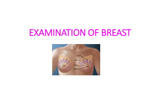
EXAMINATION OF BREAST_074455.pptx
- 2. ANATOMY OF BREAST • The breast is the tissue overlying the chest muscles (pectoral). • Components • Glandular tissue (milk production) • Fibrous tissue • Fatty tissue • The breast has no muscular tissue. Muscles lie underneath the breast. • Location – vertically the breast lies between the 2nd and 6th ribs. Horizontally between the lateral border of sternum and the mid axillary line.
- 3. CONT.. • Parts • Each breast has 8 – 10 sections that branch out from the nipples called lobes. • Inside each lobe are many smaller structures called lobules. • At the end of the each lobule has tiny hollow sacs called alveoli. • The lobes are linked by a network of thin tubes called ducts. • Ducts carry milk from the alveoli toward the dark area of skin in the center of the breast (areola).
- 4. CONT.. • Spaces around the lobules and ducts are filled with fat, ligaments and connective tissues. • The amount of fat in breasts largely determine their size. • Lymphatic system – It is a network of lymph nodes and lymph ducts that helps fight infection.
- 5. ASSESSMENT OF BREAST INSPECTION • The breast should be inspected with the patient seated in the following positions – • Hands pressing on the top of the head • Arms raised in the air • Arms at the side • Hands pressing on the hips
- 6. CONT.. INSPECTION • Size and shape of the breast • Symmetry • Skin colour (orange peel: present in edema) • Superficial veins • Areola rounded or oval and colour • Nipples: everted, inverted • Cracking or eczema • Bleeding or discharge • Retractions or dimpling
- 7. CONT.. PALPATION • Sit the patient at 45⁰ • Ask the patient to place their hands behind the head. • Retract the breast with the left hand and palpate with the right. Feel with the palmar surface of the fingers. • Palpation should be done by a clockwise rotary motion from the periphery to center. • Findings – palpable lumps or masses (size, location, mobility), tenderness, discharge.
- 8. SELF BREAST EXAMINATION PURPOSE • To detects the majority of breast abnormalities • Potentially life-saving TIME • Monthly exam, 5-7 days after menstruate ends. • Start in front of mirror.
- 9. STEPS STEP 1 • Stand before a mirror. Inspect both breasts for skin changes, redness, discharge from nipples, puckering, dimpling, visible bumps, nipple crusting.
- 10. CONT.. STEP 2 • Watching closely in the mirror, clasp hands behind your head. Note for changes in the contour and shape of the breast.
- 11. CONT.. STEP 3 • Press hands firmly on hips and bow slightly toward the mirror to pull shoulders and elbows forward looking for the same changes as step2.
- 12. CONT.. STEP 4 • Raise the left arm. • Use three or four fingers of the right hand to feel left breast firmly, carefully and thoroughly. • Do the vice versa to feel any unusual lump, mass or lymph nodes under the skin. • Middle portion of fingers is used while palpating.
- 13. CONT.. STEP 5 • Gently squeeze the nipple and look for a discharge. If discharge present seeking for medical help. • Repeat the step on another breast.
- 14. CONT.. STEP 6 • Step 4 and step 5 should be repeated on lying down position. • Lie flat on your back, right arm over the head and a pillow or folded towel under the left shoulder. • This position flattens the breast and makes it easier to examine.