
Recommended
More Related Content
What's hot
What's hot (20)
Similar to Esotropia
Similar to Esotropia (20)
Recently uploaded
Recently uploaded (20)
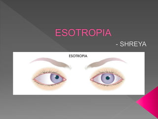
Esotropia
- 2. Derived from 2 greek words- ‘eso’ which means ‘inward and ‘trepo’ means ‘turn’.
- 3. Apparent convergent squint Angle kappa- negative Causes: 1. Telecanthus 2. Epicanthus
- 7. (A)-Symmetric central Asymmetric – (B) - Pupillary margin: 15° (C) - Close to limbus: 30° (D) -Beyond limbus: 45°
- 8. Three commonly recognized stages: Esophoria Intermittent Esotropia Esotropia
- 9. ESOPHORIA
- 10. Esodeviation that is intermittently controlled by fusion mechanisms. Manifest under certain conditions such as fatigue, illness and stress.
- 11. Esodeviation that is not controlled by fusional mechanisms so that deviation is constant
- 12. ESODEVIATION INCOMITANT PARALYTI C RESTRICTIVE SPASTIC CONCOMITANT ACCOMODATIVE PARTIALLY ACCOMODATIVE NON ACCOMODATIVE •Neurogenic •Myogenic •Musculofacial •other Refractive Non refractive •Essential infantile •Essential acquired •Acute comitant •Microtropia •Cyclic esotropia •Sensory esotropia •Nystagmus blockage syndrome
- 14. Esodeviations due to excessive convergence associated with accomodation are called accomodative esotropia. ACCOMODATIVE ESOTROPIA REFRACTIVE NON REFRACTIVE PARTIALLY ACCOMODATIVE
- 15. MOST CONSISTENT FEATURE: VARIABLE ANGLE OF ESODEVIATION WHICH INCREASES WITH THE EFFORT FOR ACCOMODATION
- 16. Uncorrected hyperopia To see clear at distance- they accommodate- esodeviation for distance fixation. Normal AC/A- therefore ET same for distance and near (within 15 PD) NO CONVERGENCE EXCESS Respond well to full cycloplegic correction of hyperopia
- 17. Usually mild to moderate hyperopia (+2 to +6D) Very high hyperopia- donot accommodate- bilateral amblyopia Apart from ET and amblyopia, they may present with asthenopia due to constant accomodative effort.
- 18. 6/6 6/6
- 20. Heterophoria method AC/A= IPD+ (∆n- ∆d)/3 Normal- 5-7.5pd/1D Gradient method AC/A= N-D/3 Normal- 3-5pd/1D
- 21. ET for distance + convergence excess (>15PD) ET for near (at 33cm) High AC/A ratio
- 22. No clinically significant hyperopia No accomodation for distance- no ET for distance High AC/A ratio >15PD ET for near ‘Hyper accomodative type’
- 23. Weak accomodative mechanism Over-accomodation Normal AC/A ratio Convergence excess type Remote NPA and NPC- whereas hyper accomodative type have a normal NPA Seen in early presbyopes or cases under mild cycloplegia
- 24. 2nd year of life Variable angle of ET, presence of convergence excess and full cycloplegic error should be looked for.
- 25. Full cycloplegic correction If convergence excess- bifocals are prescribed. The minimal plus add that corrects convergence excess ET is added.
- 26. Full cycloplegic correction/bifocals Residual esotropia Non-accomodative element Partially accomodative- require surgery for the non- accomodative part MR recession with or without retro-equatorial myopexy. (Faden)
- 27. 1. Early onset- high risk of amblyopia 2. Large angle ET(30pd) 3. Free alteration/ cross fixation in alternators and fixation preference of normal eye in amblyopes 4. No significant refractive error 5. No Neurologic deficit 6. Confirmed only after 4-6 months
- 29. 7.May be associated with: IOOA (68%) Nystagmus (33%) DVD(50%) 8. Asymmetric optokinetic nystagmus: Temporal to nasal- smooth Nasal to temporal- cogwheel
- 30. A special characteristic of congenital esotropia - OKN asymmetry Temporal to nasal (T/N) Smooth, following and rapid refixation Nasal to temporal (N/T) Jerky inaccurate movements with halting refixation OKN asymmetry is present in all infants but becomes symmetrical by 6 months. Patients with congenital ET retain OKN asymmetry
- 31. Ciancia syndrome Lang’s syndrome
- 32. Early onset ET Bilateral abduction limitation Manifest-latent jerk nystagmus (fast phase in the direction of fixating eye) - Increasing in abduction; decreasing in adduction - Face turn towards fixating eye - Fixating eye in adduction(null point in adduction)
- 33. Early onset ET DVD Nystagmus Excyclodeviation of non fixating eye May be associated with torticollis
- 34. DIFFERENTIAL DIAGNOSIS Cranial nerve 6th palsy Doll’s eye manouvere Duane’s retraction syndrome Changes in palpebral aperture, upshoot/downshoots on adduction CNS anomalies Down’s syndrome, Mobius, Cerebral Palsy, Albinism Accomodative ET/ Partially accomodative ET Cycloplegic refraction Nystagmus blockage syndrome Inverse relation between amplitude of nystagmus and degree of esotropia
- 35. Unknown Multifactorial Heritable factors- monozygotic twins showing esotropia Developmental anomaly in first 4 months
- 36. Depends on treatment of amblyopia Full cycloplegic refraction under atropine 1 % eye ointment Full hyperopic correction Occlusion
- 37. Conventional full time, fully opaque occlusion of dominant eye. At no point during the treatment, is binocular viewing allowed. Thus, patching is done for 3:1, 4:1 or 5:1 days for a 3,4 or a 5 year old child respectively. Above 6 years, the regime remains 6:1 for all ages.
- 38. Vision assessment done monthly. (fortnightly in infants). End point: free alternation of the two eyes which is equally maintained.
- 39. Large angle ET- earliest/ 4 months of age Small angle ET- proper hyperopic correction till 6 months or till examination can be done satisfactorily.
- 40. Associated inferior oblique ‘v’ phenomenon Amblyopia therapy DVD Nystagmus Eccentric fixation or uncorrected amblyopia have unpredictable results.
- 41. MR surgery is more effective LR surgery Mono-ocular recession-resection or bimedial recession MR 1mm surgery corrects 3-4.5 PD of deviation LR 1mm surgery corrects 2-3 PD of deviation
- 42. Onset after infancy No accomodative factors and no neurological cause (excluded by CT/MRI) ‘acquired essential (non-accomodative) esotropia. Basic type or convergence excess type Rarely divergence insuffiency may be present. Management- Surgery with better binocular visual potential than infantile esotropia
- 43. Ultra small angle ET Missed by ordinary methods of examination Usually have amblyopia of one eye with variable levels of binocularity 1. Primary 2. Secondary(residual deviations after surgery)
- 44. Macular scotoma Good peripheral fusion with fusional amplitudes and gross stereopsis
- 45. Small angle (<5˚) heterotropia Harmonius ARC Mild amblyopia Partial stereopsis Based on fixation pattern Cover test Type 1 Central fixation Shows tropia Type 2 Eccentric fixation without identity Shows tropia Type 3 Eccentric fixation with identity Does not pick up a tropia
- 46. Type 3- eccentric fixation with identity implies that angle of anomaly is same as the eccentricity of fixation.
- 47. CONSISTENT Amblyopia ARC Relative scotoma on fixation spot Normal or near normal fusional amplitudes Defective stereoacuity VARIABLE Size of deviation (5˚-8˚) Foveal or non-foveal fixation Relationship between degree of eccentric fixation and angle of anomaly Presence or absence of anisometropia Positive or negative cover test
- 48. Macular scotoma by Bagolini’s glasses or 4PD test. Presence of amblyopia and associated refractive error should be detected.
- 49. Treat amblyopia with occlusion therapy Good prognosis if treated in younger children.
- 50. Sudden presentation c/o diplopia 2 types: 1. Those which manifest after the fusion has been interrupted by a patch or occlusion for a short time. 2. Those which have no such interruptions to fusion but have very poor fusional control, which may be further compromised by physical or emotional stress.
- 51. First type- spontaneously resolve within 6 months. Second type- Surgery/ prismatic neutralisation in case of small angle ET.
- 52. Regular cycles of presentation Usually 24-48 hours of squint alternating with same duration of no squint. Squint days: 1. large angle ET (40-50PD) 2. sensory anomalies 3. squint is consistent.
- 53. No squint days: 1. BSV with good fusional amplitudes 2. No latent squints Lasts for a few months to years before they become fully manifest squints Surgery as per deviation on squinting days gives satisfactory results.
- 54. Infantile ET is associated with manifest-latent and latent nystagmus. However, there is a special form of nystagmus which has a dampening mechanism with eyes in adduction Inverse relationship between ET and nystagmus Nystagmus is present when eyes are straight and it disappears when eyes are locked in ET. Management: Faden with or without B/L MR recession.
- 55. Lost vision in one eye usually develops squint over a period of time. XT or ET depending on convergence tonus XT- in first year and after 8-9 years of age ET- after first year upto 8-9 years of age Refractive error and accomodative status of the straight eye needs to be evaluated before planning any cosmetic surgery
- 57. Refraction and proper prescription including use of bifocals Use of prisms Purpose: Fusion and binocular vision Maintains relationship between accomodation and convergence mechanisms
- 58. PROPER CYCLOPLEGIA PROPER PRESCRIPTION- full cycloplegic correction with no over or under correction. BIFOCALS- In case of high AC/A ratio
- 60. Glass prisms- upto 7-8 pd over each eye Fresnel prisms- upto 25-30 pd
- 62. Miotics Cyclopegics Chemodenervation by botox
- 63. Site of action- ciliary muscles Facilitates accomodation Reduces accomodative effort Hence, reduces accomodative convergence
- 64. Di-isopropyl-flourophosphate (DFP) Phospholine iodide (Echothiophate)- 0.03%, 0.06% and 0.125% Demecarium bromide (Humorsol)
- 65. Desirable end results after use of miotics is proper binocular alignment at all times, especially near work with accomodative tasks.
- 66. To make patients accept full hyperopic correction To suppress accomodative convergence mechanisms and stimulate divergence
- 67. This is still under trial by Scott and McNeer Infantile esotropes if aligned before 6 months can have good functional binocularity. While the surgical effect of botox in adult eye muscles is transient, effect in early infancy is permanent because of the changes in the developmentally immature muscles.
- 68. Initial overcorrection may restructure the ocular motility balance to yield better result ultimately. However on the negative side, associated changes of oblique overactions/ AVpatterns/ DVD may still require surgical correction.
- 69. PRE OP EVALUATION Assessment of vision and amblyopia therapy Measurement of deviation
- 70. Weakening Strengthening Aim- symmetrizing and not symmetric surgery
- 71. To split amount of surgery across two recti rather than single large surgery in single muscle alone If AV, with obliques overaction- vertical shifting of horizontal recti or slanting recession and resection can be done
- 72. Recession • Conventional • Hangback • Adjustable • Vertical transposition of horizontal recti • Slanting recession Retroequatorial myopexy
- 73. Muscle is disinserted and reinserted to a point closer to its origin. Induces a slack/laxity Muscle becomes less effective Reinsertion to be done within its arc of contact Max recession defined by functional equator- depends on arc of contact
- 74. Max recession MR- 6mm LR- 8mm in adults Children- MR 5.5mm LR 7mm Minimum MR 3mm LR 4mm
- 75. Recession is done with long ends of the suture between the site of insertion and the muscle. This is indicated if supramaximal recession is intended and its difficult to pass sutures that posteriorly Or, there’s a risk of scleral perforation in high myopes
- 76. Weakening effect is more than a conventional recession because of a central (mid width) sag. A pseudotendon may form in the intervening space around the sutures which may lead to a late under correction.
- 77. Ability to modify the position of a newly operated muscle by use of adjustable sutures. Indications: Large angle deviations where results may be inconsistent Reoperations/ previously injured muscles Incomitant starbismus with diplopia when precise post operative results are desired. Mechanical limitaions as in dysthyroid orbitopathy or musculofacial anomalies. Aberrant innervation in III nerve palsy or DRS
- 78. Posterior fixation suture Musle is sutures posterior to insertion farther than its limit of arc of contact. This shortens the lever arm and reduces the action of muscle in its field of action. No change in primary position if no recession is done along with Faden.
- 79. MR- 12-14mm LR-16-20mm SR- 14-16mm IR- 14-16mm More effective on MR; least on LR
- 80. Non accomodative convergence excess esotropia (MR) Nystagmus blockage syndrome (MR) DVD (SR) Paralytic strabismus (contralateral synergist) DRS (contralateral MR) Additional weakening effect over recessed muscle.
- 81. Resection Advancement Transpositioning of adjacent muscles
- 82. Shortens muscle length making itmore taut Improves efficiency by raising it to a higher length tension curve Excision of tendinous part of the muscle. Therefore max limit for MR- 6mm LR- 4.5mm
- 83. Strengthening procedure whereby muscle is reinserted closer to limbus. Reverse of recession Indications: To correct consecutive squint in a recessed muscle In paralytic squint, in addition to resection.
- 84. Classification and management of esotropia in children. What is faden operation. Accomodative squint- signs symptoms investigations Management of accommodative esotropia. Investigations and management of concomitant esotropia Describe the clinical features and management of partially accommodative esotropia
- 85. Management of alternating convesrgent squint Monofixation syndrome What is microtropia? Discuss the types and clinical features of microtropia What are the features and differential diagnosis of infantile esotropia? When it should be operated and its prognosis for binocular single vision (BSV)?
- 86. *Define essential infantile esotropia? Give at least four differential diagnosis of essential infantile esotropia and give at least two differentiating features among them. 2+8 25. Describe Faden’s operation as applied in management of strabismus. *Describe the Clinical Features, investigations, indications and surgical management of infantile esotropia, and its post-operative complications.
- 87. Classify and give complete management of esotropias in detail Define and classify esotropia. Management of a 6 year old patient with esotropia Classify esotropia. How wouldyou plan the management of convergneceexcess esotropia in a 5 year old child? Describe the choice of procedure and surgical planning in detail.