
Recommended
More Related Content
What's hot
What's hot (20)
Similar to Eales disease
Similar to Eales disease (20)
Recently uploaded
Recently uploaded (20)
Eales disease
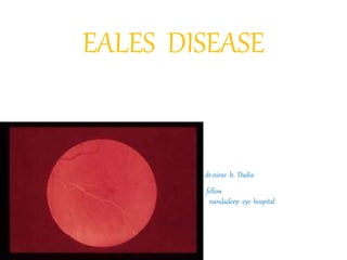
- 1. EALES DISEASE dr.nirav b. Dadia fellow nandadeep eye hospital
- 2. In 1880 and 1882, Henry Eales - “primary recurrent retinal hemorrhage”. Similar conditions of retinal and vitreous hemorrhage were described under the name of Eales’ Disease. Eales didn’t mention any inflammatory signs preceding or accompanying the hemorrhages.
- 3. • In 1887 Wadsworth reported on signs of inflammation of the retinal vasculature - Eales’ disease and periphlebitis • Elliot initially suggested that the disease be called “periphlebitis retinae”.
- 4. • Currently, Eales’ disease is considered to be an idiopathic inflammatory venous occlusion that primarily affects the peripheral retina.
- 6. • This inflammation induced vascular occlusion can lead to a proliferative vascular retinopathy, with sequelae such as recurrent vitreous hemorrhage and traction retinal detachment.
- 7. PATHOPHYSIOLOGY • Patchy perivascular or intramural infiltration of lymphocytes or granulation tissue sometimes with or without giant cells • Plasma cells are occasionally present. • Veins are primarily affected • The vascular changes are usually seen on retinal periphery
- 8. Hyalinization and thinning of vein wall Narrowing and obstruction of the lumen Endothelial cell proliferation Thrombosis and rupture of the vein Intravitreal new vessel formation
- 10. Systemic disease associated with Eales’ disease: • Tuberculosis • Hypersensitivity to tuberculoprotein • Thromboangitis obliterans • Neurologic disease • Hematological abnormalities
- 11. • The assumption of tubercular aetiology is based on active or healed tuberculosis in some patient with Eales’ disease. • Ophthalmoscopic evaluation in patient with active or healed TB showed 1.3% had Eales’ disease .
- 12. Hypersensitivity to tuberculoprotein: Allergic reaction to tuberculosis has been reported by many authors till date. Positive Mantoux reaction which is as high as 90% in some series.
- 13. Systemic disease: • Several studies have shown association between neurological and hematological disease. • bilateral hearing loss 48% (Renie et al) , 25% (William et al). • 2 pt with Eales’ disease had progressive worsening of neurological deficit (Rodier G). • Myelopathy with Eales’ disease has been described by many.
- 14. Immunological studies in Eales’ disease: Immune mediated mechanism has been suggested by many authors as a possible cause of Eales’ disease. • Acute onset, steroid responsiveness, lymphocytic infiltration and abnormal immunological parameters all indicate an immunological basis of disease.
- 15. Immunological studies in Eales’ disease: cont…d Altered immune response of type III and/or IV reaction to an infectious agent (Muthukaruppan et al). • Raised IgG and IgA levels (Johnson et al) , elevated levels of circulating immune complexes and antiretinal antibody (Kasp et al) , immunophenotyping predominant T cell CD4 • Higher frequencies of HLA B5(B51), DR1 and DR4 (Biswas et al)
- 16. Biochemical studies in Eales’ disease: • Raised alpha-globulins and reduced albumin levels in the serum samples. • PDGF, IGF1, TGFa and TGFb play a key role in neovascularisation. • Raised serum alpha1 acid glycoproteins in 27 patients of Eales’ disease
- 17. Stages of Eales’ disease Stage I: (Inflammatory stage) • Localized areas of peripheral retinal edema with sheathing of the smaller caliber vascular branches. • Minute retinal hemorrhages as well as minute vascular brackets or hooklets connecting two adjoining vessels.
- 19. • Active periphlebitis with subhyaloid hemorrhage over the macula.
- 20. Stage II (ischemic stage) • Involvement of larger vessels and extend more posteriorly . • Veins as well as arterioles may be sheathed • Widespread retinal hemorrhages and vitreous looks hazy .
- 22. • Stage III (stage of neovascularisation) • Peripheral new blood vessels with numerous vitreous and retinal hemorrhages. • The hemorrhages frequently recurs.
- 24. Sea- fan like neovascularization
- 25. • Stage IV (complicated stage) • Massive retinal proliferans associated retinal and massive vitreous hemorrhage. • With this advanced disease the neovascularization can cause tractional rhegmatogenous retinal detachment.
- 27. Differential diagnosis: • Vasculitis mimicking Eales’ disease Systemic Ocular Behcet’s disease Birdshot retinochoroidopathy Lyme Borreliosis Coat’s disease Leukemia Pars planitis Multiple sclerosis Viral retinitis Systemic lupus erythematosus Toxocariasis Toxoplasmosis Tuberculosis Wegener’s granulomatosis
- 28. • Proliferative vascular retinopathy mimicking Eales’ disease: Systemic Ocular Diabetes mellitus BRVO Sarcoidosis CRVO Sickle cell disease ROP Pars planitis Coats’ disease
- 29. Clinical features • Usually occurs in young , healthy people, with a peak incidence between the ages of 30 and 40 years. • • It occurs more frequently in males 80-90%. • • 75% cases it presents before 49 years. • Can be unilateral or bilateral.90% bilateral (Duke Elder) retinal vasculitis
- 30. Vitreous floaters or blurring of vision, symptoms attributable to recurrent vitreous hemorrhages. 80% between the age of 20-40 years and 95% were male (O.K Malla and co workers) 54.34% between 20-30 years and 94.73% male Rare in more developed countries.
- 31. • More commonly reported from Indian subcontinent. The reported incidence in India is 1 in 200-250 patient • Anterior uveitis/Vitritis. • Active perivasculitis with exudates around the veins in one or more quadrants. Arterioles may be affected.
- 33. Healed perivasculitis as sheathing of the veins Macular changes uncommon Peripheral retinal neovascularisation reported in 36-84% of cases
- 34. Recurrent vitreous hemorrhages, the hall mark of the disease Some vitreous hemorrhages resolve, some do not ( organize with multiple VR adhesions & RRD/TRD Some patient specially with multiple sclerosis are asymptomatic.
- 35. Fundus fluorescein angiography • To delineate areas of capillary nonperfusion, peripheral retinal nonperfusion is present in all patients with Eales’ disease. • Retinal or disc neovascularisation • • Macular edema • Helps in monitoring the regression and disappearance of new vessels during treatment and follow up.
- 38. TREATMENT
- 39. • Symptomatic treatment. • Treatment aim : reducing retinal perivasculitis and associated vitritis ; reducing risk of vitreous hemorrhage from new vessels by retinal ablation and surgical removal of non resolving vitreous hemorrhage and/or vitreous membranes.
- 40. Treatment of Eales’ disease: • Observation. • Medical Corticosteroids Antituberculosis drugs Immunosuppressive drugs. • Retinal ablation Photocoagulation cryotherapy • Surgical vitrectomy
- 41. Observation: • Patient with inactive retinal vasculitis • Follow up 6 months to 1 year interval. • Patient with fresh vitreous hemorrhage if retina is found to be attached. • Such vitreous hemorrhage usually clears by 6 to 8 weeks.
- 42. Medical therapy • Corticosteroids are mainstay of therapy in active perivasculitis stage of Eales’ disease. • Majority of cases 1mg/kg body weight, tapered to 10mg/week over 6 to 8 weeks. • Maintenance 15 to 20mg/day for 1 to 2 months. • Periocular depot steroid injection may be added for associated macular edema.
- 43. • Systemic and Periocular steroid useful in patient having 3 quadrants involvement with macular edema. • Systemic steroid only if less than 3 quadrant involvement. • No difference in response between Mantoux positive and negative cases.
- 44. • Immunosuppressive therapy in patient unresponsive or have unacceptable side effects. (Azathioprine and cyclosporine) • Some investigators have recommended ATT (Rifampicin and Isoniazid) for 9 months.
- 45. Photocoagulation • Mainstay of therapy in proliferative stage of Eales’ disease. • The aim Regulate the circulation To obliterate surface neovascularisation and Close leaking intraretinal microvascular abnormalities.
- 46. • Sectoral laser for capillary non perfusion and PRP for neovascularisation of disc. • Occasional massive hemorrhage can occur. • After laser, regressing neovascularisation can cause macular distortion and retinal tear. • Laser not advised in active inflammatory stage
- 48. Vitreoretinal surgery • Vitrectomy alone or combined with other vitreoretinal surgical procedures is often required. • Nonresolving vitreous hemorrhage with obscuration of central vision of 3 months duration may be subjected to vitrectomy.
- 49. • Vitrectomy done between 3 to 6 months has better results than done after 6 months (Kumar et al). • Early vitrectomy in patient with TRD, extensive vitreous membranes or epimacular membranes. • Endolaser can be given along with vitrectomy.
- 50. Tractional retinal fold after vitrectomy
- 51. Summary and conclusions: • Characteristic clinical findings and angiographic pattern. • Mimic several ocular or systemic disease presenting as retinal vasculitis or proliferative retinal vasculopathy. • Hypersensitivity to tubercular protein has been considered a prime cause of Eales’ disease .
- 52. • Probable multifactorial etiology. • HLA, retinal autoimmunity, mycobacterium genome, free radical mediated damage. • Corticosteroids in active disease and laser photocoagulation in ischemic and proliferative stage. • Results of vitrectomy in non resolving vitreous hemorrhage with or without retinal detachment are satisfactory.
- 53. • Thank u for listening