
Recommended
More Related Content
What's hot
What's hot (20)
Similar to CELL CYCLE,.pptx
Similar to CELL CYCLE,.pptx (20)
Recently uploaded
Recently uploaded (20)
CELL CYCLE,.pptx
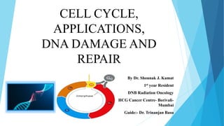
- 1. CELL CYCLE, APPLICATIONS, DNA DAMAGE AND REPAIR By Dr. Shounak J. Kamat 1st year Resident DNB Radiation Oncology HCG Cancer Centre- Borivali- Mumbai Guide:- Dr. Trinanjan Basu
- 2. Overview Cell Cycle Regulation of Cell Cycle Effect of Radiation on Cell Cycle DNA Damage DNA Repair Mechanisms
- 3. Cell Cycle A cell cycle is a precisely programmed series of events which enables a cell to duplicate its contents and divide into 2 daughter cells It has the following phases:- 1] G1 phase 2] S phase Interphase 3] G2 phase 4] M phase or the Mitosis phase 5] G0 or the resting state of cells that have withdrawn from active cell cycle
- 4. Interphase It is the phase in which cells spend most of their lives preparing for mitosis. It is divided into 3 stages:- 1] G1 phase – phase of cell growth and protein synthesis 2] S phase – phase where DNA and centrosomes are replicated 3] G2 phase – phase where energy replenishment, mitotic specific protein synthesis, cytoskeleton dismantling and additional growth take place.
- 7. Prophase In this phase the chromosomes which were invisible microscopically during interphase begin to condense and become visible Also centrosomes begin to assemble at the poles of cells
- 8. Metaphase In this phase the chromosomes align along a plane that bisects the cell and become attached to microtubule fibres of mitotic spindle Also the nuclear membrane disappears at this time
- 9. Anaphase During this phase the chromatids are pulled apart by the mitotic spindle to the opposite poles of the cell.
- 10. Telophase and Cytokinesis The chromatids cluster into two sets , they de-condense and a new nuclear membrane forms around each set of chromatids During this time the cytoplasm of the cell also divides into two thus yielding two daughter cells
- 11. Regulation of Cell Cycle
- 12. Cyclin Dependent Kinases They are serine/threonine kinases that sequentially regulate progression of cell through the cycle via phosphorylation. They do this via 4 mechanisms Association with cyclins Assosiation with CDK inhibitors Addition of phosphate groups to activate CDK activity Deletion of phosphate groups to inhibit CDK activity
- 14. Association with Inhibitors 2 Families of inhibitors are involved in regulating cyclin–cdk activity: - p16 INK 4a family - p21 Cip/Kip family INK Protein binds to cdk 4/6 and interferes with its binding to cyclin D Cip/Kip family of inhibitors interact with both cyclins and their associated cdks (mainly with cdk2 and cyclin E) and disable kinase activity ubiquitin-mediated degradation of inhibitors ensures that the inhibitors are present during a specific period of time during the cell cycle.
- 15. Regulation by Phosphorylation This involves both activation and inhibition Two steps are required for cdks to become active: 1] Dephosphorylation of the inhibitory phosphate groups by cdc25 phosphatases 2] Phosphorylation of a central threonine residue- Thr161 by cdk-activating kinase (CAK).
- 16. Cell Cycle Checkpoints Signaling pathways that sense and induce a cellular response to DNA damage. The components are DNA damage sensors, signal transducers, or effectors. Disruption of checkpoint function leads to genomic and chromosomal instability leading to mutations that can induce carcinogenesis
- 19. Role of p53 Gene p53 is a tumor suppressor gene that is critical in the pathway that arrests the G1 checkpoint. In response to DNA damage, ataxia telengectasia mutated (ATM) autophosphorylates and releases active monomer which phosphorylates p53 and activates it. Activated p53 enhances p21 gene expression which results in sustained inhibition of G1 cyclin and Cdks. This in turn inhibits Rb phosphorylation and progression from G1 to S. Mutation in this gene compromises this checkpoint function and results in damaged dna replication leading to carcinogenesis.
- 20. Effect of Radiation on the Cell Cycle Phases
- 21. In Chinese hamster cells Most sensitive cells to Radiation are the ones in M and G2 [ steep curve, no shoulder] Most radioresistent cells are in late S phase [less steep curve but very broad shoulder] Other phases G1 and Early S are intermediate between the two extremes.
- 22. In HeLa Cells Similar to the hamster cells in most aspects Major difference is length of G1 phase in HeLa cells is appreciably long and at the beginning of G1 there is a peak of radioresistance followed by a trough of radiosensitivity towards end of G1.
- 24. CELL CYCLE SPECIFIC DRUGS During M Phase:- Taxanes and Vinca Alkaloids cause a synergistic action by acting on DNA and causing its damage. During S Phase:- Drugs such as Capecitabine, Gemcitabine and 5-fluorouracil inhibit nucleotid formation and DNA replication. Since this phase is radioresistant, they cause a synergistic action with radiation. During G2-M phase:- Drugs such as Topoisomerase Inhibitors arrest the cells in G2M phase which is a radiosensitive phase thus causing increase in the effect caused by radiation.
- 25. EFFECT OF OXYGEN ON CELL CYCLE OXYGEN ENHANCEMENT RATIO (OER) Ratio of doses administered under hypoxic to aerated conditions needed to achieve same biologic effect. OER for Xrays and Gamma rays has a value between 2.5 to 3.5. With respect to cell cycle the OER in the G2 and M phase and also in the G1 phase is lower than in S phase because of the radiosensitivity of these phases. Thus maintaining an aerated state helps in increasing the efficacy of the radiation treatment.
- 26. DNA Damage
- 27. Structure of DNA DNA is made up of 2 strands and arranged in a helical pattern. The strands are formed of a Deoxy ribose sugars and phosphate molecules. On those strands are the purine and pyrimidine molecules which are bound by Hydrogen bonds.
- 28. Effect of Radiation on DNA Direct Effect Indirect Effect There are 2 effects :-
- 29. Process of free radical formation
- 30. Types of DNA damage Single strand breaks Double strand breaks DNA crosslink formations Radiation induced chromosome and chromatid aberrations.
- 31. Single strand breaks This occurs when one strand of DNA is damaged or broken. More common type of defect Is readily repaired because of availability of template on intact opposite strand. Of lesser biologic significance Approx. 1000 SSBs per cell after 1 -2 Gy
- 32. Double strand break This occurs when both the strands of DNA are damaged. Less common defect Has more significance since this takes time to be repaired and can result in mutations, carcinogenesis and cell death Approx. 40 DSBs after 1-2 Gy
- 33. Measuring DNA Strand Breaks Pulsed Field Gel Electrophoresis (PFGE) Single-cell Gel Electrophoresis (also known as the comet assay). DNA damage induced nuclear foci assay
- 34. DNA crosslink formations This occurs in oxidative stress when O2 free radicals form intermediaries which then react with DNA nucleotides and form covalent links between the nucleotides. They can be of the following types Intrastrand crosslinks- when crosslinking occurs within same strand Interstand crosslinks- when crosslinking occurs between opposite strands of DNA DNA Protein crosslink- between DNA and an oxidised protein
- 36. Radiation Induced Chromosome Aberrations Occurs when cells are irradiated in the early interphase stage before the chromosome has been duplicated. Can be of the following types 1] Dicentric chromosome 2] Ring aberration
- 37. Radiation Induced Chromatid Aberration Occurs when the cell is irradiated in the late interphase after the chromosome has duplicated Anaphase Bridge
- 38. Non Lethal Chromosome Lesions Translocations Deletions
- 39. DNA Damage Repair Mechanisms
- 40. Base Excision Repair Removal of the defective base by a Glycosylase/DNA lyase Followed by removal of the sugar residue by an apurinic endonuclease 1 (APE1) then replacement with the correct nucleotide by DNApolymerase completed by DNA ligase the complex of replication factor C (RFC)/proliferating cell nuclear antigen (PCNA)/DNApolymerase / performs the repair synthesis, the overhanging flap structure is removed by the flap endonuclease 1 (FEN1) DNA strands are sealed by ligase I Single Base Defect Multiple Base Defect
- 42. Nucleotide Excision Repair Nucleotide excision repair (NER) removes bulky adducts in the DNA such as pyrimidine dimers. Subdivided in 2 pathways:- 1] Global genome repair:- to repair DNA that encodes or does not encode for genes. 2] Transcription coupled repair:- to repair DNA strands of actively transcribed genes Post DNA damage, RNA polymerase blocks access to the site of damage. The TC-NER prevents this blockade by removing RNA polymerase from the site.
- 43. The 2 mechanisms differ only in detection of lesion, remaining pathway is same. The essential steps in this pathway are:- (1) damage recognition (2) DNA incisions that bracket the lesion usually 24 to 32 nucleotides in length. (3) removal of the region containing the adducts (4) repair synthesis to fill in the gap region (5) DNA ligation
- 44. DNA Double-Strand Break Repair Homologous Recombination Repair [HRR] This requires an undamaged DNA strand as a participant in repair as a template Error free process since repair is performed by copying information from undamaged homologoes chromatid. Primararily occurs in late S/G2 phase when undamaged sister chromatid is available to act as template Non Homologous End Joining [NHEJ] This mediates end to end joining Error prone and accounts for many premutagenic lesions induced in DNA by ionizing radiation Occurs in G1 phase when template doesn’t exist..
- 45. NHEJ NHEJ can be divided into five steps: (1) end recognition by Ku binding (2) recruitment of DNA-dependent protein kinase catalytic subunit (DNA-PKcs) (3) end processing (4) fill-in synthesis or end bridging (5) ligation
- 46. HRR In this, two of the protiens used are encoded by genes BRCA 1 and 2 Accessory factors such as Rad54, Rad 54B and Rdh54 help recognize and invade the homologous region. After D-loop formation, DNA polymerase is involved to elongate the invading strand.
- 47. Cross Linking Repair Steps Signals for repair is stalling of the DNA replication fork. The crosslink is removed in a multistep process:- 1] Crosslink is removed from one strand by a NER, resulting in a strand break and a DNA adduct. 2] The strand break requires HRR for restitution. 3] Finally, the adduct that remains is removed by NER
- 48. Miss Match Repair The mismatch repair (MMR) pathway removes base–base and small insertion mismatches that occur during replication. Steps 1] The mismatch must be identified by sensors that transduce a signal of a mismatched base repair 2] MMR factors are recruited 3] The newly synthesized strand harboring the mismatch is identified and the incorrect/ altered nucleotides are excised 4] Resynthesis and Ligation of the DNA.