
Recommended
More Related Content
What's hot
What's hot (20)
Similar to Understanding the Cerebral Cortex and its Key Functions
Similar to Understanding the Cerebral Cortex and its Key Functions (20)
Recently uploaded
Recently uploaded (20)
Understanding the Cerebral Cortex and its Key Functions
- 2. CEREBRAL CORTEX Introduction: Cerebral cortex is involved in different motor and sensory function Cerebral cortex area is about 2.2 square meter It has two cerebral hemisphere right and left The characteristic feature of cerebral cortex is Sulci and Gyri Sulci are the depression present on cerebral cortex Gyri are the elevations present on cerebral cortex
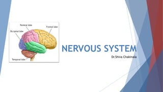
- 3. The two cerebral hemisphere are separated by a deep vertical fissure fissure These two halves separated fully at anterior and posterior and partially in the middle part due to the presence of Corpus callosum its is a connecting fibers of these two halves And each half of cerebral hemisphere divided into 4 lobes namely Frontal lobe Parietal lobe Temporal lobe Occipital lobe
- 4. The main fissures or sulci which separate these lobes are 1.Centaral sulcus is in between frontal and parietal lobe 2.Parieto occipital sulcus is in between parietal and occipital lobe 3.Lateral sulcus or sylvian fissure is in between parietal and temporal lobe 4.Callosomarginal fissure is in between temporal and limbic area of frontal lobe
- 6. Brodmann area: Brodmann areas are the regions of cerebral cortex based on its microscopic structures and organisation of cells Brodmann areas are 52 in number i.e from area no 1,2,3------to------area no 52
- 7. Frontal lobe: Frontal lobe is situated in between frontal pole and central sulcus inferiorly up to lateral sulcus It occupies 1/3 rd of cerebral cortex It has pre central gyrus,superior,middle,inferior frontal gyrus Functionally this lobe divided into two parts namely 1.Prefrontal cortex 2.Precentral cortex Precentral cortex further divided in to 3 parts 1.Primary motor area 2.Premotor area 3.Supplementary motor area
- 8. Prefrontal cortex: It is also called Orbito frontal cortex It is situated anteriorly in between anterior pole and precentaral are of frontal lobe It has lateral medial and inferior surfaces Areas present in pre frontal cortex are 9,10,11,12,13,14 on lateral surface 23,24,29,32 areas on medial surface Inferior surface has Orbital gyri 9 10 11 12
- 9. Connections of Prefrontal cortex: Afferent connections Receives fibers mainly from Dorsomedial nucleus of thalamus Hypothalamus Corpus striatum amygdala Mid brain Efferent connections Send fibers to Thalamus Hypothalamus Tegmentum Caudate nucleus Pons Temporal lobe Area 13 along with hippocampus send fibers to mammillary body via fornix this area related with Emotions
- 10. Functions of Prefrontal cortex: This area is center for higher functions like Emotions,learning,memory,social behaviour Short term memories stored Center for planned actions Center for intelligence Bilateral removal of these area leads to lack of mental alertness and lack of intiation very little changes in memory judgement and intelligence
- 12. Precentral cortex: It is posterior part of frontal lobe It is situated anteriorly in between central sulcus and prefrontal cortex ,posterior portion of middle,superior and inferior frontal gyri It extend to medial surface also It is also called excito motor cortex Precentral cortex again divided into Primary motor area Pre motor area Supplementary motor area
- 13. Primary motor area: This area present through out the area of pre central gyrus adjoining area of central sulcus Area no. 4 and 4s are present in it Connections of Primary motor area: Afferent connections Receives fibers from Dentate nucleus via red nucleus Thalamus Efferent connections Send fibers to Corticospinal tract to spinal cord of opposite side Frontopontine fibers to pontine nuclei of same side Corpus striatum Red nucleus Thalamus Subthalamus Reticular formation Association fibers to this area to other area of cortex
- 14. Functions of Primary motor area: This area is involved in voluntary movements and speech Area 4: Area situated just in front of central sulcus on pre central gyrus as a strip Area has broad end at superiorly Most of efferent fibers arised from primary motor area and these fibers involved in voluntary movements of opposite side on stimulation Area 4S: This is called suppressor area It is present in front of area no.4 on precentral gyrus It inhibited the movements created by the area no 4 so that it control exaggerated movements • Lesion in this area leads to paralysis of contralateral side(opposite side of body) • Lesion of this area both side leads to total paralysis • Recovery is late while recovering upper limb recovered first than lower limbs
- 15. 4 4s 6 8 44 45
- 16. Pre motor area: This area present anterior to primary motor area Area involved in postural movements of body sent efferent fibers to axial muscles of body Areas involved are 6,8,44 and 45 Area 6: Area no 6 divided in to 6a and 6b on frontal gyri, infront of area no 4 This area also send efferent fibers through corticospinal tract along with area 4 Functions of Area 6: Coardinate the movements intiated by area 4,that is it involved in skilled movements to perform accurate lesion of this area leads to loss of skilled movements opposite side Lesion along with area 4 leads to severe hemiplegia
- 17. Area 8: This area also called Frontal eye field Present anterior to the area no 6 It is concerned with eye ball movements Send fibers to i.e efferent connection with Occulomotor nuclei of midbrain Receive fibers from i.e afferent connection with Thalamus and Occipital lobe Functions of Area 8: this area involved in eye ball movements,open and closure of eyelids,dilatation of pupil and lacrimation of opposite side of eye lesion of this area leads to loss of eye ball movements but not effect eyelids and pupil
- 18. Area 44 and 45: It is also called as Broca’s area It is a Motor area of speech Situated on inferior frontal gyrus of frontal lobe Functions of Broca’s area: area responsible for movements of tongue,lips,and larynx which area involved in speech lesion of this area leads to Motor aphasia or Broca’s aphasia In this person has idea about his statement but he is unable to talk fluently Supplementary motor area: This area present medial surface of frontal lobe Exact function of this area not understood clearly It suggested that it is concerned with coordinate skilled movements
- 19. 4 4s 6 8 44 45
- 20. Homunculus arrangement of motor areas in cerebral cortex The muscles of various parts of body represent in motor area 4 from medial to lateral surface In this the lower part of body present at lateral surface and upper parts of body present at medial surface The order of representation from medial to laeral cortex is Toes-->ankle-->knee-->hip-->trunk-->shoulder-->arm-->elbow-->wrist-->hand-->fingers-->face From their respective are the efferent fibers send to respective muscles of opposite side except face Face receives efferents from both side cortex
- 22. Parietal lobe: Frontal lobe is situated in between frontal lobe,occipital lobe and temporal lobe separated by Central sulcus,parieto occipital sulcus and sylvian fissure respectively Parietal lobe divided into 3 areas Somesthetic area I Somesthetic area II Somesthetic association area Along with it has nother area present namely sensory motor area Parietal lobe mainly involved in sensory functions
- 23. Somesthetic area I: It is also called Primary somesthetic area or Primary sensory area It is situated posterior to the central sulcus on post central gyrus and on paracentral lobule of medial side Areas: It has three areas 3,1 and 2 Anteriorly on post central gyrus has area 3 and posteriorly has area 1 and 2 Connections: It receives afferent fibers from Thalamus as thalamic radiation
- 24. Functions of Somesthetic area I : It receives sensory impulses like pain,touch,temperature,pressure and proprioception from opposite side Area 1 involved in sensory perception and area 2 and 3 in integration of sensory impulses These area send feedback to pre motor area Apart from this it has also involved in Spatial recognition: Tactile localization and two point discrimination Recognition of intensity of stimuli Recognition of similarities and difference between stimuli If lesion in this area without involvement of thalamus leads to loss of discrimination but patient able perceive the stimuli If lesion in this area along with thalamus leads to loss of sensations opposite side
- 25. 1 2 3 7 5 Somesthetic area II Somesthetic area I Somesthetic Association area
- 26. Somesthetic area II: It is situated on post central gyrus below the somesthtic area I some part of this area present in sylvian fissure Connections: It receives afferent fibers from Thalamus and somesthetic area directly Exact role of this area not clear but it concerned with perception of sensation
- 27. 1 2 3
- 28. Somesthetic association area: It is situated posterior to post central gyrus infront of visual cortex and above auditory cortexmedial side Areas: It has areas 5 and 7 Anteriorly on post central gyrus has area 3 and posteriorly has area 1 and 2 Function: This area center for combined sesation like Stereognosis Lesion of this area leads to Astereognosis
- 30. Sensory motor area: Sensory area not limited to parietal lobe it extends in to motor area of pre central gyrus Similarly motor area not limited to frontal lobe it extends In to sensory area post central gyrus This combined area called as sensory motor area Timing and programming of skilled movents involved in Corticocerebellum are stored in sensory motor area
- 31. Homunculus arrangement of motor areas in cerebral cortex The various parts of body represent in primary sensory area from medial to lateral surface In this the lower part of body present at lateral surface and upper parts of body present at medial surface The order of representation from medial to laeral cortex is Toes-->ankle-->knee-->hip-->trunk-->shoulder-->arm-->elbow-->wrist-->hand-->fingers-- >face(eyelids-->nose-->cheek-->upper lip-->lower lip)
- 34. Temporal lobe: Temporal lobe is situated below the fronto parietal lobe Temporal includes 3 areas Primary auditory area Auditopsychic area Area for equilibrium
- 35. Primary auditory area: This area include area no 41,42 41’42areas are present on anterior transvers gyrus and anterior part of superiortemporal gyrus Connections: This area receives afferent fibers from Medial geniculate body and Pulvinar Send efferent fibers to medial geniculate body,inferior colliculus of mid brain and pulvinar Functions of primary auditory area: primary auditory area involved in perception of auditory stimulus but interpretation of sound i.e analysis of speech source of sound is done with the help of Auditopsychic area
- 36. Auditory psychic area: This area include area no 22 This is also called wernick’s area It is situated on superior temporal gyrus posterior to area no 41,42 It is helpful in interpretation of sound It is a sensory area of speech Lesion of this area leads to Sensory aphasia or Wernick’s aphasia In this patient able to talk clearly but not related Area for equilibrium: It is situated posterior to superior temporal gyrus It is helpful maintainence of equilibrium Stimulation of this area leads to dizziness,swaying,falling and feeling of rotation of body 22
- 37. 22 4241
- 38. Occipital lobe: It is posterior part of cerebral cortex Temporal include visual cortex Visual cortex includes Primary visual area----- area no.17 Visual association area--area no.18 Occipital eye field-------area no.19 Connections: It receives afferent fibers from latearal geniculate body Send efferents to superior colliculus and lateral geniculate body
- 39. Functions of Visual cortex : Primary visual are involved in perception of visual impulses Visual association area involved in interpretation of visual impulses Occipital eye field concerned with movement of eyes 17 18 19
- 41. Histology of cerebral cortex Cerebral cortex formed by nerve cells along with their processes and neuroglia it does not have uniform thickness Thickest part is at Precentral gyrus i.e 4.5cms and thinnest part is at frontal and occipital poles Cerebral cortex is formed by six layers (from out side to in side) Molecular or plexiform layer External granular layer Outer pyramidal layer Internal granular layer Internal pyramidal layer or Ganglionic layer Fusiform cell layer
- 43. THANK YOU..!