
Recommended
More Related Content
Similar to Frontal Lobe.pptx
Similar to Frontal Lobe.pptx (20)
Recently uploaded
Recently uploaded (20)
Frontal Lobe.pptx
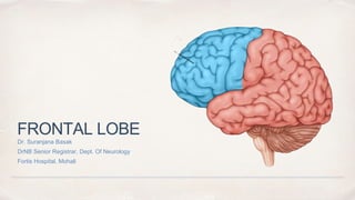
- 1. FRONTAL LOBE Dr. Suranjana Basak DrNB Senior Registrar, Dept. Of Neurology Fortis Hospital, Mohali
- 3. ✤ Precentral gyrus (motor strip) lies just anterior to central sulcus ✤ On medial surface, frontal lobe extends to cingulate sulcus ✤ Paracentral lobule = extensions of pre central + post central gyri onto the medial hemisphere above cingulate sulcus. Bladder control. Anatomy Medial Surface Lateral Surface
- 4. Important Areas ✤ Supplementary motor & premotor regions - area 6 anterior to pre central gyrus. ✤ Premotor area area - anterior to primary motor cortex ✤ Frontal eye fields in middle frontal gyrus ✤ Inferior frontal gyrus : i. Pars Orbitalis ii. Pars Triangularis iii. Pars Opercularis
- 5. Inferior surface ✤ Pars Opercularis and Triangularis (Dominant) - Motor Speech Area/ Bro’s ( Areas 44,45) ✤ Inferior surface medial to inferior frontal gyrus = Orbital Gyri ✤ Separated by olfactory sulcus from gyrus rectus = most medial structure on orbital surface ✤ Olfactory bulbs and tracts overlie the olfactory sulcus
- 6. Frontal Lobe Functional Anatomy ✤ Motor Cortex i. Primary (BA 4) ii. Premotor (BA 6 Lateral) iii. Supplementary (BA 6 Medial) iv. Frontal Eye Field (BA 8) v. Broca’s Speech Area (BA 44,45) ✤ Prefrontal cortex i. Dorsolateral prefrontal cortex (DLPFC) ii. Medial prefrontal cortex (MPC) iii. Orbitofrontal cortex (OFC)
- 7. Blood Supply of Frontal Lobe Blue- Anterior Cerebral Artery, Pink - Middle Cerebral Artery, Yellow - Posterior Cerebral Artery Lateral Medial
- 8. Primary Motor Cortex M1/Area 4 ✤ Location: Anterior wall of central sulcus & adjacent portions of pre central gyrus ✤ Lowest threshold for stimulation to cause muscle contraction of opposite side of body ✤ Cortex of M1 - Agranular, heterotypical. Most characteristic feature - presence of giant pyramidal neurone (Betz cells) in lamina V ✤ Localisation of function within the pre central gyrus is depicted by motor homunculus Area Gigantopyramidalis
- 9. Motor Homunculus ✤ Homunculus - distorted figure with size of an anatomical part proportional to the amount of cortex to which it is related. ✤ The phylogenetic acquisition of speech & complex hand function has caused expansion of cortical areas representing the tongue, mouth, lips, thumb & fingers ✤ Neurons controlling the lower extremities and perineal musculature are in the paracentral lobule, which plays an important role in bowel and bladder
- 10. Functions of Primary Motor Cortex ✤ Conscious control of voluntary movement ✤ Encodes parameters defining individual movements or simple movement sequences ✤ Encodes force, direction, extent and speed of the movement
- 11. Area 6 ✤ Similar to Area 4 except for absence of giant bets cells. But much greater developed layer 3 ✤ Regulation of autonomic and motor processes ✤ Stimulation of area 6 and area 8 cause deviation of head and eyes to opposite side and complex limb movements ✤ Depression or inhibition of motor activity at lower levels of brain ✤ Central regulation of autonomic processes ✤ Vogt and Vogt (1935) observed that extirpation of the premotorcortex does not cause permanent paralysis, but leads to disturbance of skilled movements & appearance of spasticity & rigidity of movements, grasping reflexes and vasomotor disorders. ✤ Lesions of the more anterior and medial parts of the motor cortex cause less paralysis and more spasticity and may allow the emergence of primitive reflexes, such as grasping and groping responses.
- 12. Pre Motor Cortex ✤ Location: Anterior to primary cortex between pre central gyrus and posterior border of prefrontal area (area 6) ✤ Planning & execution of movements (basis of Luria’s hand sequence/fist-edge-palm test) ✤ Receives afferents from other areas of the cortex incl. sensory cortex and projects to the motor cortex and motor thalamus ✤ Some fibres descend and make up a part of extrapyramidal system
- 13. Functions of Pre Motor Cortex ✤ Sensorimotor integration ✤ Lesions cause inability to make use of sensory feedback in the performance of smooth movements ✤ Performs more complex, task related processing than primary motor cortex ✤ Controls learned, repetitive or patterned motor skills ✤ Lesion: Inability to make use of sensory feedback in performance of smooth movements and apraxia
- 14. Supplementary Motor Area ✤ Areas of cortex lying on medial aspect of hemisphere just anterior to primary motor cortex ✤ Planning motor movements such as a sequence of actions provided from memory ✤ Crucial for temporal organization of multiple movements ✤ Lesions of SMA impair memory based sequencing of movements ✤ Also coordinates movements between hands and lesions in this area may cause Alien Hand syndrome
- 15. Syndrome of SMA ✤ Not well recognized and can easily be confused with corticospinal weakness. ✤ Patients have reduced spontaneous movements and difficulty in performing volitional motor acts to command in the contralateral limbs, although the limbs function normally in automatic motor activities, for example, dressing ✤ Lesion: Mutism, contralateral motor neglect, impairment of bibrachial coordination
- 16. Other Clinical Aspects ✤ Seizures may arise as simple partial or complex partial. Seizures arising from motor cortex typically produce focal Jacksonian epilepsy of contralateral limbs ✤ Partial complex seizures arising from frontal lobe are bizarre and likely confused with pseudoseizures ✤ M1 receives association fibres from premotor and supplementary motor areas and insula ✤ These connections are involved in preparation and planning for voluntary movements that are then executed by the primary motor cortex
- 18. Decoding a Motor Command 1. Reception and decoding of a motor command in Wernickes area 2. Transmission through the arcuate fasciculus 3. To the left premotor area 4. To the left motor cortex 5. Transmission information across the corpus callosum 6. To the right premotor area 7. To the right motor cortex Apraxic syndromes may occur with lesions along these pathways
- 19. Frontal Eye Field / Area 8 ✤ Located anterior to the premotor cortex and superior to Broca’s area in caudal part of middle frontal gyrus(BA-8) ✤ Centre for voluntary eye movements independent of visual stimuli ✤ Participates in initiation of voluntary horizontal saccades ✤ The FEF controls horizontal conjugate saccades to the opposite side ✤ Left FEF initiates a command to look right. ✤ Lesion: Eyes deviate ipsilaterally with destructive lesion & contralaterally with irritating lesions
- 20. ✤ The saccadic pathway arises in the frontal lobe and descends to the contralateral pons ✤ A left FEF-initiated command to look right is thus transmitted down to the right PPRF, which simultaneously influences the right sixth nerve to contract the lateral rectus and the left third nerve to contract the yoked medial rectus—both contract, according to Hering’s law, exactly the same amount
- 21. Broca’s Area/ Area 44 ✤ Location: Inferior frontal gyrus. ✤ In the majority of individuals, this area lies on the left or dominant hemisphere. ✤ Input: Wernicke’s area ✤ Output: primary motor cortex ✤ Function: speech production (dominant hemisphere); emotional, melodic component of speech (non-dominant) ✤ Lesions: motor aphasia
- 22. Broca’s Aphasia ✤ Speech : Nonfluent, labored, interrupted by many word-finding pauses, and usually dysarthric. ✤ It is impoverished in function words but enriched in meaning-appropriate nouns. ✤ Speech is telegraphic but quite informative. ✤ Output may be reduced ✤ In addition, fluency, naming and repetition are impaired. ✤ Comprehension of spoken language is intact except for syntactically difficult sentences with a passive voice structure or embedded clauses, indicating that Broca’s aphasia is not just an “expressive” or “motor” disorder and that it also may involve a comprehension deficit in decoding syntax. ✤ The cause is most often infarction of Broca’s area (the inferior frontal convolution and surrounding anterior perisylvian and insular cortex due to occlusion of the superior division of the MCA). Mass lesions, including tumor, intracerebral hemorrhage, and abscess, also may be responsible.
- 23. Prefrontal Cortex ✤ Location: Anterior to area 6, area 8, and the motor speech centers are areas referred to as the prefrontal cortex. It includes areas 9 to 12, 32, 45, 47, and others. ✤ These areas are connected with the somesthetic, visual, auditory, and other cortical areas by long association bundles and with the thalamus and the hypothalamus by projection fibers. ✤ It is the main projection site for the dorsomedial nucleus of the thalamus.
- 24. Clinically PFC divided to: ✤ Dorsolateral Prefrontal Cortex (DLPFC) ✤ Medial Prefrontal Cortex (MPC) ✤ Orbitofrontal Cortex (OFC)
- 25. Dorsolateral Prefrontal Cortex (DLPFC) ✤ DLPFC is important in the organization of self-ordered tasks. It plays a critical role in the neural network subserving working memory ✤ The responsibility for executive function largely resides with the DLPFC and its connections. ✤ Plan, carry out, and monitor a series of actions intended to accomplish a goal. Planning and organizational skills, the ability to benefit from experience, abstraction, motivation, cognitive flexibility, and problem solving. ✤ Role as well in the ability to predict the consequences of actions, emotional expression (affect), “go/no-go” decision making, personality, and the sense of time
- 26. ✤ The DLPFC is also important in oculomotor control, which is responsible for decision making regarding voluntary eye movements and inhibiting unwanted reflex saccades. ✤ The dorsolateral prefrontal cortical area may be involved in mechanisms responsible for inhibiting unwanted saccades. Antisaccades are voluntary saccades away from a target ✤ Patients with frontal lobe disease, progressive supranuclear palsy, Parkinson’s disease, Alzheimer’s disease, and schizophrenia, when asked to look away from a visual stimulus, may be unable to inhibit a saccade toward the target (prosaccade) and are therefore unable to make an antisaccade or make it only after a prosaccade. Dorsolateral Prefrontal Cortex (DLPFC)
- 27. Medial Prefrontal Cortex (MPC) ✤ MPC has connections with the several thalamic nuclei, particularly the dorsomedian, and with the superior temporal cortex. There are connections with other portions of the frontal lobe, including the OFC, the DLPFC and the medial motor areas. The MPC is important in auditory and visual associations. ✤ The ventrolateral prefrontal cortex is concerned with mnemonic processing of objects. ✤ The anterior cingulate gyrus circuit is associated with motivation, will power, initiation of activity
- 28. Orbitofrontal Cortex (OFC) ✤ OFC has important connections with the limbic system, including the amygdala. ✤ Emotional Behaviour ✤ Arousal ✤ Inhibition ✤ Suppression of distracting signals ✤ Patients with OFC dysfunction are also prone to display emotional lability, poor judgment and insight, and distractibility.
- 29. Frontal Subcortical Circuits ✤ Five frontosubcortical circuits described by Goldman Rakic subserve cognition, behavior and movement. 1. Motor 2. Oculomotor 3. Dorsolateral prefrontal 4. Lateral orbitofrontal 5. Anterior cingulate Information originates from cerebral cortex -> travels to basal ganglia -> thalamus -> return to cortex
- 30. ✤ Supplementary Motor & Premotor: planning, initiation & storage of motor programs; fine-tuning of movements ✤ Motor:final station for execution of the the movement according to the design SMA, Premotor,Mo tor Putamen VL Globus Pallidus VL, VA, CM Thalamus Motor Circuit
- 31. ✤ Independent of visual stimuli ✤ Voluntary scanning eye movement Frontal Eye Field Central Caudate DM Globus Pallidus Substantia Nigra VA, MD Thalamus Oculomotor Circuit
- 32. ✤ Executive functions: motor planning, deciding which stimuli to attend to, shifting cognitive sets ✤ Lesions will cause poor organizational strategies, poor memory search strategies, stimulus bound behaviour, environmental dependency Lateral Prefrontal DL Caudate DM Globus Pallidus Substantia Nigra VA Thalamus DLPFC Circuit
- 33. ✤ Connects frontal monitoring functions to the limbic system. ✤ This circuit governs appropriate responses to social cues, empathy, social judgement, and interpersonal sensitivity, ✤ Dysfunction of this circuit can lead to disinhibition, irritability, aggressive outburst and inappropriate social response, OCS, emotional incontinence VM Caudate DM Globus Pallidus Substantia Nigra MD Thalamus Orbito-frontal Orbitofrontal Circuit
- 34. ✤ Anterior cingulate gyrus involved in motivated behaviour ✤ Lesions in this circuit result in apathy, abulia, impaired motivation, akinetic mutism, poverty of speech, psychic emptiness, poor response inhibition Anterior Cingulate Gyrus Ventral Striatum RL Globus Pallidus Substantia Nigra MD Thalamus Anterior Cingulate Circuit
- 35. Executive function ✤ Defined as ‘complex set of cerebral process that operate in non-routine situations and exert top- down volitional control over cognition and behavior’. ✤ First order process – working memory, inhibition, monitoring and initiation ✤ Second order executive functions include: planning, organization, resisting interference, set shifting, affect regulation, and context appropriate behavior.
- 36. Working memory ✤ Working memory refers to the temporary maintenance of information that was just experienced or just retrieved from long term memory. ✤ Rt prefrontal cortex is engaged in spatial working memory task and left is engaged in verbal working memory task. ✤ Ventrolateral pre frontal cortex involved in the maintanace aspect of working memory (holding information online)
- 37. Monitoring ✤ Dorsolateral prefrontal cortex monitor information held with in the working memory. ✤ Anterior cingulate gyrus monitor one’s own performance. It plays an important role in performance monitoring in relation to anticipated rewards, requiring both continuous evaluation of current behaviour and future outcomes associated with behaviour.
- 38. Initiation ✤ Medial frontal structures including supplementary motor area, presupplementary motor area and anterior cingulate gyrus – play an important role in drive, motivation and the initiation of behaviour. ✤ Medial frontal damage – impairments in initiation of speech and motor movements ✤ B/L damage of ant cingulate- Akinetic mutism
- 39. Second order executive functions ✤ Planning requires heavy working memory demands. Initial step- generating and holding within the working memory one or more possible future events. ✤ Organization – ordering or manipulation of objects, concepts, or responses, such that the individual items are eventually arranged in a manner conforms to higher order logic. ✤ Set shifting- ability to intentionally disengage from, current activity and reengage in different way of thinking and activity.
- 41. ATTENTION ✤ DIGIT SPAN FORWARD ✤ repeat 3-5; 7-5-8; 3-9-4-8 ✤ Tests attention , concentration , immediate memory ✤ Expected performance – 7+/- 2forward ✤ Another test of attention and concentration is a three-step task. For instance, tear a piece of paper in half, then tear half of it in half, then tear one half in half again, so that there are three different sizes. Give the patient an instruction such as, “Give the large piece of paper to me, put the small piece on the bed, and keep the other piece.”
- 42. Working Memory ✤ BACKWARD DIGIT SPAN ✤ EP – 5+/-1 ✤ Reverse digit span should not be more than 2 digits less than forward span. ✤ SERIAL 7s ✤ SPELL WORD BACKWARDS ✤ SAY MONTHS OF THE YEAR BACKWARDS ( Normal < 30 sec)
- 43. ABSTRACT THINKING (Ex) ✤ SIMILARITIES & DIFFERENCES ✤ FIND ANALOGIES ✤ INTERPRET PROVERBS ✤ Eg: “Don’t cry over splilt milk” ✤ A stich in time saves nine
- 44. JUDGEMENT what she would do if she found a sealed, addressed, stamped letter on the sidewalk, or if she smelled smoke in a crowded theater Historical information from family members about the patient’s actual judgment in reallife situations
- 45. FAB
- 46. Lexical Fluency
- 47. LURIA 3 STEP TEST (SET SHIFTING)
- 48. Fist Edge Palm Test Gade
- 49. Conflicting Instructions (sensitivity to interference) • Tap twice when I tap once. • To ensure that the patient has understood the instruction, a series of 3 trials is run: 1-1-1. • Tap once when I tap twice. • To ensure that the patient has understood the instruction, a series of 3 trials is run: 2-2-2. • The examiner then performs the following series: 1-1-2-1- 2-2-2-1-1-2. • Score No errors: 3, 1 -2 errors: 2, > 2 errors: 1
- 50. Go – No Go (Response inhibition) • “Tap once when I tap once.” • “Do not tap when I tap twice.” • Score No errors: 3, 1 -2 errors: 2, > 2 errors: 1 • Patient taps like the examiner at least four consecutive times: 0
- 51. Prehension Behaviour • “Do not take my hands.” • The examiner is seated in front of the patient. Place the patient’s hands palm up on his knees. • Without saying anything or looking at the patient, the examiner brings his own hands close to the patient’s hands and touches the palms of both the patient’s hands, to see if he will spontaneously take them. • Patient does not take the examiner’s hands: 3 • Patient hesitates and asks what he/she has to do: 2 • Patient takes the hands without hesitation: 1 • Patient takes the examiner’s hand even after he/she has been told not to do so: 0
- 52. Interpreting Results • A cut off score of 12 on the FAB has a sensitivity of 77% and specificity of 87% in differentiating between frontal dysexecutive type dementias and AD.
- 54. Wisconsin Card Sorting Test Please sort the 60 cards under the 4 samples. I won’t tell you the rule, but I will announce every mistake. The rule will change after 10 correct placements.”
- 55. WCST ✤ After ten (or six) consecutive correct sorts, the sorting principle is changed ✤ Responding in accordance to the previous correct rule is considered a perseverative error ✤ Provides a measure of the ability to identify abstract categories and shift cognitive set
- 56. PERSEVERATION
- 58. STROOP TEST ✤ Part 1: read aloud color names printed in black ink ✤ Part 2: the patient has to name the color of dots ✤ Part 3: presented with color names printed in color ink. The color of the ink does not correspond to the color name. Subject must name the color of the ink. ✤ ( response inhibition or ability to overcome cognitive interference )
- 59. PURPLE RED BLUE YELLOW GREEN BLACK YELLOW BLACK GREEN PURPLE BLUE RED _______________________________________________ XXXXX XXXXX XXXXX XXXXX XXXXX XXXXX XXXXX XXXXX XXXXX XXXXX XXXXX XXXXX _______________________________________________ RED BLACK GREEN BLUE PURPLE YELLOW BLUE PURPLE RED BLACK YELLOW GREEN _____________________________________________ One must resist automatic tendency to read target word (red) and instead engage in the required behaviour of naming the ink colour
- 60. FRONTAL RELEASE SIGNS ✤ The grasp reflex is obtained when the examiner's hand is gently inserted into the palm of the patient's hand. The grasp reflex is fairly specific for a lesion of the supplementary motor area on the medial surface of the contralateral frontal lobe ✤ The snout reflex is brought about by tapping the upper lip lightly. The contraction of the muscles causes the mouth to resemble a snout ✤ The palmomental reflex occurs when a disagreeable stimulus is drawn from the thenar eminence at the wrist up to the base of the thumb. There is ipsilateral contraction of the orbicularis oris and mentalis muscles
Editor's Notes
- Area 32 - Dorsal anterior cingulate cortex Area 33 - Part of anterior cingulate cortex Area 24 - Ventral Anterior cingulate cortex.